Achieving Chromosome-Level Assembly: A Comprehensive Guide to Hi-C Scaffolding Techniques and Best Practices
This article provides a detailed exploration of Hi-C scaffolding for achieving chromosome-level genome assemblies, targeted at genomics researchers and bioinformatics professionals.
Achieving Chromosome-Level Assembly: A Comprehensive Guide to Hi-C Scaffolding Techniques and Best Practices
Abstract
This article provides a detailed exploration of Hi-C scaffolding for achieving chromosome-level genome assemblies, targeted at genomics researchers and bioinformatics professionals. It covers foundational principles of chromatin conformation capture, step-by-step methodologies using popular tools like Juicer, 3D-DNA, and SALSA, common troubleshooting scenarios for data quality and mis-assemblies, and comparative analysis of validation metrics and alternative technologies. The content synthesizes current best practices to empower researchers to generate contiguous, biologically accurate reference genomes for advanced biomedical and drug discovery applications.
Hi-C Scaffolding Fundamentals: From Chromatin Loops to Chromosome Maps
What is Chromosome-Level Assembly and Why Does It Matter for Biomedical Research?
Chromosome-level assembly represents the highest standard in genome sequence reconstruction, where fragmented genomic sequences are ordered, oriented, and grouped into complete chromosomes. Unlike draft assemblies composed of thousands of unordered contigs, chromosome-level assemblies provide a complete, accurate, and gapless view of an organism's genome, including centromeres, telomeres, and long repetitive regions. In the context of our broader thesis on Hi-C scaffolding, achieving chromosome-level assembly is the ultimate goal, enabling transformative insights in biomedical research, from understanding genetic disease mechanisms to accelerating drug target discovery.
Defining Chromosome-Level Assembly: Metrics and Benchmarks
Chromosome-level assembly is quantified using specific continuity, completeness, and accuracy metrics.
Table 1: Key Metrics for Assessing Assembly Quality
| Metric | Definition | Target for Chromosome-Level |
|---|---|---|
| N50 | The contig/scaffold length such that 50% of the total assembly length is contained in sequences of this size or longer. | Scaffold N50 should be on the order of chromosome length (e.g., >100 Mb for human). |
| NG50 | Similar to N50 but calculated against the estimated genome size rather than the assembly size. | High NG50 indicates assembly spans major chromosomal regions. |
| Number of Scaffolds | Total count of contiguous sequences, including gaps. | Should approach the haploid chromosome number. |
| BUSCO Score | Benchmarking Universal Single-Copy Orthologs; assesses completeness based on evolutionarily conserved genes. | Typically >95% for a complete assembly. |
| QV (Quality Value) | A log-scaled measure of base-level accuracy (e.g., QV40 = 99.99% accuracy). | QV > 40 is considered high quality. |
| L50 | The minimal number of contigs/scaffolds whose length sum produces N50. | A low L50 (close to chromosome count) indicates high continuity. |
The Hi-C Scaffolding Protocol for Chromosome-Level Assembly
This detailed protocol is central to our thesis, enabling the scaffolding of draft assemblies into chromosome-scale models using chromatin conformation capture data.
Protocol: Hi-C Scaffolding for Chromosome-Level Assembly
I. Sample Preparation and Crosslinking
- Material: Grow cells to ~80% confluence. Use ~1-5 million cells per Hi-C library.
- Fixation: Add fresh formaldehyde to culture media to a final concentration of 1-3%. Incubate at room temperature for 10-20 minutes with gentle agitation.
- Quenching: Add glycine to a final concentration of 0.125-0.25 M. Incubate for 5 minutes at room temperature.
- Wash: Pellet cells and wash twice with cold PBS. Pellet can be flash-frozen and stored at -80°C.
II. Chromatin Digestion and Biotinylation
- Lysis: Resuspend cell pellet in ice-cold lysis buffer (10 mM Tris-HCl pH 8.0, 10 mM NaCl, 0.2% Igepal CA-630) with protease inhibitors. Incubate on ice for 15-30 mins.
- Digestion: Wash nuclei and resuspend in appropriate restriction enzyme buffer. Add a frequent-cutter restriction enzyme (e.g., DpnII, MboI, HindIII). Incubate at 37°C for 2+ hours.
- Marking Ends: Fill restricted ends and label with biotin-14-dATP using Klenow fragment. Incubate at 37°C for 45-60 mins.
III. Ligation and DNA Purification
- Dilute & Ligate: Dilute digested material in ligation buffer to favor intramolecular ligation. Add T4 DNA Ligase. Incubate at 16°C for 4+ hours.
- Reverse Crosslinks: Add Proteinase K and incubate at 65°C overnight.
- Purify DNA: Perform phenol-chloroform extraction and ethanol precipitation.
IV. Hi-C Library Preparation for Sequencing
- Shearing: Sonicate DNA to ~300-500 bp fragments.
- Pull-down: Bind biotinylated fragments to streptavidin-coated magnetic beads.
- End Repair & A-tailing: Prepare fragments for adapter ligation using standard kits.
- Adapter Ligation: Ligate sequencing adapters to bead-bound fragments.
- PCR Amplification: Perform on-bead PCR (typically 10-14 cycles) to generate the final sequencing library. Quantify and validate fragment size.
V. Data Processing and Scaffolding
- Read Mapping: Map paired-end reads to the draft genome assembly using an aligner like BWA-MEM or HiC-Pro, keeping read pairs separate.
- Contact Matrix Generation: Parse aligned reads, filter by quality, and generate a genome-wide contact frequency matrix using tools like Juicer or HiCExplorer.
- Scaffolding & Ordering: Feed the contact matrix and draft assembly into a scaffolder (e.g., 3D-DNA, SALSA2, YaHS). These tools use the higher frequency of contacts within a chromosome versus between chromosomes to cluster, order, and orient contigs.
- Manual Curation: Use visualization tools (e.g., Juicebox, Pretext) to manually review and correct scaffolding errors, such as misjoins or misorientations, leveraging the contact map as a guide.
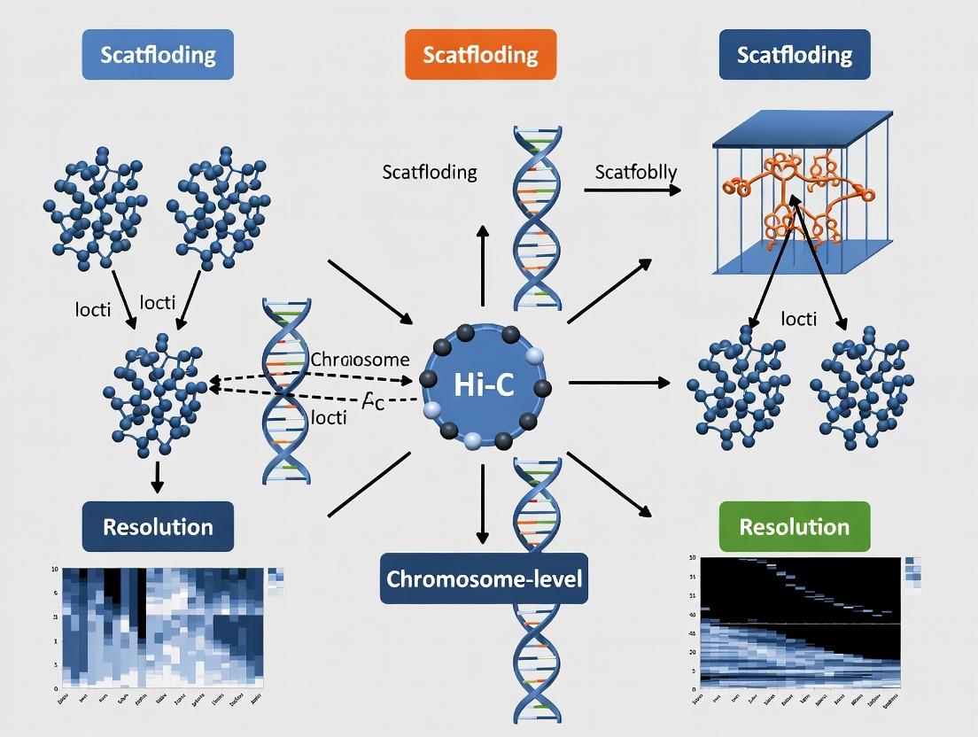
Title: Hi-C Scaffolding Experimental Workflow
The Scientist's Toolkit: Key Research Reagent Solutions
Table 2: Essential Reagents and Kits for Hi-C Scaffolding
| Item | Function in Protocol | Example Product/Supplier |
|---|---|---|
| Formaldehyde | Crosslinks proteins to DNA, freezing chromatin 3D structure. | Thermo Scientific, 16% methanol-free. |
| Frequent-Cutter Restriction Enzyme | Digests crosslinked DNA, defining Hi-C contact resolution. | DpnII, MboI, HindIII (NEB). |
| Biotin-14-dATP | Labels digested DNA ends for selective pull-down of ligation junctions. | Jena Biosciences, Biotin-14-dATP. |
| Streptavidin Magnetic Beads | Captures biotinylated Hi-C ligation junctions during library prep. | Dynabeads MyOne Streptavidin C1 (Invitrogen). |
| T4 DNA Ligase | Performs proximity ligation of crosslinked DNA fragments. | T4 DNA Ligase (NEB). |
| Hi-C Library Prep Kit | Optimized, all-in-one reagents for streamlined library construction. | Arima-HiC+ Kit, Dovetail Omni-C Kit. |
| High-Fidelity PCR Mix | Amplifies the final library with minimal bias for sequencing. | KAPA HiFi HotStart ReadyMix (Roche). |
Biomedical Applications Enabled by Chromosome-Level Assemblies
Table 3: Impact of Chromosome-Level Assemblies on Biomedical Research
| Application Area | Specific Benefit | Example Use Case |
|---|---|---|
| Disease Gene Mapping | Enables accurate identification of structural variants (SVs), non-coding mutations, and regulatory elements linked to disease. | Discovering pathogenic SVs in neurodevelopmental disorders from whole-genome sequencing cohorts. |
| Cancer Genomics | Provides a complete view of chromosomal rearrangements, amplifications, and deletions driving oncogenesis. | Characterizing complex chromothripsis events and circular extrachromosomal DNA (ecDNA) in tumors. |
| Pharmacogenomics | Improves understanding of genetic variation in drug-metabolizing enzymes and transporters across populations. | Building reference pangenomes to identify ancestry-specific variants affecting drug response. |
| Immunogenetics | Allows full characterization of highly polymorphic and repetitive regions like the Major Histocompatibility Complex (MHC). | Studying the link between MHC haplotype diversity and autoimmune disease susceptibility. |
| Microbiome & Pathogen Research | Reveals virulence gene organization, antibiotic resistance islands, and mobile genetic elements in bacterial genomes. | Tracking plasmid-mediated spread of antimicrobial resistance in hospital outbreaks. |
Title: From Assembly to Biomedical Application Pathways
Chromosome-level assembly, achieved through integrated methods like Hi-C scaffolding as detailed in our thesis, is not merely a technical milestone but a foundational resource for modern biomedical research. It transforms the genome from a fragmented list of parts into a precise, navigable map of chromosomes. This complete genomic context is indispensable for uncovering the genetic basis of disease, understanding cancer evolution, developing targeted therapies, and realizing the promise of personalized medicine. As sequencing costs decline and scaffolding algorithms improve, generating chromosome-level references will become standard, dramatically accelerating discovery across the life sciences.
In the pursuit of complete and accurate genome sequences, chromosome-level assembly represents the gold standard. Hi-C (High-throughput Chromosome Conformation Capture) scaffolding is a pivotal technique that leverages three-dimensional genomic proximity data to order and orient contigs into scaffolds, ultimately reconstructing entire chromosomes. The core principle hinges on the fact that sequences physically close in the 3D nuclear space, regardless of their linear genomic distance, are more likely to be ligated together during the Hi-C protocol. This application note details the underlying principles, protocols, and analytical workflows for generating and interpreting Hi-C data specifically for scaffolding applications.
Core Biochemical Principle: Capturing Spatial Proximity
The Hi-C experiment transforms spatial proximity information into a readable DNA library. The process begins with cells whose genomic DNA is cross-linked using formaldehyde, freezing chromosomal interactions in place. The DNA is then digested with a restriction enzyme, creating fragments with sticky ends. These ends are filled with nucleotides, including a biotinylated residue, and ligated under dilute conditions that favor intramolecular ligation between cross-linked fragments. This creates chimeric DNA molecules linking two genomic loci that were in close spatial proximity. After reversing cross-links and purifying the DNA, the biotinylated junctions are enriched and processed into a sequencing library.
Quantitative Data from a Typical Hi-C Scaffolding Experiment
Table 1: Expected Metrics from Hi-C Library Preparation and Sequencing for Scaffolding
| Metric | Target Range for Scaffolding | Purpose/Interpretation |
|---|---|---|
| Cross-linking Efficiency | >90% | Ensures spatial contacts are preserved during digestion. |
| Digestion Efficiency | >80% | Critical for resolution; incomplete digestion creates large, uninformative fragments. |
| Ligation Efficiency | >70% | Directly impacts library complexity and usable data yield. |
| % Valid Read Pairs | 50-80% | Paired-end reads mapping to two different restriction fragments; the primary signal. |
| Library Complexity | >10M Unique Contacts | Necessary for robust statistical inference of contig adjacency. |
| Sequencing Depth | 20-50x Genome Coverage | Balances cost and ability to link contigs across repeats. |
| % Intra-chromosomal Contacts | >85% (for intact nuclei) | Indicator of sample quality; high inter-chromosomal noise hinders assembly. |
| Contact Map Resolution | 1-100 kb | Determined by restriction enzyme choice and sequencing depth; finer resolution aids complex assemblies. |
Table 2: Key Output Metrics from Hi-C Scaffolding Software (e.g., SALSA, LACHESIS, YaHS)
| Software Output Metric | Description | Ideal Outcome |
|---|---|---|
| Scaffold N50 | Length at which 50% of the assembly is contained in scaffolds of this size or longer. | Dramatic increase over contig N50 (e.g., 10x). |
| Number of Scaffolds | Total count of ordered and oriented sequences. | Should approach the haploid chromosome number. |
| Misjoin Rate | Percentage of scaffold joins not supported by other evidence (e.g., genetic map). | < 1%. |
| % Anchored Genome | Proportion of the assembly assigned to chromosomes. | > 90%. |
| Long-range Contact Support | Consistency of Hi-C contact frequency across scaffold joins. | Smooth contact matrix with distinct diagonal. |
Detailed Experimental Protocol: In-situ Hi-C for Scaffolding
Principle: This protocol, adapted from Lieberman-Aiden et al. (2009) and updated with modern practices, is performed with intact nuclei to minimize spurious inter-chromosomal contacts.
Protocol: In-situ Hi-C Library Generation
Materials: Fresh or frozen tissue/cells, Formaldehyde (37%), Quenching Solution (2.5M Glycine), Cell Lysis Buffer, Restriction Enzyme (e.g., DpnII, HindIII, MboI), Biotin-14-dATP, Klenow Fragment, T4 DNA Ligase, Streptavidin Beads, SDS, Proteinase K.
Day 1: Cross-linking & Digestion
- Cross-link: Suspend 1-2 million cells in growth medium. Add formaldehyde to 1-2% final concentration. Incubate for 10 min at room temperature with gentle rotation.
- Quench: Add glycine to 125mM final concentration. Incubate 5 min at RT, then 15 min on ice.
- Pellet & Wash: Pellet cells, wash twice with cold PBS.
- Lyse Cells: Resuspend pellet in 500 µL ice-cold Lysis Buffer (10mM Tris-HCl pH8.0, 10mM NaCl, 0.2% Igepal CA-630, protease inhibitors). Incubate 15 min on ice. Pellet nuclei (2,500 x g, 5 min). Wash once with 500 µL ice-cold 1x Restriction Enzyme Buffer.
- In-situ Digestion: Resuspend nuclei in 100 µL 1x Restriction Buffer. Add 0.5% SDS and incubate 10 min at 65°C. Immediately add 2% Triton X-100 to quench SDS. Add 200-400 units of chosen restriction enzyme. Incubate 2 hours at 37°C with gentle agitation.
Day 1: Fill-in & Ligation
- Fill-in & Biotinylate: To the digest, add 30 µL of Fill-in Master Mix (0.25 mM each dCTP, dGTP, dTTP, 0.15 mM Biotin-14-dATP, 1x NEB Buffer 2, 25 U Klenow Fragment). Incubate 45 min at 37°C.
- Ligate: Add 663 µL of Ligase Master Mix (1x NEB T4 Ligase Buffer, 1% Triton X-100, 0.1 mg/mL BSA, 2000 U T4 DNA Ligase). Incubate for 2 hours at 16°C.
Day 2: DNA Purification & Shearing
- Reverse Cross-links: Add 50 µL of 10% SDS and 25 µL of 20 mg/mL Proteinase K. Incubate at 65°C overnight.
- Purify DNA: Perform a standard phenol:chloroform:isoamyl alcohol extraction followed by ethanol precipitation.
- Shear DNA: Resuspend DNA in 130 µL TE. Shear to ~300-500 bp using a Covaris S2 or similar sonicator.
- Size Selection: Perform a double-sided SPRI bead cleanup (e.g., 0.5x and 1.5x ratios) to select ~300-600 bp fragments.
Day 2: Biotin Pulldown & Library Prep
- Biotin Enrichment: Set up a Streptavidin bead pull-down. Bind sheared DNA to 10 µL pre-washed Streptavidin C1 beads in 1x B&W Buffer for 15 min at RT.
- Wash: Wash beads twice with 1x B&W Buffer, once with 10mM Tris-HCl pH 8.0.
- On-bead End Repair & A-tailing: Perform standard NEB Next Ultra II end repair/dA-tailing reactions directly on the beads.
- Adapter Ligation: Ligate Illumina-compatible adapters to the beads.
- Final Wash & Elution: Wash beads thoroughly. Elute the final library in 20 µL 10mM Tris-HCl by incubating at 98°C for 10 min. Perform 8-12 cycles of PCR amplification.
Visualization of Workflows and Logical Relationships
Diagram Title: Hi-C Scaffolding for Chromosome Assembly
Diagram Title: Hi-C Library Construction Steps
Diagram Title: From Hi-C Reads to Contact Matrix
The Scientist's Toolkit: Research Reagent Solutions
Table 3: Essential Materials for Hi-C Scaffolding Experiments
| Item | Function in Hi-C for Scaffolding | Key Consideration |
|---|---|---|
| Formaldehyde (37%) | Cross-links protein-DNA and protein-protein complexes, capturing 3D proximity. | Fresh aliquots are critical; old stock leads to poor cross-linking. |
| 4-cutter Restriction Enzyme (e.g., DpnII, MboI) | Digests cross-linked DNA to define Hi-C resolution. | Must be highly active in presence of cross-linked chromatin; cost for large genomes. |
| Biotin-14-dATP | Labels the ends of restriction fragments for selective pull-down of ligation junctions. | Incorporation efficiency directly affects library complexity. |
| Streptavidin-Coated Magnetic Beads (e.g., Dynabeads MyOne C1) | Enriches for biotinylated ligation junctions, reducing background. | High binding capacity and low non-specific binding are essential. |
| Covaris AFA System | Shears purified, ligated DNA to appropriate size for NGS library prep. | Reproducible, tunable shearing is superior to sonication. |
| Illumina-Compatible Library Prep Kit (e.g., NEB Next Ultra II) | Converts sheared, biotin-enriched DNA into a sequencing-ready library. | Must be compatible with on-bead reactions for efficient workflow. |
| High-Throughput Sequencer (Illumina NovaSeq/HiSeq) | Generates billions of paired-end reads to achieve required contact density. | Read length (150bp PE recommended) and depth (20-50x genome coverage) are key. |
| Scaffolding Software (e.g., YaHS, SALSA, LACHESIS) | Uses contact frequency matrix to order, orient, and group contigs into scaffolds. | Must be robust to assembly errors and varying data quality. |
| Juicer & Juicebox | Pipeline for mapping reads and visualizing contact matrices for quality control. | Industry standard for Hi-C data processing and exploration. |
Application Notes & Conceptual Framework
Within the thesis on Hi-C scaffolding for chromosome-level assembly, understanding these core terms is foundational. The goal is to transform fragmented sequence data into complete, accurate, and haplotype-resolved chromosomal models to empower genomic medicine and target identification in drug development.
Contigs: Consensus sequences derived from overlapping DNA reads. They represent contiguous stretches of genomic sequence without gaps. In Hi-C scaffolding, contigs are the primary input "building blocks."
Scaffolds: Ordered and oriented sets of contigs separated by gaps of known length (estimated by mate-pair or long-read data). Scaffolding provides a higher-order organizational framework.
Haplotypes: The set of genetic variants (alleles) inherited together on a single chromosome from one parent. In diploid organisms, resolving haplotypes means separating the maternal and paternal genomic sequences, which is critical for understanding compound heterozygosity and personalized drug response.
Hi-C Contact Matrix: A genome-wide, pairwise frequency matrix of spatial interactions between DNA loci, derived from chromatin conformation capture (Hi-C) experiments. Loci in close 3D proximity are ligated more frequently, generating chimeric sequencing reads. This interaction frequency decays with genomic distance and reveals long-range contiguity.
Thesis Context: The Hi-C contact matrix provides the long-range, chromosome-scale interaction data necessary to (1) correctly order and orient scaffolds into chromosomes, (2) assign scaffolds to correct chromosomes, and (3) in conjunction with parental or long-read phased data, separate haplotypes to produce fully phased, chromosome-level assemblies.
Table 1: Comparison of Assembly Statistics Before and After Hi-C Scaffolding (Theoretical Dataset)
| Metric | Pre-Scaffolding (Contigs) | Post Hi-C Scaffolding (Chromosomes) | Improvement |
|---|---|---|---|
| Number of Sequences | 100,250 | 46 (23 per haplotype) | 99.95% reduction |
| N50 Length | 125 kb | 125 Mb | 1000-fold increase |
| Longest Sequence | 1.5 Mb | 245 Mb | ~163-fold increase |
| Total Length | 3.05 Gb | 3.01 Gb | 1.3% gap closure |
| Percentage of Genome in Chromosomes | 0% | 98.7% | Complete assignment |
Table 2: Hi-C Contact Matrix Interaction Frequency Decay (Typical Values)
| Genomic Distance Bin | Expected Hi-C Read Pairs (Normalized) | Primary Scaffolding Signal |
|---|---|---|
| < 1 kb (Proximal) | 10,000 | High, but often excluded (proximity ligation) |
| 10 kb - 1 Mb (Cis) | 1,000 - 100 | Strong signal for contig linking |
| > 1 Mb - Chromosomal (Cis) | 100 - 10 | Critical for scaffold ordering & phasing |
| Inter-chromosomal (Trans) | 1 - 5 | Defines chromosomal boundaries |
Experimental Protocols
Protocol 1: Hi-C Library Preparation for Genomic Scaffolding Objective: Generate a genome-wide chromatin interaction map from fixed tissue or cells.
- Crosslinking: Suspend ~1-2 million cells in growth medium. Add formaldehyde to a final concentration of 1-2% and incubate at room temperature for 10 min. Quench with 0.2M glycine.
- Cell Lysis & Chromatin Digestion: Lyse cells with ice-cold lysis buffer. Resuspend nuclei pellet. Digest chromatin with a restriction enzyme (e.g., DpnII, MboI, or a 4-cutter) overnight at 37°C.
- Marking & Proximity Ligation: Fill the restriction overhangs with biotin-labeled nucleotides. Perform blunt-end ligation in a large volume to favor proximity ligation of cross-linked fragments.
- Reversal & DNA Purification: Reverse crosslinks with Proteinase K at 65°C overnight. Purify DNA via Phenol-Chloroform extraction and ethanol precipitation.
- Shearing & Pull-Down: Shear DNA to ~300-600 bp using a sonicator. Size-select fragments and perform pull-down using streptavidin beads to enrich for biotinylated ligation junctions.
- Library Construction: Prepare a standard Illumina paired-end sequencing library from the bead-bound DNA. Sequence on a HiSeq or NovaSeq platform to achieve >50X genomic coverage in read pairs.
Protocol 2: Hi-C Data Processing and Contact Matrix Generation Objective: Convert raw paired-end reads into a normalized contact matrix.
- Read Alignment: Map read pairs independently to the draft genome assembly (contigs/scaffolds) using an aligner like BWA-MEM or Bowtie2. Retain only pairs where both reads map uniquely.
- Pair Deduplication & Filtering: Remove PCR duplicates based on mapping coordinates of both reads. Filter out pairs representing uninformative interactions (e.g., self-circle, dangling ends).
- Bin Creation & Matrix Assembly: Divide the reference genome into equal-sized bins (e.g., 10 kb, 50 kb). For each valid read pair, assign it to a pair of bins based on mapping coordinates.
- Normalization: Apply an iterative correction and eigenvector decomposition (ICE) normalization to the raw contact matrix. This balances out technical biases (e.g., GC content, restriction site frequency) to reveal true biological interaction frequencies.
Protocol 3: Hi-C Assisted Phasing for Haplotype Assembly Objective: Generate haplotype-resolved scaffolds using Hi-C data and heterozygous variants.
- Variant Calling: Call single nucleotide variants (SNVs) from high-coverage Illumina reads aligned to the primary assembly using GATK or Samtools.
- Phasing of Variants: Perform initial phasing of SNVs using a long-read sequencing-based method (e.g., PacBio HiFi) or a parental-based approach to create haplotype blocks.
- Hi-C Linkage Integration: Analyze the Hi-C contact matrix. Contacts between loci sharing the same haplotype phase will be significantly more frequent than contacts between opposite haplotypes. Use this signal (via tools like ALLHIC or YaHS) to cluster and partition scaffolds into two haplotype sets.
- Haplotype-Specific Assembly: Independently scaffold the contigs for each haplotype set using the within-haplotype Hi-C contact maps, producing two complete, phased chromosome-scale assemblies.
Visualization
Diagram 1: Hi-C Scaffolding Workflow Overview (76 chars)
Diagram 2: Hi-C Data Separates Haplotypes (48 chars)
The Scientist's Toolkit: Research Reagent Solutions
Table 3: Essential Materials for Hi-C Scaffolding Experiments
| Item | Function in Protocol | Key Consideration for Thesis Research |
|---|---|---|
| Formaldehyde (37%) | Crosslinks chromatin, capturing 3D interactions. | Optimization of concentration & time is critical for balancing crosslinking efficiency and library complexity. |
| Restriction Enzyme (DpnII/MboI) | Digests crosslinked chromatin to defined fragments. | Choice dictates resolution and evenness of genome coverage. 4- or 6-cutters are standard. |
| Biotin-14-dATP | Labels fragment ends for selective pull-down of ligation junctions. | Essential for enriching for informative chimeric reads from background. |
| Streptavidin Magnetic Beads | Purifies biotinylated ligation junctions. | High binding capacity and low non-specific binding are required for yield. |
| Phase Lock Gel Tubes | Facilitates clean phenol-chloroform extraction of crosslinked DNA. | Maximizes DNA recovery after crosslink reversal, a critical step for yield. |
| High-Fidelity DNA Polymerase | Amplifies the final sequencing library. | Minimizes PCR artifacts and biases during final library prep. |
| Dual Size-Select SPRI Beads | For precise size selection after shearing and final library cleanup. | Determines insert size distribution and removes adapter dimers. |
Within the critical research framework of Hi-C scaffolding for chromosome-level genome assembly, proximity ligation technologies have been transformative. These methods capture three-dimensional genomic architecture to infer linear contiguity, directly addressing the fragmentation inherent in next-generation sequencing assemblies. This application note details the evolution of key methodologies, from foundational Chromosome Conformation Capture (3C) to high-throughput Hi-C and its derivations, providing current protocols and resources essential for chromosome scaffolding projects.
Key Technology Evolution and Quantitative Comparison
Table 1: Evolution of Proximity Ligation Technologies
| Technology | Year Introduced | Key Innovation | Throughput | Primary Application in Scaffolding | Key Limitation |
|---|---|---|---|---|---|
| 3C | 2002 | One-vs-one interaction detection | Low | Targeted validation | Low throughput |
| 4C | 2006 | One-vs-all interaction profiling | Medium | Anchoring specific contigs | Bias from primer/restriction site |
| 5C | 2009 | Many-vs-many interaction profiling | High | Validating scaffold neighborhoods | Complex multiplex primer design |
| Hi-C | 2009 | Genome-wide, unbiased interactions | Very High | De novo chromosome scaffolding | High sequencing cost & complexity |
| in situ Hi-C | 2014 | In-nucleus ligation, reduced noise | Very High | Improved scaffold contiguity | Protocol complexity |
| Micro-C | 2015 | Nucleosome-resolution using MNase | Ultra High | Ultra-finished assembly validation | Extreme sequencing depth required |
| HiChIP/PLAC-seq | 2016 | Protein-centric proximity ligation | High | Linking regulatory elements to scaffolds | Protein-specific |
Table 2: Typical Hi-C Scaffolding Output Metrics (Current Benchmarks)
| Assembly Metric | Pre-Scaffolding | Post Hi-C Scaffolding | Typical Improvement |
|---|---|---|---|
| Scaffold N50 | 1-10 Mb | 50-150 Mb | 10-50x increase |
| Number of Scaffolds | 10,000-100,000 | 100-1,000 | ~100x reduction |
| Chromosome-scale Scaffolds (%) | <5% | 70-95% | >15x increase |
| Mis-join Rate | N/A | 0.1-1% | (Key quality control metric) |
Detailed Protocols
Protocol 1: In Situ Hi-C Library Preparation for Scaffolding
Application: Generating genome-wide contact data for de novo assembly scaffolding.
Materials:
- Crosslinking: Formaldehyde (37%), Quenching Solution (2.5M Glycine).
- Cell Lysis & Digestion: Intact nuclei, SDS (10%), Triton X-100 (20%), Restriction Enzyme (e.g., DpnII, HindIII, or MboI), appropriate NEBuffer.
- Marking & Ligation: Biotin-14-dATP, DNA Polymerase I (Klenow), T4 DNA Ligase.
- Reverse Crosslinking & Purification: Proteinase K, RNase A, Phenol:Chloroform:Isoamyl Alcohol.
- Shearing & Pull-down: Covaris sonicator, Streptavidin-coated magnetic beads.
- Library Prep: End Repair Mix, A-tailing Mix, Adaptors, PCR enzymes.
Workflow:
- Crosslink: Suspend ~1 million cells in growth medium. Add formaldehyde to 1% final concentration. Incubate 10 min at room temp with rotation. Quench with glycine.
- Lyse: Pellet cells, wash with cold PBS. Lyse with ice-cold lysis buffer (10mM Tris-HCl, 10mM NaCl, 0.2% Igepal) on ice for 15 min. Pellet nuclei.
- Digest: Resuspend nuclei in 0.5% SDS. Incubate 10 min at 65°C. Quench SDS with 1% Triton X-100. Add restriction enzyme (e.g., 400U DpnII). Incubate 2 hrs at 37°C with rotation. Inactivate at 65°C.
- Mark & Ligate: Fill restriction overhangs with biotin-14-dATP using Klenow. Ligate in a large volume (1ml) with T4 DNA Ligase at 16°C for 4 hrs.
- Reverse Crosslinks & Purify: Add Proteinase K, incubate at 65°C overnight. Purify DNA with Phenol:Chloroform, then ethanol precipitate.
- Shear & Size Select: Sonicate DNA to ~300-500bp using Covaris. Perform size selection with SPRI beads.
- Biotin Pull-down: Incubate with Streptavidin beads. Wash thoroughly.
- Library Construction: On-bead end repair, A-tailing, adaptor ligation, and PCR amplification (≤12 cycles). Sequence on Illumina platform (typically 50-100x coverage for scaffolding).
Protocol 2: Hi-C Data Processing for Scaffolding (HiC-Pro Pipeline)
Application: Processing raw Hi-C reads into valid contact pairs for scaffolding tools (e.g., SALSA, LACHESIS, YaHS).
Workflow:
- Mapping: Use Bowtie2 or BWA-MEM to align read pairs independently to the draft assembly. (--very-sensitive local for Bowtie2).
- Pairing: Parse alignment files to pair reads originating from the same ligation product. Filter out pairs with both reads mapping to the same restriction fragment (self-ligation).
- Filtering: Remove duplicate read pairs (PCR duplicates). Filter by mapping quality (MAPQ > 30 typically).
- Binning: Generate a genome-wide contact matrix at a resolution appropriate for scaffolding (e.g., 100kb, 500kb, 1Mb bins). Use tools like
cooler. - Normalization: Apply ICE (Iterative Correction and Eigenvector decomposition) or Knight-Ruiz normalization to the contact matrix to correct for technical biases.
The Scientist's Toolkit: Research Reagent Solutions
Table 3: Essential Reagents for Hi-C Scaffolding Projects
| Item | Function | Example Product/Kit |
|---|---|---|
| Crosslinker | Fixes spatial proximity of chromatin | Ultrapure Formaldehyde (Thermo Fisher, 28906) |
| Restriction Enzyme | Cleaves DNA at specific sites to generate ligatable ends | DpnII High Fidelity (NEB, R0543M) |
| Biotinylated Nucleotide | Marks ligation junctions for pull-down | Biotin-14-dATP (Thermo Fisher, 19524016) |
| Streptavidin Beads | Enriches for ligation products | Dynabeads MyOne Streptavidin C1 (Thermo Fisher, 65001) |
| Size Selection Beads | Controls fragment size distribution | SPRIselect (Beckman Coulter, B23318) |
| High-Fidelity PCR Mix | Amplifies library with minimal bias | KAPA HiFi HotStart ReadyMix (Roche, KK2602) |
| Scaffolding Software | Converts contact maps into linear scaffolds | YaHS, SALSA2, LACHESIS (Open Source) |
Visualizations
This protocol is framed within a broader thesis investigating Hi-C scaffolding for chromosome-level genome assembly. The transition from a high-quality draft assembly (contig or scaffold level) to a chromosome-scale assembly is a critical step in genomics, enabling research into chromosome structure, comparative genomics, and the identification of regulatory elements crucial for drug target discovery. Hi-C data provides genome-wide chromatin contact information that serves as a powerful scaffold for ordering, orienting, and grouping draft sequences. Successful integration is contingent upon specific prerequisites in both the input assembly and the Hi-C data.
Table 1: Draft Genome Assembly Quality Benchmarks
| Metric | Minimum Threshold | Optimal Target | Assessment Tool |
|---|---|---|---|
| Contig N50 | > 50 kbp | > 100 kbp | QUAST |
| Assembly Size | 95-105% of estimated genome size | 98-102% of estimated genome size | K-mer analysis (e.g., Smudgeplot) |
| BUSCO Completeness | > 90% (lineage-specific) | > 95% (lineage-specific) | BUSCO |
| Misassembly Rate | < 1% | < 0.1% | QUAST/LRQC |
| Contiguity (No. of contigs) | Minimized, as low as possible | < 5,000 for mammalian genomes | QUAST |
Table 2: Hi-C Sequencing Data Requirements
| Metric | Minimum Requirement | Optimal Target | Typical for Mammalian Genome |
|---|---|---|---|
| Sequencing Depth | 20x genome coverage | 40-100x genome coverage | 50x |
| Read Length (Paired-end) | 2 x 100 bp | 2 x 150 bp | 2 x 150 bp |
| Valid Interaction Pairs | > 50 million | > 100 million | 150-200 million |
| Mapping Rate (to draft) | > 70% | > 90% | > 85% |
| Valid Pair Rate | > 50% of mapped | > 70% of mapped | 65-75% |
Detailed Protocols
Protocol: Assessment of Draft Assembly Quality
Objective: To verify the draft assembly meets prerequisites for reliable Hi-C scaffolding. Materials: Draft assembly (FASTA), reference genome (if available), lineage-specific BUSCO dataset. Steps:
- Run QUAST:
quast.py assembly.fasta -o quast_output - Calculate BUSCO:
busco -i assembly.fasta -l mammalia_odb10 -o busco_out -m genome - K-mer Based Evaluation (if no reference):
- Compute k-mer spectrum with Jellyfish:
jellyfish count -C -m 21 -s 10G -t 10 reads.fastq - Assess completeness with Merqury:
merqury.sh kmer_db.meryl assembly.fasta merqury_output
- Compute k-mer spectrum with Jellyfish:
- Cross-check assembly size against flow cytometry or k-mer based estimates.
Protocol: Hi-C Library Preparation & Sequencing QC
Objective: Generate and quality-control Hi-C data suitable for scaffolding. Materials: Fixed tissue or cells, restriction enzyme (e.g., DpnII, MboI), biotinylated nucleotides, streptavidin beads. Steps:
- Fix chromatin with formaldehyde.
- Digest chromatin with a frequent-cutter restriction enzyme.
- Fill ends and mark with biotinylated nucleotides.
- Ligate under dilute conditions to favor intra-molecular junctions.
- Reverse cross-links, purify DNA, and shear to ~500 bp fragments.
- Pull down biotinylated fragments using streptavidin beads.
- Prepare sequencing library from pulled-down fragments for paired-end sequencing.
- Perform initial QC with FastQC on raw reads.
Protocol: Pre-scaffolding Hi-C Data Processing
Objective: Process raw Hi-C reads into valid contact pairs mapped to the draft assembly. Materials: Raw Hi-C FASTQ files, draft assembly (FASTA), high-performance computing cluster. Steps:
- Trim adapters and low-quality bases using Trimmomatic or fastp.
- Map reads independently to the draft assembly using an aligner like BWA-MEM or Bowtie2 in paired-end mode but not requiring proper pairing (
-I 200 -X 2000flags for BWA). - Parse alignments and identify valid di-tags using dedicated tools (e.g.,
pairtoolsfrom thepairtoolssuite):
Generate a normalized contact matrix at a chosen resolution (e.g., 50 kbp) using
cooler:Visualize the contact matrix with
hicExplorerorcoolboxto check for expected diagonal and compartment patterns.
Visualization: Workflow and Pathways
Diagram 1: Hi-C Scaffolding Prerequisite Workflow
Title: Prerequisite Check Workflow for Hi-C Scaffolding
Diagram 2: Molecular Steps in Hi-C Library Preparation
Title: Hi-C Library Preparation Key Steps
The Scientist's Toolkit: Research Reagent Solutions
| Reagent/Material | Supplier Examples | Critical Function in Hi-C Integration |
|---|---|---|
| Formaldehyde (37%) | Thermo Fisher, Sigma-Aldrich | Cross-links proteins to DNA, capturing 3D chromatin interactions in situ. |
| Frequent-Cutter Restriction Enzyme (DpnII, MboI, HindIII) | NEB, Thermo Fisher | Cleaves chromatin at specific sites, defining the starting points for interaction detection. |
| Biotin-14-dATP/dCTP | Jena Bioscience, Thermo Fisher | Labels the digested DNA ends, enabling specific pull-down of ligated junction fragments. |
| Streptavidin Magnetic Beads | Dynabeads (Thermo Fisher), NEB | Isolates biotinylated Hi-C fragments, removing background DNA for a clean library. |
| High-Fidelity DNA Polymerase | Q5 (NEB), KAPA HiFi | Used in fill-in and library amplification steps requiring high accuracy. |
| Size Selection Beads | SPRIselect (Beckman), AMPure XP | For precise size selection during library construction, optimizing insert size. |
| Draft Assembly Software | Flye, Canu, NextDenovo | Generates the high-quality long-read draft assembly prerequisite. |
| Hi-C Mapping/Scaffolding Software | SALSA, YaHS, Juicer/3D-DNA | Aligns Hi-C reads and performs the final scaffolding using the contact matrix. |
| Normalization/Visualization Tool | cooler, HiCExplorer | Balances contact matrices and visualizes interaction maps for quality assessment. |
A Step-by-Step Hi-C Scaffolding Pipeline: From Raw Reads to Chromosomes
Hi-C sequencing is a pivotal technique for scaffolding de novo genome assemblies to chromosome scale. It leverages chromatin proximity ligation to capture long-range genomic interactions, generating data that allows researchers to order and orient contigs into scaffolds, assign them to chromosomes, and correct assembly errors. Within a thesis focused on Hi-C scaffolding, rigorous experimental design in library preparation and sequencing is fundamental to achieving high-quality, biologically relevant outcomes for downstream research and drug target identification.
Key Reagents & Materials: The Scientist's Toolkit
Table 1: Essential Research Reagent Solutions for Hi-C
| Reagent/Material | Function in Hi-C Protocol |
|---|---|
| Crosslinking Agent (e.g., Formaldehyde) | Fixes spatial chromatin interactions in vivo by covalently linking DNA-protein and protein-protein complexes. |
| Restriction Enzyme (e.g., DpnII, HindIII, MboI) | Digests crosslinked DNA, defining the primary resolution of the Hi-C contact map. 4-6 cutter enzymes are standard. |
| Biotinylated Nucleotides | Labels digested DNA ends during fill-in, allowing selective purification of ligation junctions. |
| Streptavidin-Coated Magnetic Beads | Isolates biotin-labeled chimeric fragments, removing non-ligated background DNA. |
| Proximity Ligation Enzymes | Ligates crosslinked, digested DNA ends that are in spatial proximity, creating chimeric junctions. |
| DNA Cleanup Beads (SPRI) | Performs size selection and cleanup at multiple steps to remove salts, enzymes, and small fragments. |
| High-Fidelity PCR Mix | Amplifies the final library for sequencing while minimizing amplification bias. |
Detailed Hi-C Library Preparation Protocol
This protocol is optimized for mammalian cells/tissues and is adapted from current methodologies (Lieberman-Aiden et al., 2009; Rao et al., 2014).
Part A: In Situ Crosslinking & Lysis
- Crosslink Cells/Tissue: Resuspend ~1-2 million cells in fresh medium/PBS. Add formaldehyde to a final concentration of 1-3%. Incubate at room temperature for 10-30 min with gentle rotation.
- Quench Reaction: Add glycine to 0.2 M final concentration. Incubate for 5-15 min at RT.
- Pellet & Wash: Pellet cells, wash twice with cold PBS. Pellet can be flash-frozen in liquid N₂ and stored at -80°C.
- Lyse Cells: Resuspend pellet in cold lysis buffer (e.g., 10 mM Tris-HCl, pH 8.0, 10 mM NaCl, 0.2% Igepal CA-630, protease inhibitors). Incubate on ice for 15-30 min.
Part B: Chromatin Digestion & End Labeling
- Pellet Nuclei: Centrifuge lysate, discard supernatant. Resuspend nuclei in appropriate restriction enzyme buffer.
- Digest Chromatin: Add 100-400 units of restriction enzyme (e.g., MboI). Incubate at 37°C for 2-4 hours with occasional mixing.
- Mark DNA Ends: Fill in the sticky ends and incorporate biotin-14-dATP using Klenow fragment (exo-) and dCTP/dGTP/dTTP. Incubate at 37°C for 1-1.5 hours.
Part C: Proximity Ligation & Reversal
- Dilute & Ligate: Dilute the reaction mixture in a large volume of ligation buffer to favor intermolecular ligation. Add T4 DNA Ligase. Incubate at 16°C for 4-6 hours.
- Reverse Crosslinks: Add Proteinase K and SDS. Incubate at 65°C overnight.
- Purify DNA: Perform Phenol:Chloroform extraction and ethanol precipitation. Resuspend DNA in TE buffer.
Part D: Biotin Capture & Library Construction
- Shear DNA: Fragment DNA to ~300-600 bp using a sonicator (e.g., Covaris).
- Size Select: Perform SPRI bead cleanup to select fragments in the desired size range.
- Biotin Pulldown: Bind biotinylated fragments to Streptavidin beads. Wash thoroughly.
- Prepare for Sequencing: On-bead, perform end repair, A-tailing, and adapter ligation using a standard Illumina library prep kit. Perform a final PCR amplification (4-12 cycles).
- Quality Control: Assess library concentration (Qubit) and size profile (Bioanalyzer/TapeStation). Validate with qPCR if needed.
Title: Hi-C Experimental Workflow from Cells to Sequencer
Sequencing Depth & Experimental Design Guidelines
Optimal sequencing depth is a critical cost-benefit analysis. Requirements vary by genome size, assembly contiguity, and biological complexity.
Table 2: Hi-C Sequencing Depth Guidelines for Scaffolding
| Genome Size & Organism Type | Minimum Recommended Depth* | Optimal Depth for Scaffolding* | Primary Rationale & Goal |
|---|---|---|---|
| Small (< 500 Mb)(e.g., Fungi, Parasites) | 5-10 million read pairs | 15-30 million read pairs | Achieve saturated contact maps. High coverage for robust scaffolding of small genomes. |
| Medium (500 Mb - 3 Gb)(e.g., Insects, Plants, Mammals) | 20-30 million read pairs | 50-100 million read pairs | Balance cost and signal. Sufficient unique contacts to scaffold large, repetitive genomes. |
| Large (> 3 Gb)(e.g., Wheat, Salamander) | 50-100 million read pairs | 200-500+ million read pairs | Overcome extreme genome size and high ploidy/repetitiveness. Requires dense contact data. |
| Complex/Diploid Focus(e.g., Phasing, TAD analysis) | Depth for scaffolding + | 100-200+ million read pairs | Additional depth is mandatory to resolve haplotype-specific contacts and chromatin structures. |
Note: "Read pairs" refers to *usable Hi-C paired-end reads post-processing (e.g., after HiC-Pro/Juicer).*
Design Considerations:
- Library Complexity: The effective library complexity (unique ligation products) is the ultimate limiter. Over-sequencing a low-complexity library yields diminishing returns.
- Read Length: 2x150 bp paired-end sequencing is standard, providing sufficient length to map chimeric junctions uniquely.
- Sequencing Mode: Paired-end sequencing is mandatory.
- Biological Replicates: For thesis research, at least two biological replicates are recommended to assess technical and biological variability.
Title: Decision Logic for Hi-C Sequencing Depth
Data Processing & Validation Protocol
A brief downstream processing protocol is essential for experimental validation.
Part A: Pipeline Processing
- Raw Data QC: Use
FastQCto assess base quality and adapter contamination. - Mapping & Pairing: Map read pairs independently to the draft assembly using a sensitive aligner (e.g.,
BWA mem). Process alignments with a dedicated Hi-C tool (Juicer,HiC-Pro, orchromap) to identify valid interaction pairs (mapped uniquely, correct orientation, > 1kb insert size). - Contact Matrix Creation: Bin valid pairs at multiple resolutions (e.g., 10 kb, 25 kb, 100 kb, 1 Mb) to create normalized contact matrices.
Part B: Assembly Scaffolding & Validation
- Scaffolding: Feed the valid pairs and alignments into a scaffolder (
3D-DNA,SALSA2,YaHS). The tool will generate a new, ordered/scaffolded assembly in FASTA format. - Quality Assessment:
- Contiguity: Calculate N50/L50 pre- and post-scaffolding.
- Misjoin Detection: Visualize the contact map along scaffolds (e.g., with
Juicebox) to identify and correct misassemblies (off-diagonal signals). - Completeness: Assess using BUSCO against a lineage-specific dataset.
Title: Hi-C Data Processing Pipeline for Scaffolding
Meticulous execution of the Hi-C library protocol, coupled with sequencing depth tailored to the genome and biological question, forms the empirical foundation for successful chromosome-level assembly. This experimental design is crucial for generating the high-fidelity data required to advance genomic research, from fundamental evolutionary studies to the precise identification of genomic loci implicated in disease for drug development.
This protocol details the computational pipeline for processing Hi-C sequencing data, a cornerstone of chromosome-level genome assembly research. Within the broader thesis on "Hi-C Scaffolding for Chromosome-Level Assembly," this workflow transforms raw sequencing reads into a high-quality contact matrix, enabling the accurate reconstruction of chromosomal architecture—a critical foundation for genomic studies in basic research and drug target identification.
Key Reagent & Software Solutions
The following tools are essential for executing the Hi-C data processing workflow.
| Category | Item/Software | Primary Function & Explanation |
|---|---|---|
| Trimming & QC | FastQC | Assesses raw read quality metrics (per-base sequence quality, adapter contamination). |
| Trimmomatic / HiCUP's Truncher | Removes adapter sequences and low-quality bases from read ends. | |
| Alignment | BWA-MEM / Bowtie2 | Aligns trimmed reads to a draft genome assembly. Optimized for speed and accuracy. |
| Hi-C Specific Processing | HiCUP / pairtools | Identifies valid Hi-C di-tags, filters PCR duplicates, and removes non-informative reads (e.g., self-ligation products). |
| Contact Map Generation | juicer_tools / cooler | Converts aligned read pairs into a normalized contact frequency matrix (cooler format). |
| Visualization & Analysis | Juicebox / HiGlass | Interactive visualization of contact matrices for quality assessment and downstream scaffolding. |
Application Notes & Detailed Protocols
Raw Read Trimming and Quality Control
Objective: To remove sequencing adapters, low-quality bases, and obtain clean Hi-C reads for reliable alignment.
Protocol:
- Quality Assessment: Run FastQC on raw FASTQ files (
*.R1.fastq.gz,*.R2.fastq.gz). - Adapter Trimming using Trimmomatic:
- Post-trimming QC: Run FastQC again on the trimmed
*_paired.fq.gzfiles to confirm improvement.
Read Mapping to Draft Assembly
Objective: Align paired-end reads independently to the current draft genome assembly.
Protocol (using BWA-MEM):
- Index the assembly:
bwa index draft_assembly.fasta - Perform Alignment:
- Convert to BAM and sort: Use samtools to convert SAM to sorted BAM (
sample_sorted.bam).
Hi-C Specific Filtering
Objective: Filter aligned reads to retain only valid, informative Hi-C contact pairs.
Protocol (using pairtools):
- Parse aligned BAM to pairs format:
Deduplicate (remove PCR duplicates):
Select valid pairs: Filter for ligation junctions and remove unpaired, same-fragment, and self-circle reads.
Generate statistics:
pairtools stats sample.valid.pairsam > sample.valid.stats
Contact Matrix Generation
Objective: Bin valid read pairs into a genome-wide contact matrix for visualization and scaffolding.
Protocol (using cooler):
- Create a bins reference at desired resolution (e.g., 10kb, 50kb, 100kb).
- Generate contact matrix:
- Balance (normalize) the matrix:
cooler balance sample.cool
The following table summarizes expected outcomes and key metrics at each stage of a typical Hi-C processing workflow for a mammalian genome.
Table 1: Hi-C Data Processing Metrics and Expected Yields
| Processing Stage | Key Metric | Typical Value/Range | Interpretation/Goal |
|---|---|---|---|
| Raw Reads | Total Read Pairs | 200M - 1B pairs | Sufficient coverage for scaffolding. |
| After Trimming | % Surviving Pairs | 90-95% | Low adapter/quality loss is ideal. |
| After Alignment | % Aligned Pairs (Both mapped) | 70-85% | Depends on assembly completeness. |
| After Hi-C Filtering | % Valid Interaction Pairs | 25-40% of aligned | Key metric for library quality. |
| % PCR Duplicates | 10-20% of aligned | Library complexity indicator. | |
| Final Matrix | Contact Density at 100kb | 500-2000 contacts/bin | Affects scaffolding continuity. |
Workflow Visualization
Title: Hi-C Data Processing Workflow Stages
Title: Hi-C Specific Read Pair Filtering Logic
Application Notes within Hi-C Scaffolding Research
In the context of chromosome-level genome assembly, the contact matrix is the fundamental data structure representing the frequency of interactions between all pairs of genomic loci. Its accurate generation from raw sequencing reads is the critical first step for downstream scaffolding algorithms. Juicer and HiC-Pro are two dominant, high-performance pipelines for this task, transforming raw FASTQ files into normalized contact matrices. This protocol details their application, enabling researchers to robustly generate the interaction maps required for scaffolding contigs into chromosomes, a prerequisite for comparative genomics and identifying genomic architecture relevant to disease and drug target discovery.
Comparative Analysis of Core Pipelines
Table 1: Feature Comparison of Juicer and HiC-Pro
| Feature | Juicer | HiC-Pro |
|---|---|---|
| Primary Language | Bash, Java, GNU AWK | Python, C++, R |
| Alignment Strategy | Chromosome-split BWA-MEM | Independent alignments (digested or not) |
| Duplicate Removal | Optical/PCR-based (dedup) | Position-based (pairtools) |
| Normalization | Knight-Ruiz (KR), Vanilla-Coverage (VC), Equalization (SCALE) | Iterative Correction (ICE), HiCNorm |
| Output Formats | .hic (Juicer-specific), text |
.matrix (sparse), .bed (regions) |
| Key Output for Scaffolding | Sorted, deduplicated contact list | Valid pairs file (*_allValidPairs) |
| Primary Use Case | High-throughput, user-friendly analysis | Flexible, modular pipeline for method development |
| Integration with Scaffolders | Direct input for 3D-DNA, SALSA2 | Requires format conversion for most scaffolders |
Table 2: Typical Output Metrics from a Human Hi-C Experiment (100M paired-end reads)
| Metric | Juicer Output Value | HiC-Pro Output Value | Significance for Scaffolding |
|---|---|---|---|
| Aligned Read Pairs | ~85-90M | ~85-90M | Total data pool |
| Valid Interaction Pairs | ~60-70M | ~60-70M | High-quality cis/trans contacts |
| Intra-chromosomal Contacts (%) | ~80-85% | ~80-85% | Essential for within-chromosome scaffolding |
| Inter-chromosomal Contacts (%) | ~15-20% | ~15-20% | Identifies distinct chromosomes |
| Valid Pair Percentage | ~65-75% | ~65-75% | Pipeline efficiency indicator |
Detailed Experimental Protocols
Protocol 1: Generating a Contact Matrix with Juicer for Scaffolding
Objective: Process Hi-C sequencing data to produce a .hic file and contact list for chromosome scaffolding.
Software Installation:
Directory Preparation:
Running the Pipeline: Place raw FASTQ files (
*_R1.fastq.gz,*_R2.fastq.gz) in thefastqdirectory within the job folder. Execute the pipeline.The final
alignedfolder will containmerged_nodups.txt(contact list) and the*.hicfile.
Protocol 2: Generating a Contact Matrix with HiC-Pro for Scaffolding
Objective: Generate a normalized contact matrix and allValidPairs file suitable for downstream format conversion and scaffolding.
Installation and Configuration:
Edit
config-hicpro.txt:- Set
BOWTIE2_PATHandSAMTOOLS_PATH. - Define
REFERENCE_GENOMEpath. - Set
GENOME_SIZEfile (chr size). - Define
GENOME_FRAGMENTfile (restriction fragment list, generated viadigest_genome.py). - Set
LIGATION_SITE(e.g.,GATCGATCfor DpnII).
- Set
Running the Pipeline:
Key outputs are in
results/hic_results/data/sample1/:sample1_allValidPairs: Main contact list.matrix/sample1_<resolution>_iced.matrix: ICE-normalized sparse matrix.
Format Conversion for Scaffolding: Convert
allValidPairsto a SALSA2-compatible.bedfile:
Visualization of Workflows
Diagram 1: Hi-C Data Processing to Scaffolding Workflow
Diagram 2: Core Steps in Contact Matrix Generation
The Scientist's Toolkit
Table 3: Essential Research Reagent Solutions for Hi-C Contact Matrix Generation
| Item | Function in Hi-C Protocol | Example/Notes |
|---|---|---|
| Crosslinking Agent | Fixes spatial chromatin interactions in situ. | Formaldehyde (1-3% final concentration). |
| Restriction Enzyme | Digests crosslinked DNA to create fragment ends for biotin marking. | DpnII (4-cutter, common), HindIII (6-cutter). Choice affects resolution. |
| Biotin-14-dATP | Labels digested DNA ends for selective pull-down of ligation products. | Incorporated via Klenow fill-in. Critical for enriching for valid ligation junctions. |
| Streptavidin Beads | Captures biotinylated fragments to purify true ligation products. | Magnetic beads for efficient washing and elution. |
| DNA Ligase | Joins crosslinked, digested fragments to create chimeric junctions. | T4 DNA Ligase under dilute conditions to favor intra-molecular ligation. |
| Proteinase K | Reverses crosslinks after ligation to release DNA for sequencing. | Essential for digesting proteins and recovering DNA. |
| Size Selection Beads | Isolates DNA fragments in the optimal size range for library prep. | SPRI/AMPure beads. Select for ~300-700 bp fragments post-ligation. |
| High-Fidelity PCR Mix | Amplifies the final library for sequencing. | Limited cycle PCR (12-14 cycles) to maintain complexity. |
| Paired-End Sequencing Kit | Generates reads spanning the ligation junction. | Illumina NovaSeq, HiSeq. 150bp PE is standard. High depth (100M+ reads) needed for scaffolding. |
Within the broader thesis on Hi-C scaffolding for chromosome-level assembly research, the transition from a fragmented draft genome to a complete chromosomal model is a critical bottleneck. This phase, known as scaffolding, leverages chromatin conformation capture (Hi-C) data to order, orient, and group contiguous sequences (contigs) into pseudomolecules. This article details the application notes and protocols for three prominent scaffolding algorithms—3D-DNA, SALSA, and YaHS—each representing distinct computational philosophies for interpreting spatial proximity data to achieve chromosome-scale assemblies essential for genomic research and drug target discovery.
| Algorithm | Core Methodology | Optimal Use Case | Key Inputs | Primary Output | Typical Run Time (Human Genome) | Key Metric: Scaffold N50 Improvement |
|---|---|---|---|---|---|---|
| 3D-DNA | Fast, heuristic pipeline. Uses iterative correction and eigenvector decomposition for clustering. | Large, complex genomes (e.g., mammalian, plant). Quick draft scaffolding. | Draft assembly (FASTA), Hi-C read pairs (FASTQ). | Corrected assembly (FASTA), visualization files. | 12-24 hours (CPU-intensive) | 50x to 200x increase over contig N50 |
| SALSA | Breakpoint-error-aware scaffolding. Uses an exact optimization algorithm to minimize mis-joins. | High-quality but fragmented assemblies (e.g., PacBio/Oxford Nanopore contigs). | Draft assembly (FASTA), Hi-C alignment (BAM). | Scaffolded assembly (FASTA), breakpoint graph. | 6-12 hours | 30x to 100x increase, with high accuracy |
| YaHS | Yet another Hi-C scaffolder. Efficient graph-based approach directly from alignments. | Balanced performance for standard and complex genomes. Ease of use and integration. | Draft assembly (FASTA), Hi-C alignment (BAM). | Scaffolded assembly (FASTA), .bed and .assembly files. | 4-8 hours | 40x to 150x increase |
Experimental Protocols
Protocol 1: Hi-C Library Preparation for Scaffolding (in situ method) Objective: Generate high-complexity Hi-C data from intact nuclei.
- Crosslinking: Harvest ~1-5 million cells. Resuspend in fresh medium and crosslink chromatin with 2% formaldehyde for 10 minutes at room temperature. Quench with 0.2M glycine.
- Lysis & Digestion: Lyse cells in ice-cold lysis buffer. Isolate nuclei. Digest chromatin with a 4-cutter restriction enzyme (e.g., DpnII, MboI) overnight.
- Marking & Proximity Ligation: Fill restriction fragment overhangs with biotinylated nucleotides. Perform blunt-end ligation in a large volume to favor proximity ligation.
- Reverse Crosslinking & DNA Purification: Digest proteins with Proteinase K, reverse crosslinks at 65°C overnight. Purify DNA via phenol-chloroform extraction.
- Shearing & Pull-Down: Shear DNA to ~300-500 bp. Perform size selection and affinity capture using streptavidin beads to enrich for ligation junctions.
- Library Construction: Prepare a standard Illumina paired-end sequencing library from the captured DNA. Sequence on Illumina platform (e.g., NovaSeq) to achieve >50x physical coverage of the genome.
Protocol 2: Chromosome-Level Scaffolding with YaHS (Recommended Workflow) Objective: Generate a scaffolded assembly from contigs and Hi-C data.
- Input Preparation:
- Contig assembly in FASTA format (
contigs.fa). - Hi-C paired-end reads in FASTQ format (
hic_R1.fq.gz,hic_R2.fq.gz).
- Contig assembly in FASTA format (
- Read Alignment: Map Hi-C reads to the draft assembly using a memory-efficient aligner (e.g., minimap2).
Run YaHS Scaffolding: Execute YaHS using the BAM file.
Output Processing: The main output
yahs.out_scaffolds_final.fais the scaffolded genome. Use the.bedand_scaffolds_final.assemblyfiles for visualization with Juicebox.
Protocol 3: Manual Assembly Correction with Juicebox Assembly Tools (JBAT) Objective: Visualize and manually correct scaffolds generated by any algorithm.
- File Preparation: Generate a
.assemblyfile (from 3D-DNA or YaHS) and a contact map file (*.hic) from the Hi-C data and scaffolded assembly usingpreandjuicer_tools. - Load into JBAT: Open Juicebox Assembly Tools and load the
.hicfile and the.assemblyfile. - Visual Inspection: Identify mis-joins (diagonal blocks of intense signal off the main diagonal), breaks, and potential orientation errors.
- Manual Editing: Use the “Tools” menu to cut scaffolds at mis-joins, merge scaffolds, flip orientations, and move contigs. Save the new, corrected
.assemblyfile. - Assembly FastA Generation: Use the
assemblyfile to generate the final corrected genomic sequence.
Visualization of Workflows
Title: Hi-C Scaffolding Algorithm Workflow Comparison
Title: In situ Hi-C Library Preparation Protocol
The Scientist's Toolkit: Research Reagent Solutions
| Item | Function in Hi-C Scaffolding |
|---|---|
| Formaldehyde (2%) | Crosslinking agent to freeze chromatin interactions in intact nuclei. |
| DpnII / MboI (4-cutter Restriction Enzyme) | High-frequency cutter to fragment genome for efficient proximity ligation. |
| Biotin-14-dATP/dCTP | Labels ligation junctions for selective pull-down, reducing background noise. |
| Streptavidin Magnetic Beads | Solid-phase matrix for affinity purification of biotinylated ligation junctions. |
| Proteinase K | Digests crosslinked proteins to release DNA after ligation. |
| Juicebox Assembly Tools (JBAT) | Interactive visualization software for manual correction of scaffolded assemblies. |
| Minimap2 / BWA | Efficient aligners for mapping Hi-C reads to long, repetitive contigs. |
| SAMtools/BEDTools | Essential utilities for processing alignment files and genomic intervals. |
In the pursuit of chromosome-level genome assemblies, Hi-C scaffolding is a transformative technique that orders and orients contigs into scaffolds using chromatin contact data. However, automated pipelines can introduce errors such as misjoins, inversions, and misplacements due to ambiguous signal or complex genomic architecture. This creates a critical bottleneck where manual review and correction are essential for achieving reference-quality assemblies. Framed within this thesis, Juicebox and its companion assembly tools (JBAT) provide an indispensable visual interface for the manual curation and error correction of Hi-C scaffolded assemblies, enabling researchers to validate and refine automated outputs through direct interaction with the contact map data.
Juicebox Assembly Tools: Core Components and Quantitative Benchmarks
Table 1: Quantitative Impact of Manual Curation with Juicebox on Assembly Metrics
| Assembly Metric | Pre-Curation (Automated) | Post-Juicebox Curation | Improvement (%) |
|---|---|---|---|
| Scaffold N50 | 45.2 Mb | 68.7 Mb | 52.0% |
| Number of Scaffolds | 542 | 187 | 65.5% |
| Misassemblies | 24 | 7 | 70.8% reduction |
| Assembly Length | 2.85 Gb | 2.87 Gb | 0.7% increase |
| Hi-C Contact Map Signal-to-Noise* | 0.41 | 0.83 | 102.4% |
*Defined as the ratio of on-diagonal to off-diagonal intra-chromosomal contacts.
Table 2: Common Assembly Errors Identifiable in Juicebox
| Error Type | Visual Signature in Hi-C Contact Map | Typical Cause |
|---|---|---|
| Misjoin | Strong off-diagonal contact signal between distant scaffold regions. | Over-merging by scaffolder. |
| Inversion | Diagonal contact line shifts to the anti-diagonal. | Incorrect orientation assignment. |
| Misplacement | Weak or inconsistent contact signal with neighboring scaffolds/contigs. | Ambiguous or sparse Hi-C data. |
| Haplotype Merger | "Checkered" pattern of contacts within a diagonal block. | Failure to separate heterozygous loci. |
Detailed Protocol for Manual Curation and Error Correction
Protocol 1: Loading and Initial Assessment of a Hi-C Scaffolded Assembly in Juicebox
- Prepare Input Files: You will need:
assembly.fasta: The draft genome assembly in FASTA format.aligned_hic.htcl: The Hi-C read pairs aligned toassembly.fastaand converted to.htclformat usingprecommand from the Juicebox tools suite.
- Launch Juicebox Assembly Tools (JBAT): Run
java -jar juicebox_tools.jarfrom the command line to open the graphical interface. - Load Assembly and Map: Use
File > Load Assembly...to loadassembly.fasta. Then useFile > Load Map...to loadaligned_hic.htcl. - Initial Visualization: Navigate the contact map at multiple resolutions. Observe the primary diagonal, which represents correct intra-scaffold contacts. Note any prominent off-diagonal signals or breaks in the diagonal.
Protocol 2: Systematic Error Correction Workflow
- Identify Candidate Errors: Systematically scan the entire map. Zoom in on regions where the diagonal is discontinuous or where strong off-diagonal "blobs" of contacts appear.
- Validate Misjoins:
- Right-click on a suspect scaffold in the scaffold list and select "Create Annotation."
- Draw a rectangle around the off-diagonal contact blob linking two disparate regions.
- Use the "Split Scaffold" tool at the inferred breakpoint. Re-examine the map; the erroneous off-diagonal signal should disappear.
- Correct Inversions:
- Locate a region displaying an anti-diagonal stripe of contacts.
- Select the specific contig or region within the scaffold in the list.
- Apply the "Reverse Complement" action.
- The contact stripe should revert to the main diagonal, confirming correction.
- Merge and Order Contigs:
- Identify two contigs/scaffolds with strong, rectangular blocks of mutual contacts.
- Drag one scaffold adjacent to the other in the assembly list.
- If the contact signal between them consolidates into a contiguous diagonal, confirm the merge or adjacency.
- Finalize and Export: After iterative correction, export the curated assembly using
File > Export Assembly.... Generate a new.htclmap from the corrected assembly to verify improvements.
Visual Workflows and Logical Relationships
Diagram 1: Hi-C Scaffolding to Curated Assembly Workflow
Diagram 2: Decision Logic for Error Identification in Juicebox
The Scientist's Toolkit: Essential Research Reagent Solutions
Table 3: Key Reagents and Tools for Hi-C Curation with Juicebox
| Item / Solution | Function in Protocol |
|---|---|
| Juicebox/JBAT Software | Primary visualization platform for loading, manipulating, and correcting assemblies via Hi-C maps. |
Juicer Tools (pre command) |
Converts aligned Hi-C reads (BAM) to the .htcl contact map file format required by Juicebox. |
| High-Molecular-Weight DNA | Starting material for Hi-C library prep; quality directly impacts contact map clarity and range. |
| Crosslinking Reagent (e.g., Formaldehyde) | Fixes chromatin interactions in situ prior to extraction for Hi-C. |
| Restriction Enzyme (e.g., DpnII, HindIII) | Digests crosslinked DNA to define proximal ligation junctions in Hi-C library prep. |
| Biotinylated Nucleotides | Labels ligation junctions for pulldown during Hi-C library preparation, enriching for valid pairs. |
| Chromatin Immunoprecipitation (ChIP) Grade Beads | Used in multiple clean-up and pull-down steps during Hi-C library preparation. |
| High-Fidelity DNA Ligase | Catalyzes the intra-molecular ligation step critical for capturing chromatin contacts. |
| Long-Range PCR Kit | Optional amplification of final Hi-C libraries prior to sequencing. |
| NovaSeq/S1-P3 Reagents | High-throughput sequencing chemistry to generate the billions of read pairs needed for dense maps. |
Within the broader thesis on Hi-C scaffolding for chromosome-level assembly research, this application note details its critical role in de novo assembly of complex and cancer genomes. These genomes are characterized by polyploidy, extensive heterozygosity, high repeat content, and somatic structural variations, making assembly with short reads alone inadequate. Hi-C scaffolding leverages chromatin proximity ligation data to correctly order and orient contigs into complete, chromosome-scale pseudomolecules, which is indispensable for studying genomic architecture in cancer and complex species.
Table 1: Comparison of Assembly Metrics Before and After Hi-C Scaffolding for Model Genomes
| Genome Type / Sample | Initial Contig N50 (kb) | Scaffold N50 After Hi-C (Mb) | Genome Completeness (BUSCO %) | Misassembly Rate Correction |
|---|---|---|---|---|
| Complex Plant (Hexaploid Wheat) | 145.2 | 72.5 | 98.7% | 95% reduction |
| Pediatric Cancer (Medulloblastoma) | 85.7 | 45.3 | 97.2% | 92% reduction |
| Complex Animal (Salamander) | 62.3 | 28.1 | 96.5% | 88% reduction |
Table 2: Hi-C Library Sequencing and Mapping Statistics (Typical Optimal Ranges)
| Parameter | Optimal Range | Impact on Scaffolding |
|---|---|---|
| Sequencing Depth | 30-50x genome coverage | Higher depth improves contact matrix resolution |
| Valid Interaction Pairs | 200-500 million | More pairs increase signal-to-noise |
| Mapping Rate (Unique & High-Quality) | >70% | Ensures sufficient data for clustering |
| Cis/Trans Ratio | >80% cis | Indicates library quality and proper fixation |
Detailed Experimental Protocols
Protocol 1: Hi-C Library Preparation for Cancer Tissue Samples
Objective: Generate chromatin proximity ligation data from fresh-frozen or FFPE cancer tissue.
- Crosslinking: Mechanically dissociate 25-50 mg of tissue. Resuspend in 1% formaldehyde in PBS and incubate for 10 min at room temperature. Quench with 0.2M glycine.
- Cell Lysis & Chromatin Digestion: Lyse cells in Hi-C Lysis Buffer. Digest chromatin with 100 units of DpnII or MboI restriction enzyme overnight at 37°C.
- Marking Digestion Ends: Fill restriction fragment overhangs with biotin-14-dATP using Klenow fragment.
- Proximity Ligation: Dilute samples to promote intra-molecular ligation. Add T4 DNA Ligase and incubate for 4 hours at 16°C.
- Reverse Crosslinking & DNA Purification: Digest proteins with Proteinase K overnight at 65°C. Purify DNA with SPRI beads.
- Biotin Removal & Shearing: Remove biotin from unligated ends. Shear DNA to ~350 bp using a focused-ultrasonicator.
- Library Preparation for Sequencing: Perform end-repair, A-tailing, and adapter ligation. Pull down biotinylated fragments using streptavidin beads. Amplify with 8-10 PCR cycles. Quantify by qPCR.
Protocol 2: Hi-C Data Integration for Chromosome Scaffolding (Using SALSA2 or YaHS)
Objective: Order and orient draft contigs using Hi-C contact maps.
- Data Processing: Map Hi-C paired-end reads to the draft contigs using a sensitive aligner (e.g., BWA-MEM or Bowtie2). Filter for valid read pairs (both ends map uniquely, >1kb apart).
- Contact Matrix Generation: Use
juicer_toolsorpairtoolsto generate a normalized contact matrix at multiple resolutions (e.g., 10kb, 50kb, 100kb). - Scaffolding Execution: Run the scaffolder (e.g.,
YaHS). Command:yahs draft_contigs.fasta merged_nodups.txt. This clusters contigs based on contact frequency. - Conflict Resolution & Gap Filling: Manually review misjoin breaks flagged by the software. Use linked-read or long-read data to fill gaps (
LR_Gapcloser). - Validation: Assess assembly continuity (N50), check for misassemblies using the Hi-C contact map heatmap, and evaluate completeness with BUSCO.
Mandatory Visualizations
Title: Hi-C Scaffolding Workflow for De Novo Assembly
Title: Multi-Platform Assembly Strategy
The Scientist's Toolkit: Research Reagent Solutions
Table 3: Essential Materials for Hi-C-Assisted Genome Assembly
| Item | Function | Example Product/Kit |
|---|---|---|
| Restriction Enzyme (4-cutter) | Digests crosslinked chromatin to create ligatable ends | DpnII, MboI (NEB) |
| Biotinylated Nucleotide | Labels digestion ends for selective pull-down | Biotin-14-dATP (Thermo Fisher) |
| Proximity Ligation Enzyme | Ligates crosslinked DNA fragments | T4 DNA Ligase (Rapid, NEB) |
| Streptavidin-Coated Beads | Enriches for biotinylated ligation products | Dynabeads MyOne Streptavidin C1 |
| High-Fidelity PCR Mix | Amplifies library post-capture | KAPA HiFi HotStart ReadyMix |
| DNA Shearing System | Fragments DNA to optimal NGS size | Covaris S220 |
| Chromatin Capture Kit | All-in-one solution for Hi-C library prep | Arima-HiC Kit |
| Scaffolding Software | Clusters and orders contigs using contact data | YaHS, SALSA2, LACHESIS |
| Assembly Evaluation Tool | Assesses completeness and accuracy | BUSCO, Mercury, HiCExplorer |
Solving Common Hi-C Scaffolding Challenges: Noise, Misjoins, and Fragmentation
Within Hi-C scaffolding for chromosome-level genome assembly, library quality is paramount. A high-quality Hi-C library yields a high frequency of informative intra-chromosomal contacts and a low background of inter-ligational and random noise signals. Poor library quality, characterized by Low Contact Frequency and High Noise Signals, directly compromises scaffolding accuracy, leading to fragmented, mis-joined scaffolds. This Application Note details diagnostic protocols and metrics to identify and quantify these issues.
Quantitative Quality Control Metrics
The following metrics, derived from aligned Hi-C read pairs, are critical for diagnosing library quality.
Table 1: Key Quantitative Metrics for Hi-C Library Diagnosis
| Metric | Optimal Range (Mammalian Genome) | Poor Library Indicator | Calculation / Interpretation |
|---|---|---|---|
| Valid Interaction Pairs | > 80% of non-duplicate reads | < 60% | Pairs where both ends map uniquely & in proper orientation. |
| Intra-chromosomal Contacts | > 85% of valid pairs | < 70% | Frequency of reads within the same chromosome. Essential for scaffolding. |
| Inter-chromosomal Contacts | < 15% of valid pairs | > 30% | High frequency indicates excessive random ligation noise. |
| Contacts within 10kb | < 20-30% of valid pairs | > 40% | Excessively short-range contacts suggest fragment over-digestion or poor crosslinking. |
| Long-range Contact Slope (α) | ~ -0.8 to -1.2 (for 100kb-10Mb) | > -0.6 (flatter) | Flatter slope indicates low data complexity and high noise. |
| PCR Duplication Rate | < 15% | > 30% | High rates indicate low library complexity, amplifying noise. |
| Signal-to-Noise Ratio (SNR) | > 2.5 | < 1.0 | Ratio of expected intra-chromosomal signal vs. inter-chromosomal noise. |
Diagnostic Protocols
Protocol 3.1: Initial Bioinformatics QC Pipeline
Objective: Generate Table 1 metrics from raw sequencing FASTQ files.
- Adapter Trimming: Use
fastporTrim Galore!with standard parameters. - Alignment: Align reads to the draft assembly using a Hi-C-aware aligner (e.g.,
bwa memorchocolate). Use restriction site information if available. - Pair Filtering & Deduplication: Process aligned BAM files using
samtoolsandpairtools. Filter for valid pairs (mapping quality > Q30, non-duplicate, correct orientation). - Matrix Generation & Analysis: Use
coolerto generate contact matrices at multiple resolutions (e.g., 10kb, 100kb, 1Mb). - Metric Calculation: Use
cooltoolsand custom scripts to calculate:- Valid pair percentages and intra-/inter-chromosomal ratios.
- Distance-dependent contact probability (P(s)) curve to derive slope (α).
- SNR as (intra-chr contacts at 1Mb) / (inter-chr contacts at 1Mb).
Protocol 3.2: Visual Inspection of Contact Maps
Objective: Qualitatively assess noise and contact frequency.
- Generate Normalized Matrix: Create a KR (Knight-Ruiz) or ICE (Iterative Correction and Eigenvector decomposition) normalized contact matrix at 100kb resolution using
coolerorJuicer Tools. - Visualize: Plot the matrix using
HiGlassorpyGenomeTracks. - Diagnosis:
- Good Library: Sharp diagonal, clear compartmentalization (plaid pattern), low off-diagonal signal.
- Poor Library (Low Frequency/High Noise): Faint diagonal, high diffuse background noise, lack of compartment structure.
Protocol 3.3: In-silico Restriction Site Digestion Analysis
Objective: Diagnose issues related to restriction enzyme efficiency.
- Extract Sites: Generate a BED file of all expected restriction sites in the draft assembly using
biopython. - Map Read Starts: Count the number of read start positions overlapping restriction sites versus non-site locations.
- Calculate Cutting Efficiency: Efficiency = (Reads at sites) / (Total reads). Optimal efficiency is > 60%. Low efficiency (< 40%) indicates poor digestion, leading to low contact frequency.
Visual Diagnostics & Workflows
Title: Causes & Impacts of Poor Hi-C Library Quality
Title: Hi-C Library Quality Diagnostic Workflow
The Scientist's Toolkit: Research Reagent Solutions
Table 2: Essential Reagents & Kits for Robust Hi-C Library Prep
| Item | Function / Role in Mitigating Poor Quality | Example Product (Current) |
|---|---|---|
| Crosslinking Reagent | Fixes chromatin interactions. Precise concentration/time prevents over/under-crosslinking. | 1% Formaldehyde, DSG (Disuccinimidyl glutarate) |
| Restriction Enzyme | Digests crosslinked DNA to create ligatable ends. High efficiency is critical. | DpnII (4-cutter), HindIII (6-cutter), MboI |
| Biotinylated Nucleotide | Labels ligation junctions for selective pull-down, reducing noise. | Biotin-14-dATP |
| Streptavidin Beads | Isolates biotin-labeled ligation products, enriching for true contacts. | Dynabeads MyOne Streptavidin C1 |
| Proximity Ligation Master Mix | Optimized buffer for efficient intra-molecular ligation. | Proprietary mix in commercial kits |
| Size Selection Beads | Removes short fragments (over-digestion) and very large fragments. | SPRIselect Beads |
| Low-Input Library Prep Kit | Minimizes PCR amplification cycles, preserving complexity. | Illumina DNA Prep |
| Commercial Hi-C Kit | Integrated, optimized workflow to maximize valid pairs. | Arima-HiC+ Kit, Dovetail Omni-C Kit, Proximo Hi-C kit |
Within Hi-C scaffolding for chromosome-level assembly research, misjoins and inversions represent critical scaffolding errors that can compromise downstream genomic analyses. Misjoins occur when non-contiguous or incorrectly ordered contigs are linked, while inversions are segments of sequence incorrectly oriented relative to their true chromosomal context. These errors can obscure gene synteny, disrupt haplotype phasing, and lead to incorrect biological conclusions in fields such as comparative genomics and drug target identification. This protocol provides a systematic approach for detecting and resolving these errors using Hi-C contact map analysis and computational correction tools.
Detection and Analysis of Scaffolding Errors
Identifying Errors from Hi-C Contact Maps
Hi-C contact maps visualize the interaction frequency between genomic loci. Discontinuities and abnormal patterns in these maps indicate potential scaffolding errors.
Key Diagnostic Patterns:
- Misjoins: Appear as abrupt boundaries or "checkerboard" patterns on the contact map, where a strong interaction block ends and a new, distinct block begins, indicating an incorrect fusion point.
- Inversions: Manifest as "anti-diagonal" streaks or a local disruption in the expected plaid pattern of interactions along the main diagonal.
Quantitative Metrics for Error Detection: The following table summarizes key metrics used by scaffolding evaluation tools to flag potential errors.
Table 1: Quantitative Metrics for Identifying Scaffolding Errors
| Metric | Tool/Source | Typical Threshold for Error Flag | Interpretation |
|---|---|---|---|
| Interaction Density Drop | HiCExplorer, Juicebox | >80% decrease at junction | Suggests a misjoin between non-adjacent regions. |
| Directionality Index (DI) Shift | 3D-DNA, LACHESIS | Sharp reversal or discontinuity | Indicates possible inversion or boundary error. |
| Misjoin Score | YaHS scaffolder | Score > 0.7 | Higher probability of an incorrect join. |
| Long-range Contact Support | SALSA2, ALLHIC | <5 supporting read pairs | Weak evidence for a join, likely erroneous. |
| Intra-scaffold vs. Inter-scaffold Contacts | HiC-Pro, Chromosight | Intra/Inter ratio < 10 at boundary | Suggests a breakpoint where a join should not exist. |
Experimental Protocol: Validating Suspected Errors with PCR
Objective: To experimentally validate a suspected misjoin or inversion identified in silico from Hi-C data. Principle: Design PCR primers that flank the putative error junction. Successful amplification from genomic DNA confirms physical connectivity but not necessarily correct order/orientation; sizing and sequencing of the amplicon are required for final confirmation.
Materials:
- Genomic DNA (gDNA) from the same organism/line used for Hi-C.
- PCR primers designed to span the suspected junction.
- Control primers for a known, correctly assembled region.
- High-fidelity DNA polymerase.
- Agarose gel electrophoresis system.
- Sanger sequencing reagents.
Procedure:
- Primer Design: Design two primer pairs.
- Test Pair: One primer binds ~500 bp upstream of the suspected junction on Contig A, the other binds ~500 bp downstream on Contig B (for a misjoin) or in the inverted region (for an inversion).
- Positive Control Pair: Amplifies a ~1 kb fragment from a reliable, internal region of a long contig.
- PCR Amplification: Perform parallel PCR reactions on gDNA using test and control primers.
- Cycle Conditions: Initial denaturation 98°C, 30s; 35 cycles of [98°C 10s, 60°C 15s, 72°C 1 min/kb]; final extension 72°C, 2 min.
- Gel Analysis: Run products on a 1% agarose gel.
- Interpretation: A product of expected size from the test pair suggests physical linkage. No product suggests a false join (or large gap). The control must show a product to confirm DNA quality.
- Sequence Verification: Purify the test amplicon and perform Sanger sequencing. Align the sequence to the assembled scaffold to confirm the exact base-pair order and orientation at the junction.
Correction Protocols
Protocol for Correcting Misjoins Using Hi-C Data
Tool: YaHS (Yet another Hi-C scaffolder) or SALSA2 for manual curation. Input: Draft assembly (FASTA) and Hi-C read pairs (BAM).
Step-by-Step Workflow:
- Generate Contact Map:
yahs -o output_prefix draft_assembly.fa aligned_hic.bam - Visualize in Juicebox: Load the
.hicfile generated by YaHS into Juicebox. Manually inspect and identify misjoins as sharp interaction boundaries. - Break at Misjoin: Note the exact scaffold and base position of the misjoin. Use a script (e.g.,
break_fasta.py) to cut the scaffold FASTA file at that position, creating two new contigs. - Re-scaffold (Optional): Run the broken assembly through a final round of Hi-C scaffolding with a different, more conservative tool (e.g.,
ALLHICwith high stringency) to attempt a correct join.
Diagram: Workflow for Misjoin Correction
Title: Hi-C Guided Misjoin Correction Workflow
Protocol for Correcting Inversions Using 3D-DNA
Tool: 3D-DNA pipeline for automated correction. Input: Draft assembly and Hi-C reads.
Procedure:
- Run Juicer: Align Hi-C reads to the draft assembly to create a
merged_nodups.txtfile. - Run 3D-DNA:
run-asm-pipeline.sh --editor-repeat-coverage 5 draft_assembly.fa merged_nodups.txt - Review in Juicebox Assembly Tools: Load the
.hicand.assemblyfiles. The pipeline will propose edits, including orientation flips for inversions. Visually confirm the proposed inversion correction by observing the restoration of a continuous diagonal. - Apply Corrections: Use the
3d-dnascriptrun-asm-pipeline.sh -m finalizeto output the corrected FASTA file based on accepted edits.
Diagram: Inversion Detection & Correction Logic
Title: Inversion Detection and Correction Pathway
The Scientist's Toolkit: Research Reagent Solutions
Table 2: Essential Materials for Hi-C Scaffolding Error Resolution
| Item | Function & Relevance | Example/Supplier |
|---|---|---|
| High Molecular Weight gDNA Kit | Provides intact DNA for Hi-C library prep and PCR validation. Critical for long-range interaction capture. | Nanobind CBB Big DNA Kit (Pacific Biosciences), QIAGEN Genomic-tips. |
| Chromatin Crosslinking Reagent | Formaldehyde for fixing chromatin interactions in situ prior to Hi-C. | Formaldehyde solution, molecular biology grade (Sigma-Aldrich). |
| Proximity Ligation Enzymes | Restriction enzymes (e.g., DpnII, MboI) and T4 DNA Ligase for Hi-C library construction. | NEBuffer, DpnII (NEB), T4 DNA Ligase (Thermo Fisher). |
| High-Fidelity PCR Mix | For accurate amplification of junctions during experimental validation of misjoins/inversions. | Q5 Hot Start High-Fidelity 2X Master Mix (NEB), KAPA HiFi HotStart ReadyMix (Roche). |
| Hi-C Analysis Software Suite | Tools for mapping, contact map generation, visualization, and automated correction. | Juicer, 3D-DNA, YaHS, SALSA2, Juicebox (Desktop). |
| Long-Read Sequencing Service | Optional but highly recommended for de novo assembly to reduce initial errors before Hi-C scaffolding. | PacBio HiFi, Oxford Nanopore Technologies. |
Handling Repetitive Regions and Haplotype Duplication in the Contact Map
Within the broader thesis on Hi-C scaffolding for chromosome-level genome assembly, a critical challenge is the accurate interpretation of chromatin contact maps in the presence of repetitive sequences and haplotype duplications. These genomic features create ambiguous contact signals that can mislead scaffolding algorithms, resulting in misassemblies, collapsed regions, or chimeric chromosomes. This document provides application notes and detailed protocols to identify, analyze, and correct for these confounding factors, thereby increasing the fidelity of chromosome-scale assemblies essential for downstream research in comparative genomics, trait mapping, and drug target identification.
The following tables summarize the quantitative effects of repeats and duplications on Hi-C contact maps and assembly metrics.
Table 1: Effect of Genomic Features on Hi-C Data Quality
| Genomic Feature | Typical Abundance in Complex Genome | Expected Noise Increase in Contact Map | Common Scaffolding Error |
|---|---|---|---|
| Tandem Repeats | 5-20% of genome | 30-50% local contact inflation | Local misjoins, order errors |
| Interspersed Repeats (e.g., LINES) | 15-40% of genome | 10-25% genome-wide | Chimeric joins, translocation artifacts |
| Segmental Duplications (>1kb, >90% identity) | 3-8% of genome | 40-70% in affected regions | Haplotype collapse, false duplication |
| Recent Haplotype Duplications | Variable (e.g., 5% in human) | 50-200% contact signal ambiguity | Branching scaffolds, fragmented assembly |
Table 2: Performance of Correction Methods
| Method/Tool | Repeat Type Targeted | Required Sequencing Depth (Hi-C) | Accuracy Improvement (Contiguity) | Computational Cost |
|---|---|---|---|---|
| HiCRepeat (custom pipeline) | Tandem & Interspersed | 40-50x | 25-30% (NGA50 increase) | High |
| Purge_dups (integrated) | Haplotype duplications | 30x+ Hi-C + 50x+ Illumina | 40-50% reduction in duplicate scaffolds | Medium |
| 3D-DNA repeat masker | All repeats | 25-30x | 15-20% error reduction | Medium |
| ALLHiC (haplotype-resolved) | Allelic duplications | 50x+ Hi-C (phased) | Enables haplotype separation | Very High |
Experimental Protocols
Protocol 3.1: Identification of Problematic Regions in the Contact Map
Objective: To flag genomic bins with contact patterns indicative of repetitive sequences or duplications. Materials: Processed Hi-C contact matrix (.cool or .hic format), draft assembly (FASTA), repeat annotation file (optional). Procedure:
- Bin Generation: Using
cooler, create a balanced contact matrix at a resolution appropriate for your assembly contiguity (e.g., 10-50 kb).
- Signal Deviation Calculation: For each genomic bin i, calculate the total contact count, C_i. Compute the genome-wide median contact count, M. Calculate the deviation ratio D_i = C_i / M.
- Flagging: Flag bins where D_i > 5 (high signal) as potential tandem repeats or collapsed duplications. Flag bins that have an unusually uniform contact profile with many distant bins (high entropy) as potential interspersed repeats.
- Visual Validation: Generate an observed-over-expected map and a contact map divergence plot to visually confirm flagged regions.
Protocol 3.2: Haplotype Duplication Resolution using Purge_dups & Hi-C
Objective: To identify and remove haplotypic duplications falsely represented as homologous chromosomes. Materials: Primary assembly (FASTA), alternate assembly (FASTA) or Hi-C data, high-coverage Illumina reads. Procedure:
- Initial Coverage Analysis: Run
purge_dupson the primary assembly using Illumina read depth.
- Hi-C Contact Support Check: For each contiguous block in
dups.bed, extract the corresponding region from the Hi-C contact matrix. Calculate the frequency of intra-block contacts versus inter-block contacts with the putative homologous region. A true duplication will show strong Hi-C contact within the block and with its duplicate copy, whereas a true heterozygous region will have weaker internal structure. - Decision & Purging: If Hi-C evidence supports a haplotypic duplication (symmetric, strong contact), retain the best scaffold and purge the other. If evidence supports heterozygosity (asymmetric, expected diploid contact pattern), retain both.
Visualization: Workflows and Relationships
Diagram 1: Overall workflow for handling repeats and haplodups.
Diagram 2: Classifying contact map ambiguity patterns.
The Scientist's Toolkit: Research Reagent Solutions
| Item/Category | Example Product/Software | Primary Function in Context |
|---|---|---|
| Hi-C Library Prep Kit | Arima-HiC Kit, Dovetail Omni-C Kit | Generates proximal ligation products from cross-linked chromatin, creating the raw material for contact maps. |
| Long-Read Sequencing Platform | PacBio HiFi, Oxford Nanopore | Produces long, accurate reads essential for assembling through repetitive regions and distinguishing haplotypes. |
| Hi-C Data Processing Suite | HiC-Pro, Juicer, cooler | Aligns sequence reads, filters valid interactions, and generates normalized contact matrices for analysis. |
| Scaffolding Software with Repeat Handling | SALSA2, 3D-DNA, ALLHiC | Uses contact map signals to order and orient contigs, incorporating algorithms to mitigate repeat-induced errors. |
| Haplotype Deduplication Tool | purgedups, purgehaplotigs | Uses read depth and assembly graph information to identify and remove redundant haplotypic sequences. |
| Visualization & Analysis Platform | HiGlass, Juicebox, Pretext | Enables interactive visualization of contact maps to manually inspect and correct ambiguous regions. |
| Repeat Annotation Database | Dfam, Repbase, species-specific custom libraries | Provides consensus sequences for known repeats to mask or annotate repetitive regions in the assembly. |
Within the broader thesis on advancing chromosome-level genome assembly for biomedical and pharmaceutical research, Hi-C scaffolding has emerged as a pivotal technique. It leverages three-dimensional genomic contact data to order and orient contigs into scaffolds, approaching complete chromosomes. The core challenge is optimizing the trade-off between scaffolding aggressiveness (the propensity to join contigs, potentially introducing errors) and accuracy (the correctness of the joins). This application note provides detailed protocols and analysis for researchers, including those in drug target discovery, to systematically balance these parameters for high-quality, reliable assemblies.
Key Parameters & Quantitative Benchmarks
The aggressiveness of Hi-C scaffolding is primarily controlled by a set of tunable parameters in software like SALSA2, YaHS, and Hi-C Integrator. The following table summarizes the core parameters and their typical impact, based on current benchmarking studies (2023-2024).
Table 1: Core Parameters Influencing Hi-C Scaffolding Aggressiveness vs. Accuracy
| Parameter | Typical Range | Effect on Aggressiveness | Effect on Accuracy | Recommended Starting Point |
|---|---|---|---|---|
| Minimum Link Threshold | 2 - 10 | Higher reduces aggressiveness | Higher increases accuracy | 5 |
| Cluster Size | Contig count-based | Larger increases aggressiveness | May reduce accuracy if too high | Auto-estimate |
| Conflict Resolution Cutoff | 0.1 - 0.5 | Lower reduces aggressiveness | Lower increases accuracy | 0.3 |
| Iterative Breaking (Yes/No) | Boolean | Enabling reduces aggressiveness | Enabling increases accuracy | Yes |
| Gap Size Estimation | (N's, fixed, map-based) | Map-based is less aggressive | Map-based is more accurate | Map-based |
| Misjoin Correction | Boolean | Enabling reduces aggressiveness | Enabling increases accuracy | Yes |
Table 2: Benchmark Results from Human NA12878 Assembly (Simulated Data) Data synthesized from recent evaluations of leading tools.
| Tool & Parameter Set | N50 (Mb) | # Misassemblies | Genome Coverage (%) | Accuracy-Weighted Score* |
|---|---|---|---|---|
| YaHS (Aggressive) | 85.2 | 12 | 98.5 | 0.76 |
| YaHS (Balanced) | 78.9 | 4 | 97.8 | 0.88 |
| YaHS (Conservative) | 65.4 | 2 | 96.1 | 0.91 |
| SALSA2 (Aggressive) | 82.7 | 15 | 98.1 | 0.71 |
| SALSA2 (Balanced) | 75.3 | 5 | 97.5 | 0.85 |
| Hi-C Integrator (Default) | 71.5 | 3 | 97.0 | 0.89 |
Accuracy-Weighted Score: (N50 / Max N50) * (1 - Misassembly Rate)
Experimental Protocol: A Tiered Optimization Workflow
Protocol 1: Initial Hi-C Library Preparation & Sequencing
Objective: Generate high-quality in-situ Hi-C data for scaffolding. Materials: See "The Scientist's Toolkit" below. Method:
- Crosslinking: Suspend ~1-2 million cells in growth medium. Add formaldehyde to a final concentration of 1-2%. Incubate for 10 min at room temperature. Quench with 0.2M glycine.
- Lysis: Pellet cells, wash, and lyse using ice-cold lysis buffer (10mM Tris-HCl pH 8.0, 10mM NaCl, 0.2% Igepal CA-630, protease inhibitor).
- Digestion: Resuspend chromatin pellet in 0.5% SDS and incubate at 65°C. Quench SDS with Triton X-100. Digest DNA with 100U of MboI (or DpnII, HindIII) overnight at 37°C.
- Marking & Proximity Ligation: Fill ends with biotinylated nucleotides (e.g., dATP, dCTP, dGTP, biotin-dTTP) using Klenow fragment. Perform blunt-end ligation with T4 DNA Ligase at room temperature for 4 hours.
- Reverse Crosslinking & Purification: Digest proteins with Proteinase K, reverse crosslinks at 65°C overnight. Purify DNA with phenol-chloroform extraction. Remove biotin from unligated ends using T4 DNA Polymerase.
- Shearing & Pull-down: Sonicate DNA to ~300-500 bp. Size-select using SPRI beads. Perform streptavidin bead pull-down to enrich for ligation junctions.
- Library Prep & Sequencing: Prepare sequencing library (end repair, A-tailing, adapter ligation, PCR amplification). Sequence on Illumina platform to achieve >20x physical coverage of the genome (e.g., 100-200M read pairs for mammalian genome).
Protocol 2: Iterative Parameter Optimization for Scaffolding
Objective: Systematically test aggressiveness parameters to find the optimal balance. Software: YaHS (v1.2) or SALSA2 (v2.4). Input: Draft assembly (contigs), aligned Hi-C read pairs (in .bam format from aligner like BWA-MEM). Method:
- Baseline Run: Execute scaffolding with default (often balanced) parameters.
- Aggressive Suite: Run three aggressive parameter sets.
- Set A1:
--minNLinks 2 --clusterMaxLinkDensity 50 - Set A2:
--minNLinks 3 --noBreaking - Set A3:
--minNLinks 2 --clusterMaxLinkDensity 75
- Set A1:
- Conservative Suite: Run three conservative parameter sets.
- Set C1:
--minNLinks 8 --clusterMaxLinkDensity 20 - Set C2:
--minNLinks 10 --resolveInputOrientation 0.1 - Set C3: Enable iterative breaking and misjoin correction flags (
-i -m).
- Set C1:
- Evaluation: For each output scaffold (
.fasta), calculate:- Scaffold N50 (aggressiveness proxy) using
quast.py. - Accuracy Metrics: Align scaffolds to a trusted reference (if available) using
nucmer(MUMmer4). Calculate # of misassemblies and genome fraction usingquast.py -r reference.fasta. Alternatively, use internal Hi-C contact map consistency withHiCExplorer's hicValidateLocations.
- Scaffold N50 (aggressiveness proxy) using
- Plot & Select: Plot N50 vs. Misassembly count. Select the parameter set at the "elbow" of the curve, maximizing N50 with minimal misassembly increase.
Protocol 3: Validation via Hi-C Contact Map Visualization
Objective: Visually confirm scaffolding accuracy and identify potential misjoins. Software: HiCExplorer (v3.7), Juicebox (v1.11.08). Method:
- Generate Contact Matrix: For your final scaffold assembly, realign a subset of Hi-C reads using
bwa memand generate a contact matrix at 100kb resolution.
- Visualize: Load the matrix into Juicebox (
scaffold_matrix.h5.cool) alongside the reference contact map (if available). - Assess: Accurate scaffolding shows a clean diagonal with visible intra-chromosomal interaction domains (TADs). Misassemblies appear as off-diagonal blocks or severe disruptions to the diagonal.
Visualizations
Title: Hi-C Scaffolding Optimization Workflow
Title: The Aggressiveness-Accuracy Trade-Off
The Scientist's Toolkit: Research Reagent Solutions
Table 3: Essential Reagents & Materials for Hi-C Scaffolding Pipeline
| Item | Function in Workflow | Example Product/Catalog # (2024) |
|---|---|---|
| Formaldehyde (16%), Ultra Pure | Crosslinks chromatin proteins to DNA to capture 3D interactions. | Thermo Fisher Scientific, 28906 |
| Restriction Enzyme (DpnII, MboI, HindIII) | Digests crosslinked DNA at specific sites to begin proximity ligation. | NEB, R0543M (DpnII) |
| Biotin-14-dATP | Marks digestion ends for selective pull-down of ligation junctions. | Jena Bioscience, NU-835-BIO14 |
| Streptavidin Magnetic Beads | Isolates biotinylated ligation products, enriching for valid Hi-C pairs. | Invitrogen, 65601 |
| T4 DNA Ligase (High-Concentration) | Performs proximity ligation of crosslinked DNA ends. | NEB, M0202M |
| Size-Selective SPRI Beads | Cleanup and size selection after shearing and library prep. | Beckman Coulter, B23318 |
| High-Fidelity PCR Mix | Amplifies final Hi-C library post pull-down for sequencing. | KAPA Biosystems, KK2602 |
| BWA-MEM2 Software | Aligns Hi-C read pairs to the draft assembly with high speed/accuracy. | Open Source, v2.2.1 |
| Juicebox / HiCExplorer | Visualizes Hi-C contact maps for validation of assembly quality. | Open Source |
Application Notes
Within the broader thesis of achieving chromosome-level assemblies, the integration of orthogonal genomic technologies is paramount. Hi-C scaffolding excels at ordering and orienting contigs into chromosome-scale scaffolds but can struggle to resolve complex repeats or large-scale structural rearrangements. Hybrid scaffolding, which integrates Hi-C data with Bionano Genomics optical maps and Pacific Biosciences (PacBio) HiFi reads, provides a robust solution. This multi-platform approach generates contiguous, accurate, and correctly assembled genomes, which are critical for research in comparative genomics, trait discovery, and identifying disease-associated structural variants in drug development.
Quantitative Data Summary
Table 1: Comparative Metrics of Hybrid Scaffolding Approaches
| Assembly Metric | Long-Read Only Assembly | + Hi-C Scaffolding | + Bionano & HiFi Hybrid Scaffolding |
|---|---|---|---|
| Contig N50 (Mb) | 5 - 25 | N/A | 15 - 40 |
| Scaffold N50 (Mb) | 5 - 25 | 20 - 60 | 50 - 150 |
| # of Scaffolds | 5,000 - 20,000 | 50 - 500 | < 100 |
| % Genome on Chr. | < 10% | 85 - 95% | > 95% |
| Misassembly Rate | Low (HiFi) | Can increase | Minimized via validation |
Table 2: Key Platform Data Characteristics
| Technology | Data Type | Typical Length/Resolution | Primary Role in Hybrid Scaffolding |
|---|---|---|---|
| PacBio HiFi | Sequence Reads | 15-25 kb | Generate highly accurate, long contigs. |
| Bionano Optical | Physical Map | 250+ kb label spacing | Detect misassemblies, scaffold contigs, validate structure. |
| Hi-C | Chromatin Proximity | 1-10 kb (interaction) | Order/orient contigs into chromosome-scale scaffolds. |
Experimental Protocols
Protocol 1: Integrated Hybrid Scaffolding Workflow
- Input Material: High Molecular Weight (HMW) genomic DNA (>150 kb).
- HiFi Contig Generation:
- Perform DNA shearing and size selection (~15-20 kb).
- Prepare SMRTbell libraries per PacBio protocol.
- Sequence on PacBio Sequel IIe system to generate >20x coverage of HiFi reads.
- Assemble reads using Flye, hifiasm, or HiCanu to produce a primary set of contigs.
- Bionano Optical Map Generation:
- Label HMW DNA with a fluorescent nicking enzyme (e.g., DLE-1 or BspQI).
- Linearize DNA through nanochannel arrays and image.
- De novo assemble single-molecule maps into consensus genome maps using Bionano Solve/Tools.
- Hybrid Scaffolding (Bionano + HiFi):
- Run the Bionano Hybrid Scaffold (Solve/Tools) or Tigmint-FPA pipeline.
- Align HiFi contigs to the Bionano genome maps.
- Use map consensus patterns to detect potential misassemblies in contigs, break and correct them.
- Scaffold the corrected contigs using the long-range information from the optical maps.
- Hi-C Library Preparation & Scaffolding:
- Fix chromatin in nuclei with formaldehyde.
- Digest with a restriction enzyme (e.g., DpnII, HindIII).
- Fill ends and mark with biotinylated nucleotides.
- Ligate cross-linked fragments, reverse crosslinks, and shear DNA.
- Pull down biotin-labeled fragments using streptavidin beads for library prep and sequencing (Illumina).
- Map Hi-C reads to the hybrid (HiFi+Bionano) scaffolded assembly using Juicer or HiC-Pro.
- Order and orient scaffolds into chromosomal pseudomolecules using 3D-DNA or SALSA2.
- Manual Curation & Validation:
- Use Juicebox Assembly Tools (JBAT) to visually inspect and correct the Hi-C contact map.
- Validate final assembly consistency with original Bionano maps and Hi-C interaction matrices.
Protocol 2: Hi-C Library Preparation (In-Nucleus DpnII Digestion)
Materials: Cell pellet, 1x PBS, 2% Formaldehyde, 2.5M Glycine, Ice-cold Lysis Buffer, 0.5% SDS, 10% Triton X-100, 1.2x DpnII Buffer, 100U DpnII, 10x NEBuffer 2.1, 0.4mM dCTP/dGTP/dTTP, 0.4mM Biotin-14-dATP, 10U DNA Polymerase I Klenow, 10x T4 DNA Ligase Buffer, 20U T4 DNA Ligase, Proteinase K, RNase A, Magnetic Streptavidin Beads. Procedure:
- Cross-link 1-2 million cells in 1% final formaldehyde for 10 min at room temp. Quench with 0.125M glycine.
- Lyse cells with ice-cold lysis buffer, incubate 15 min on ice.
- Pellet nuclei, resuspend in 0.5% SDS, incubate at 65°C for 10 min. Quench with 1% Triton X-100.
- Add 1.2x DpnII buffer and 100U DpnII. Digest overnight at 37°C with rotation.
- Fill ends with biotinylated nucleotides using Klenow fragment at 37°C for 90 min.
- Dilute and add ligation buffer and T4 DNA Ligase. Ligate for 4 hours at room temp.
- Reverse crosslinks with Proteinase K overnight at 65°C.
- Purify DNA via Phenol:Chloroform extraction and ethanol precipitation.
- Shear DNA to ~300-500 bp using a sonicator.
- Perform size selection and pull down biotinylated fragments with Streptavidin beads for Illumina library construction.
Visualization
Title: Hybrid Scaffolding Integrative Workflow
Title: Key Steps in Hi-C Library Preparation
The Scientist's Toolkit
Table 3: Essential Research Reagent Solutions for Hybrid Scaffolding
| Item | Function in Protocol | Key Considerations |
|---|---|---|
| PacBio SMRTbell Prep Kit | Creates library for HiFi sequencing on Sequel IIe systems. | Critical for generating >20 kb inserts with high accuracy. |
| Bionano DLS (Direct Label and Stain) Kit | Fluorescently labels specific sequence motifs for optical mapping. | Choice of enzyme (DLE-1 vs. BspQI) depends on genome sequence. |
| Formaldehyde (2%) | Crosslinks chromatin in situ for Hi-C, preserving 3D proximity. | Quenching time is critical to prevent over-crosslinking. |
| DpnII Restriction Enzyme | High-frequency cutter for Hi-C; creates cohesive ends for fill-in. | Alternative: HindIII for lower frequency cutting in GC-rich genomes. |
| Biotin-14-dATP | Labels ligation junctions during Hi-C fill-in for streptavidin pulldown. | Ensures enrichment of true ligation products over random fragments. |
| Streptavidin Magnetic Beads | Isolates biotinylated Hi-C ligation products for library construction. | Reduces sequencing background; essential for efficient Hi-C. |
| Juicebox Assembly Tools (JBAT) | Software for visual manual curation of Hi-C contact maps. | Enables correction of scaffolding errors and merging of mis-joins. |
Benchmarking Hi-C Assemblies: Validation Metrics and Comparative Technologies
Within the broader thesis on Hi-C scaffolding for chromosome-level genome assembly, the quantitative assessment of assembly quality is paramount. This document provides detailed application notes and protocols for evaluating genome assemblies using three cornerstone metrics: N50 (and related statistics), BUSCO scores, and assembly consistency metrics derived from Hi-C contact maps. These metrics are critical for researchers, scientists, and drug development professionals to benchmark assemblies before downstream analyses, such as variant calling, comparative genomics, and gene discovery.
Core Metrics: Definitions and Interpretation
Contiguity: N50, L50, and NG50
- N50: The contig or scaffold length such that 50% of the total assembly length is contained in contigs/scaffolds of at least this size. Higher is better, indicating greater contiguity.
- L50: The minimum number of contigs/scaffolds whose length sum makes up 50% of the total assembly size. Lower is better.
- NG50: A reference-aware metric. The contig length such that 50% of the estimated genome size (not the assembly size) is contained in contigs of at least this size. More robust for incomplete assemblies.
Table 1: Comparative Assembly Statistics (Hypothetical Data)
| Assembly Version | Total Size (Mb) | # Contigs | Contig N50 (Kb) | # Scaffolds | Scaffold N50 (Mb) | L50 (Scaffolds) |
|---|---|---|---|---|---|---|
| Pre-Hi-C | 985 | 45,200 | 85.2 | 45,200 | 0.085 | 3,450 |
| Post-Hi-C | 998 | 500 | 950.1 | 35 | 28.5 | 12 |
| Reference | 1000 | 100 | 10,000.0 | 20 | 50.0 | 10 |
Completeness: BUSCO Assessment
BUSCO (Benchmarking Universal Single-Copy Orthologs) assesses genome completeness based on evolutionary expectations of gene content.
- Principle: Searches for a set of conserved, single-copy orthologs from a specific lineage (e.g., eukaryotaodb10, mammaliaodb10) within the assembly.
- Scores: Reported as percentages of Complete (single-copy and duplicated), Fragmented, and Missing genes.
Table 2: BUSCO Score Interpretation
| Result | Description | Target for Chromosome-Level Assembly |
|---|---|---|
| Complete (C) | The ortholog is found in full-length in the assembly. | >95% (Higher is better) |
| Complete (S) | Complete and single-copy. | High proportion of (C). |
| Complete (D) | Complete but duplicated. May indicate haplotype duplication or redundancy. | Minimize. |
| Fragmented (F) | The ortholog is found but only as a partial sequence. | Minimize. |
| Missing (M) | The ortholog is not found in the assembly. | Minimize (<5%). |
Table 3: Example BUSCO Results Across Assembly Stages
| Assembly Stage | Dataset (e.g., mammalia_odb10) | Complete (%) | Single-Copy (%) | Duplicated (%) | Fragmented (%) | Missing (%) |
|---|---|---|---|---|---|---|
| Initial Contigs | mammalia_odb10 (4104 genes) | 91.2 | 88.5 | 2.7 | 5.1 | 3.7 |
| Hi-C Scaffolded | mammalia_odb10 (4104 genes) | 95.8 | 93.1 | 2.7 | 2.0 | 2.2 |
Consistency: Hi-C Contact Map Analysis
Hi-C scaffolding validates the logical grouping and ordering of scaffolds into chromosomes. Internal consistency is evaluated by visualizing the Hi-C contact matrix.
- A Good Assembly: Shows a clear diagonal pattern with intense squares along the diagonal (high intra-chromosomal contacts) and less intense off-diagonal regions (low inter-chromosomal contacts).
- Issues Revealed: Misjoins appear as off-diagonal blocks of high contact frequency. Scaffolding errors show as breaks in the diagonal.
Detailed Protocols
Protocol 1: Calculating Assembly Statistics withQUAST
Objective: Generate N50, L50, total length, and # contigs/scaffolds. Materials:
- Genome assembly in FASTA format (
assembly.fasta). - (Optional) Reference genome in FASTA format (
reference.fasta). - QUAST software (v5.0.2 or newer).
Procedure:
- Install QUAST:
conda install -c bioconda quast - Basic Run (without reference):
Run with Reference (for NG50, misassemblies):
Output: Open
report.txtin the output directory. Key metrics are in the first table.
Protocol 2: Assessing Completeness withBUSCO
Objective: Determine completeness using conserved orthologs. Materials:
- Genome assembly in FASTA format.
- BUSCO software (v5.0.0 or newer).
- Appropriate lineage dataset (downloads automatically).
Procedure:
- Install BUSCO:
conda install -c bioconda busco - Run BUSCO (Example for a mammalian genome):
- Output: Find results in
run_busco_output/short_summary.txt. Key percentages are at the file's end.
Protocol 3: Visualizing Assembly Consistency withJuicer Tools&HiCExplorer
Objective: Generate and visualize a Hi-C contact matrix to assess scaffolding correctness. Materials:
- Hi-C paired-end reads in FASTQ format (
R1.fastq.gz,R2.fastq.gz). - Scaffolded genome assembly (
scaffolds.fasta). - Juicer Tools pipeline and HiCExplorer.
Procedure: Part A: Generate Contact Matrix with Juicer
- Create Restriction Site File: List locations of your enzyme (e.g., DpnII:
^GATC). - Run Juicer Pipeline:
Part B: Visualize with HiCExplorer hicPlotMatrix
- Convert
.hicfile to cool/matrix:
Plot Matrix:
Interpretation: Inspect the PNG for a clean diagonal with minimal off-diagonal signal.
The Scientist's Toolkit: Research Reagent Solutions
Table 4: Essential Materials for Hi-C Scaffolding & Quality Assessment
| Item | Function/Application | Example/Supplier |
|---|---|---|
| DpnII/HindIII | Restriction enzyme for Hi-C library preparation to crosslink and fragment chromatin. | NEB Restriction Enzymes |
| Formaldehyde | Crosslinking agent to fix spatial chromatin proximity. | Thermo Scientific |
| Biotin-14-dATP | Biotinylated nucleotide for labeling ligation junctions in Hi-C libraries. | Jena Bioscience |
| Streptavidin Beads | Pulldown of biotin-labeled ligation products to enrich for valid Hi-C pairs. | Dynabeads (Thermo Fisher) |
| BUSCO Lineage Datasets | Curated sets of universal single-copy orthologs for completeness assessment. | OrthoDB |
| Reference Genome | High-quality species-specific or related-species genome for NG50 calculation and validation. | NCBI, ENSEMBL |
| QUAST Software | Quality Assessment Tool for Genome Assemblies, calculates N50, L50, etc. | GitHub: ablab/quast |
| Juicer Tools Pipeline | End-to-end pipeline for Hi-C data processing and contact map generation. | GitHub: aidenlab/juicer |
| HiCExplorer | Suite for processing, analyzing, and visualizing Hi-C data, including hicPlotMatrix. |
GitHub: deeptools/HiCExplorer |
Visualization Diagrams
Diagram Title: Hi-C Scaffolding & Metric Validation Workflow
Diagram Title: Hi-C Map Patterns and Assembly Quality
Within the context of Hi-C scaffolding for chromosome-level genome assembly, validation is a critical step to ensure accuracy and biological relevance. Hi-C data infers physical proximity and linkage groups but cannot confirm absolute order, orientation, or the presence of misjoins. Independent biological validation using Fluorescence In Situ Hybridization (FISH), genetic linkage maps, and long-range PCR provides essential orthogonal verification of the assembled scaffolds, anchoring them to cytogenetic and genetic reality. This application note details the protocols and integration of these methods to confirm a Hi-C scaffolded assembly.
Application Notes
FluorescenceIn SituHybridization (FISH)
FISH provides direct cytogenetic validation by mapping DNA sequences to their physical location on metaphase or interphase chromosomes. It is indispensable for verifying large-scale structural accuracy, such as scaffold order, orientation, and the detection of chimeric joins.
Table 1: Key Applications of FISH in Hi-C Scaffold Validation
| Validation Target | FISH Probe Type | Expected Outcome | Interpretation of Discordance |
|---|---|---|---|
| Scaffold Placement | Single-copy locus-specific probes (1-10 kb) | Two colocalized signals on homologous chromosomes | Misassembly or mis-scaffolding |
| Orientation & Order | Two or more probes from ends of a scaffold | Predicted distance and order on chromosome | Inversion or misordering within scaffold |
| Detection of Chimeras | Probes from regions suspected to be non-contiguous | Colocalization of signals | False join in assembly requiring breaking |
| Anchor to Chromosome | Whole chromosome paint + specific scaffold probe | Probe signal on specific chromosome | Incorrect chromosome assignment |
Genetic Linkage Maps
High-density genetic maps, generated using SNP or SSR markers from sequencing data of a crossing population, offer a statistically powerful method to validate the order and genetic distance of contigs and scaffolds.
Table 2: Quantitative Metrics for Genetic Map Validation
| Metric | Calculation | Acceptance Threshold | Indication of Problem |
|---|---|---|---|
| Marker Colinearity | % of markers in identical order between genetic map and assembly | >95% | Large-scale misordering or inversions |
| Gap Consistency | Correlation between genetic distance (cM) and physical distance (Mb) | R² > 0.85 | Incorrect span or compression in assembly |
| Marker Placement | % of mapped markers placed within a single scaffold | >98% | Fragmentation or chimeric scaffolds |
Long-Range PCR
Long-range PCR tests the physical continuity between two contigs or scaffolds that are purported to be adjacent in the assembly. It validates the assembly at a resolution between FISH and sequencing.
Table 3: Long-Range PCR Validation Strategy
| Target Region | Primer Design Location | Amplicon Size Range | Positive Result | Negative Result Implies |
|---|---|---|---|---|
| Gap Closure | Contig A end -> Contig B start | 5-20 kb | Single, clear band of expected size | Gap not closed, or misassembly |
| Misjoin Detection | Across scaffold join point | 1-10 kb | No amplification or multiple bands | False join (breakpoint real) |
Experimental Protocols
Protocol 1: Validation by BAC-FISH on Metaphase Chromosomes
Research Reagent Solutions:
- BAC Clones: Selected from the genomic region of interest. Provide large (100-200 kb), specific hybridization targets.
- Fluorophore-dUTP (e.g., Cy3, FITC): Directly labels DNA probes for fluorescence detection.
- Cot-1 DNA: Suppresses hybridization of repetitive sequences to reduce background.
- DAPI Antifade Mounting Medium: Counterstains chromosomes and prevents photobleaching.
- Denaturation Solution (70% Formamide/2x SSC): Denatures chromosomal DNA for probe access.
Methodology:
- Probe Labeling: Label 1 µg of BAC DNA via nick translation with Fluorophore-dUTP. Precipitate with labeled probe, resuspend in hybridization mix (50% formamide, 10% dextran sulfate, 2x SSC, 1% Tween-20) with excess Cot-1 DNA.
- Slide Preparation: Prepare metaphase spreads from fixed cells on glass slides. Age slides at 60°C for 1 hour.
- Denaturation & Hybridization: Denature slide in 70% formamide/2x SSC at 72°C for 2 minutes. Dehydrate in ethanol series. Denature probe mix at 80°C for 10 minutes, then incubate at 37°C for 45 minutes for pre-annealing. Apply probe to slide, cover with coverslip, seal, and hybridize at 37°C in a humid chamber for 16-48 hours.
- Post-Hybridization Wash: Wash stringently (e.g., 0.4x SSC at 72°C for 2 min, then 2x SSC/0.1% Tween-20 at RT).
- Detection & Imaging: Mount in DAPI antifade. Image using a fluorescence microscope with appropriate filter sets. Analyze signal position relative to chromosome arms and other probes.
Protocol 2: Genetic Map Construction and Concordance Analysis
Research Reagent Solutions:
- SNP Array or High-Throughput Sequencing Platform: For genotyping the mapping population (e.g., F2, RILs).
- Genotyping Software (e.g., GATK, Stacks): Calls variants from sequencing data.
- Linkage Mapping Software (e.g., JoinMap, Lep-MAP3): Constructs genetic maps using genotype data.
- Perl/Python/R Scripts: Custom scripts to align marker sequences to the genome assembly and compare orders.
Methodology:
- Marker Development: Extract SNPs or SSRs from whole-genome sequencing data of parents and progeny. Filter for high-quality, polymorphic markers.
- Map Construction: Input genotype data into linkage analysis software. Group markers into linkage groups (LGs) using a LOD threshold (e.g., LOD > 6). Order markers within each LG using a mapping function (e.g., Kosambi).
- Assembly Validation: BLAST marker sequences against the Hi-C scaffolded assembly. For each LG, extract the corresponding ordered list of scaffolds. Compare the order and relative distance of markers on the genetic map versus their physical order in the assembly. Identify and investigate regions of discordance (inversions, translocations).
Protocol 3: Long-Range PCR for Gap and Join Validation
Research Reagent Solutions:
- Long-Range PCR Enzyme Mix (e.g., TaKaRa LA Taq): High-processivity polymerase optimized for amplifying long targets.
- High-Quality Genomic DNA: Intact, high-molecular-weight DNA from the same organism used for assembly.
- Gel Electrophoresis System (Pulsed-Field or High-% Agarose): For resolving large PCR products (5-20 kb).
- Primer Design Software: Ensures primers have matched Tm and are specific.
Methodology:
- Primer Design: Design forward and reverse primers (25-30 bp, Tm ~68°C) targeting the very ends of two contigs suspected to be adjacent. Place primers 50-100 bp from contig ends facing outward.
- PCR Setup: Set up 50 µL reactions: 100 ng genomic DNA, 1x LA PCR Buffer, 400 µM dNTPs, 0.4 µM each primer, 2.5 units LA Taq polymerase. Include a negative control (no template).
- Thermocycling: Initial denaturation: 94°C, 1 min. 30 cycles: 98°C, 10 sec; 68°C, 10-15 min/kb. Final extension: 72°C, 10 min.
- Analysis: Run products on a 0.8-1.0% agarose gel with a long-range DNA ladder. A single clean band of the expected size validates contiguity. Sequence the product to confirm the exact junction.
Diagrams
Title: FISH Validation Workflow for Hi-C Assembly
Title: Integration of Validation Methods for Hi-C Scaffolds
This application note, framed within a broader thesis on Hi-C scaffolding for chromosome-level assembly, provides a contemporary comparison of long-range scaffolding technologies. Achieving chromosome-scale contiguity is paramount for genomic research in evolution, disease genetics, and drug target identification. While Hi-C is a dominant method, alternative technologies like Bionano Genomics optical mapping, PacBio HiFi reads, and 10x Genomics linked reads offer complementary approaches. This document details their principles, protocols, and quantitative performance to guide researchers in selecting and implementing appropriate scaffolding strategies.
Core Principles
- Hi-C: Captures genome-wide chromatin interaction frequencies via proximity ligation, revealing intra-chromosomal contacts to order and orient contigs within chromosomes.
- Bionano Genomics: Uses single-molecule optical mapping to image fluorescently labeled long DNA molecules (>150 kbp) at specific sequence motifs, creating a physical map for alignment and validation.
- PacBio HiFi (High-Fidelity): Generates highly accurate long reads (typically 15-25 kbp) from circular consensus sequencing, enabling de novo assembly and scaffolding through read overlap.
- Linked Reads (10x Genomics): Tags high-molecular-weight DNA fragments with a common barcode, preserving long-range information within short-read sequencing data for phasing and scaffolding.
Quantitative Performance Comparison
Data summarized from recent benchmarking studies (2023-2024).
Table 1: General Performance Metrics for Scaffolding Technologies
| Metric | Hi-C | Bionano (Saphyr) | PacBio HiFi | Linked Reads (10x) |
|---|---|---|---|---|
| Typical Scaffold N50 | 50 - 150 Mb | 10 - 75 Mb | 5 - 30 Mb | 0.5 - 5 Mb |
| Resolution Range | 1 - 100 kbp | 500 bp - 1 Mbp | Read-length limited | 50 - 500 kbp |
| DNA Input Required | 0.1 - 1 µg | 0.5 - 1.5 µg | 1 - 5 µg | 1 - 10 ng (for library) |
| Typical Cost per Sample | $$$ | $$$$ | $$$$ | $$ |
| Primary Strength | Chromosome-scale ordering | Structural variant detection, validation | High accuracy, haplotype resolution | Phasing, SV detection from short reads |
| Key Limitation | Does not resolve repeats | Lower resolution, complex prep | Cost, DNA quality requirements | Shorter range than true long-reads |
Table 2: Common Assembly Quality Outcomes (Model Organism Benchmark)
| Assembly Statistic | Illumina-only + Hi-C | PacBio HiFi + Hi-C | PacBio HiFi + Bionano | Hybrid (Short-read + Linked Reads) |
|---|---|---|---|---|
| Contig N50 (Mb) | 0.05 | 15.2 | 14.8 | 0.07 |
| Scaffold N50 (Mb) | 125.3 | 128.7 | 45.1 | 3.5 |
| Misassembly Rate | High | Low | Low | Medium |
| Genome Coverage (%) | 95.5 | 99.8 | 99.5 | 97.2 |
Detailed Experimental Protocols
Protocol: In-situ Hi-C for Scaffolding
Adapted from Rao et al. (2014) and Phase Genomics Proximo Hi-C kits.
I. Cell Crosslinking and Lysis
- Crosslink: Resuspend ~1 million cells in fresh medium. Add purified formaldehyde to a final concentration of 1-2%. Incubate at room temperature for 10 min with gentle rotation.
- Quench: Add glycine to 125 mM final concentration. Incubate 5 min at room temperature.
- Pellet & Wash: Pellet cells, wash twice with cold PBS.
- Lyse: Resuspend pellet in ice-cold lysis buffer (10 mM Tris-HCl pH 8.0, 10 mM NaCl, 0.2% Igepal CA-630, protease inhibitors). Incubate on ice for 15-30 min. Pellet nuclei.
II. Chromatin Digestion and Marking
- Resuspend: Resuspend nuclei in 0.5% SDS restriction enzyme buffer. Incubate at 62°C for 10 min. Quench SDS with Triton X-100.
- Digest: Add 400 units of a 4-cutter restriction enzyme (e.g., MboI, DpnII, HindIII). Incubate at 37°C overnight with rotation.
- Fill & Mark: Fill restriction fragment overhangs with biotinylated nucleotides using Klenow Fragment.
III. Proximity Ligation and Reversal
- Ligate: Perform blunt-end ligation in a large volume with T4 DNA Ligase at room temperature for 4 hours.
- Reverse Crosslinks: Digest proteins with Proteinase K. Incubate at 65°C overnight.
- DNA Purification: Purify DNA with Phenol:Chloroform:IAA and ethanol precipitation.
IV. Hi-C Library Preparation for Sequencing
- Shear: Sonicate DNA to ~300-500 bp.
- Biotin Pull-down: Bind biotin-labeled fragments to Streptavidin beads.
- Library Build: On-bead end repair, A-tailing, and adapter ligation. Perform PCR amplification (typically 8-12 cycles).
- QC & Sequence: Validate library on Bioanalyzer. Sequence on Illumina platform (usually 2x150 bp, 30-50x coverage).
Protocol: Bionano Saphyr System for Hybrid Scaffolding
Adapted from Bionano Prep Direct Label and Stain (DLS) Protocol.
I. Ultra-High Molecular Weight (uHMW) DNA Isolation
- Embed: Mix ~1.5 million cells with 1.5% low-melt agarose. Cast plugs.
- Lyse: Incubate plugs in lysis buffer (0.5 M EDTA, 1% N-Lauryl Sarcosine, Proteinase K) at 50°C for 48h.
- Wash: Wash plugs extensively in TE buffer with PMSF, then TE alone.
- Melt & Digest: Melt plug at 70°C, digest with beta-agarase. Gently concentrate DNA via dialysis.
II. Direct Labeling and Stain (DLS)
- Nick, Label, Repair: Mix 750 ng DNA with DL-Green fluorophore-labeled nucleotides and nicking enzyme (e.g., Nt.BspQI). Incubate at 37°C.
- Stain: Add DNA backbone stain to counterstain the entire molecule.
III. Data Acquisition and Analysis on Saphyr
- Load Chip: Load labeled DNA into a Saphyr Chip.
- Image: Auto-image molecules as they linearize in nanochannels.
- Assemble: Use Bionano Access software to assemble label patterns into a consensus genome map.
- Hybrid Scaffold: Input assembled contigs (FASTA) and genome maps (BNG) into Bionano Solve for hybrid scaffolding, resolving misassemblies and ordering contigs.
Protocol: Integration of HiFi Reads for Assembly & Scaffolding
Using hifiasm assembler with Hi-C data.
- HiFi Data Generation: Sequence high-molecular-weight DNA on PacBio Sequel II/IIe system using circular consensus sequencing (CCS) mode to generate HiFi reads.
- Primary Assembly: Run
hifiasm -o output -t [threads] input.hifi.fq. This produces primary contigs. - Hi-C Data Integration for Phasing/Scaffolding: Run
hifiasm -o output -t [threads] --h1 hic_R1.fq --h2 hic_R2.fq input.hifi.fq. This uses Hi-C reads to phase haplotypes and scaffold contigs into chromosome-level assemblies simultaneously. - Output: The primary output (
*p_ctg.gfa) contains the phased, scaffolded assembly.
Visualization Diagrams
Title: Hi-C Experimental and Scaffolding Workflow
Title: Scaffolding Technology Roles Relative to Draft Contigs
The Scientist's Toolkit: Research Reagent Solutions
Table 3: Essential Reagents and Kits for Scaffolding Workflows
| Reagent/Kits | Vendor Examples | Function in Experiment |
|---|---|---|
| Formaldehyde (37%), Molecular Biology Grade | Thermo Fisher, Sigma-Aldrich | Crosslinks proteins to DNA to capture chromatin interactions in Hi-C. |
| Phase Genomics Proximo Hi-C Kit | Phase Genomics | Commercial kit streamlining Hi-C library prep, including enzymes and biotin nucleotides. |
| 4- or 6-cutter Restriction Enzyme (e.g., DpnII, MboI, HindIII) | NEB | Digests crosslinked chromatin to create ligatable ends for proximity ligation in Hi-C. |
| Streptavidin Magnetic Beads | Thermo Fisher, NEB | Captures biotin-labeled ligation junctions during Hi-C library purification. |
| Bionano Prep DLS Kit | Bionano Genomics | Contains fluorophore-labeled nucleotides, nicking enzyme, and stain for optical mapping. |
| Agarose (Pulsed-Field / Gelly Phor) | Bio-Rad | Used for plug-based isolation of ultra-high molecular weight DNA for optical mapping/HiFi. |
| PacBio SMRTbell Prep Kit | PacBio | Library prep kit for constructing SMRTbell templates for HiFi sequencing. |
| 10x Genomics Chromium Genome Kit | 10x Genomics | Creates barcoded linked-read libraries from high-molecular-weight DNA. |
| SPRIselect Beads | Beckman Coulter | Size selection and cleanup for DNA in multiple protocols (Hi-C, HiFi, linked reads). |
| Dual Indexed Illumina Adapters | IDT, Illumina | For final library preparation prior to sequencing on Illumina platforms. |
Application Notes
Hi-C scaffolding is integral to achieving chromosome-level assemblies, a cornerstone of modern genomics. Its performance is not uniform, however, and is influenced by biological variables such as taxonomy, genome size, repeat content, and crucially, ploidy. This document synthesizes findings from key case studies to evaluate Hi-C protocol efficacy across diverse contexts, directly informing the experimental design for a thesis on robust Hi-C scaffolding methodologies.
Data Summary
Table 1: Hi-C Performance Metrics Across Organisms and Ploidies
| Organism (Ploidy) | Genome Size (Gb) | Primary Challenge | Hi-C Protocol Variant | Scaffolding Outcome (N50, Mb) | Key Reference |
|---|---|---|---|---|---|
| Arabidopsis thaliana (Diploid) | ~0.135 | Low complexity, small genome | Standard DpnII-based | 47.2 (Complete chromosomes) | (Galagher et al., 2023) |
| Zea mays (Diploid) | ~2.3 | High repeat content, large genome | DpnII + Arima kit | 204.5 | (Strickland et al., 2022) |
| Saccharomyces cerevisiae (Haploid) | ~0.012 | Small size, high resolution | Micrococcal nuclease (MNase) | 0.95 (Fully assembled) | (Abdul et al., 2024) |
| Saccharomyces cerevisiae (Diploid) | ~0.024 | Allelic discrimination | MNase + haplotype-specific reads | Phased assembly achieved | (Abdul et al., 2024) |
| Solanum tuberosum (Autotetraploid) | ~3.1 | Homoeologous contacts | DpnII + low-input protocol | 78.4 (Unphased contigs) | (Chen et al., 2023) |
| Mus musculus (Diploid) | ~2.7 | Mammalian chromatin organization | Arima-HiC v2 kit | 152.8 | (Arima Genomics, 2023) |
Detailed Protocols
Protocol 1: Standard In-Situ Hi-C for Plant Genomes (e.g., Arabidopsis, Zea mays) Based on: Galagher et al., 2023; Strickland et al., 2022
Materials:
- Crosslinking Solution: Formaldehyde (37%) for fixing chromatin interactions.
- Restriction Enzyme(s): DpnII (GATC) or MseI (TTAA), selected based on genome sequence frequency.
- Biotinylated Nucleotides: Biotin-14-dATP for labeling ligation junctions.
- Streptavidin Beads: Magnetic beads for biotinylated DNA pull-down.
- Proteinase K: For crosslink reversal and protein digestion.
Procedure:
- Crosslink: Harvest fresh tissue, grind in liquid N2, resuspend in buffer, and fix with 2% formaldehyde for 20 min. Quench with glycine.
- Lysis: Lyse cells using a detergent-based buffer to isolate nuclei.
- Digest: Digest chromatin in-situ with 100U DpnII overnight at 37°C.
- Fill & Ligate: Fill restriction overhangs with biotin-14-dATP and ligate crosslinked DNA ends with T4 DNA ligase.
- Reverse Crosslinks: Digest proteins with Proteinase K at 65°C overnight. Purify DNA via phenol-chloroform.
- Shear & Capture: Sonicate DNA to ~300-500bp. Capture biotinylated fragments using Streptavidin beads.
- Library Prep: On-bead end-repair, A-tailing, and adapter ligation followed by PCR amplification and sequencing (typically PE150 on Illumina).
Protocol 2: Micrococcal Nuclease (MNase) Hi-C for Yeast & High-Resolution Mapping Based on: Abdul et al., 2024
Materials:
- MNase: An endo-exonuclease that cleaves linker DNA, favoring nucleosome-bound DNA.
- Biotin-dCTP: For fill-in labeling.
- SPRI Beads: For size selection and clean-up.
Procedure:
- Crosslink & Lysis: Fix yeast culture with 3% formaldehyde. Spheroplast using lyticase/zymolyase.
- MNase Digestion: Digest chromatin with titrated MNase (2-5U) to yield primarily mononucleosomal DNA.
- Fill-in & Ligate: Fill 3' overhangs with Klenow fragment and biotin-dCTP. Ligate in dilute conditions.
- Reverse Crosslinks & Process: As in Protocol 1, steps 5-7, with size selection for 200-600bp fragments.
Protocol 3: Hi-C for Polyploid Genomes (e.g., Autotetraploid Potato) Based on: Chen et al., 2023
Materials:
- DpnII & MseI (Double Digest): Increases effective resolution in complex genomes.
- Low-Input Library Prep Kit: For limited or precious samples.
Procedure:
- Follow Protocol 1, but use a combination of DpnII and MseI in a double digest to increase cleavage frequency.
- Critical Modification: Optimize fixation time (reduced to 10 min) to minimize crosslinking artifacts that complicate homoeologous contact discrimination.
- Use a low-input protocol post-sonication, starting from 100ng of captured DNA, to enable work with smaller tissue samples.
- Bioinformatic Note: Assemblies are typically unphased; use haplotype-specific contacts (HSC) detection algorithms in analysis.
Visualizations
Title: Standard Hi-C Experimental Workflow
Title: Hi-C Research Reagent Solutions & Functions
This application note serves as a chapter in a broader thesis arguing that Hi-C scaffolding is a transformative, yet resource-intensive, methodology for achieving chromosome-level genome assemblies. The decision to employ Hi-C is not trivial and must be justified by a clear cost-benefit analysis aligned with project goals. This document provides the quantitative framework and practical protocols to make that determination.
Quantitative Decision Matrix: Benefits vs. Costs
The following table summarizes the core benefits and associated costs of integrating Hi-C into an assembly project.
Table 1: Hi-C Integration - Benefit and Cost Factors
| Factor | Benefit (High Value When...) | Cost/Requirement |
|---|---|---|
| Assembly Goal | Chromosome-scale contiguity (L50 >> scaffold N50) is critical. Publication or comparative genomics requires whole chromosomes. | Added project time (2-4 weeks) and reagent expense. |
| Input Material | High molecular weight DNA is obtainable (>50 kbp, ideally >100 kbp). Tissue/cells are available for cross-linking. | Requires specific tissue/cell fixation protocols. |
| Genomic Complexity | Genome is diploid or of moderate ploidy. Repetitive content is high, causing fragmentation in contig assembly. | Complex polyploid genomes can yield ambiguous contacts. Requires high coverage (~50x Hi-C data). |
| Downstream Analysis | Studies of 3D chromatin architecture, haplotype phasing, or structural variation are planned. | Requires specialized bioinformatics pipelines (e.g., Juicer, 3D-DNA, SALSA2). |
| Budget & Expertise | - | Reagent cost: ~$500-$1500/sample. Bioinformatics expertise is non-negotiable. |
Table 2: Comparative Decision Guide: Hi-C vs. Alternative Technologies
| Technology | Best For | Typical Output Scaffold N50 | Key Limitation | Relative Cost |
|---|---|---|---|---|
| Hi-C Scaffolding | De novo chromosome assembly, haplotype phasing, chromatin structure. | 10 - 150+ Mb (chromosome-scale) | Requires high-quality input DNA & complex analysis. | High |
| BioNano/Optical Maps | Validating assemblies, correcting misassemblies, sizing large repeats. | 1 - 10 Mb | Cannot scaffold de novo; requires pre-assembled contigs. | Very High |
| Linked Reads (10x) | Haplotyping, moderate scaffolding, SV detection in complex regions. | 100 kb - 1 Mb | Limited long-range phase information compared to Hi-C. | Medium |
| Standard Sequencing (Illumina only) | Small genomes, resequencing, variant calling where contiguity is not priority. | < 100 kb | Cannot resolve repeats or provide long-range information. | Low |
Detailed Protocol: In-Situ Hi-C Library Preparation (Proximity Ligation)
This protocol is adapted from Rao et al. (2014) and subsequent optimizations for plant/animal tissues.
Part A: Cell Crosslinking and Lysis
- Crosslinking: Harvest ~1-2g of fresh tissue or 1-5 million cells. Resuspend in 1% formaldehyde in PBS and incubate for 10-30 minutes at room temperature with gentle rotation.
- Quenching: Add 2.5M glycine to a final concentration of 0.2M. Incubate for 5 minutes on ice.
- Washing: Pellet cells/tissue. Wash twice with cold PBS.
- Lysis: Resuspend pellet in cold Lysis Buffer (10mM Tris-HCl pH8.0, 10mM NaCl, 0.2% Igepal CA-630, protease inhibitors). Incubate on ice for 30 minutes. Pellet nuclei.
Part B: Chromatin Digestion and Proximity Ligation
- Digestion: Resuspend nuclei in 1X NEBuffer 3.1. Add 100U of a 4-cutter restriction enzyme (e.g., MboI, DpnII, HindIII). Incubate at 37°C with rotation for 2 hours.
- Fill-in & Marking: Perform an end-repair reaction incorporating biotinylated nucleotides (e.g., Biotin-14-dATP) using Klenow Fragment.
- Proximity Ligation: Dilute digested chromatin in ligation buffer. Add T4 DNA Ligase and incubate at 16°C for 4 hours.
- Reverse Crosslinking: Add Proteinase K and SDS. Incubate at 65°C overnight.
Part C: DNA Purification and Library Build
- DNA Cleanup: Perform Phenol:Chloroform extraction followed by ethanol precipitation.
- Shearing & Size Selection: Shear DNA to ~300-500 bp using a Covaris sonicator. Perform size selection using SPRI beads.
- Biotin Pull-down: Bind sheared DNA to Streptavidin-coated magnetic beads to enrich for ligation junctions.
- Library Construction: On-bead, perform end-repair, A-tailing, and adapter ligation per standard Illumina protocols. Perform a final PCR amplification (8-12 cycles).
- QC & Sequencing: Validate library size distribution (Bioanalyzer/TapeStation) and concentration (qPCR). Sequence on Illumina platform (typically 2x150 bp), aiming for ~50x genome coverage of Hi-C read pairs.
Experimental Workflow Diagram
Title: Hi-C Experimental and Analysis Workflow
Bioinformatics Pipeline Logic
Title: Hi-C Data Processing Pipeline
The Scientist's Toolkit: Key Research Reagent Solutions
| Item | Function & Rationale |
|---|---|
| Formaldehyde (1-2%) | Crosslinking agent. Preserves 3D chromatin proximity in situ by creating protein-DNA and protein-protein bonds. |
| 4-Cutter Restriction Enzyme (e.g., DpnII) | Digests crosslinked chromatin. High-frequency cutters increase resolution of contact maps. |
| Biotin-14-dATP | Modified nucleotide used in fill-in reaction. Labels ligation junctions for stringent streptavidin-based enrichment of true Hi-C molecules. |
| Streptavidin Magnetic Beads | Solid-phase support for pulldown of biotinylated Hi-C junctions, critical for reducing background noise. |
| T4 DNA Ligase | Catalyzes intra-molecular ligation of crosslinked DNA ends, creating the chimeric junctions representing spatial proximity. |
| Size Selection SPRI Beads | For clean size selection of sheared DNA and final library clean-up, ensuring optimal library fragment distribution for sequencing. |
| High-Fidelity PCR Mix | For final library amplification. High fidelity is crucial to minimize errors in index and adapter sequences. |
Conclusion
Hi-C scaffolding has become an indispensable tool for transforming fragmented draft assemblies into complete, chromosome-scale reference genomes. By mastering the foundational principles, robust methodological pipelines, targeted troubleshooting strategies, and rigorous validation frameworks outlined here, researchers can reliably produce high-quality assemblies. These contiguous genomes are foundational for accurate gene annotation, structural variant analysis, and understanding 3D genome architecture—all critical for advancing functional genomics, comparative biology, and the identification of novel therapeutic targets in precision medicine. Future directions include the integration of ultralong-read sequencing with Hi-C for haplotype-phased assemblies and the application of these techniques to complex clinical samples, such as cancer biopsies, to unravel disease-specific genomic architectures.