AlphaFold2 for Antibody Design: A Practical Guide to Accelerating Therapeutic Development
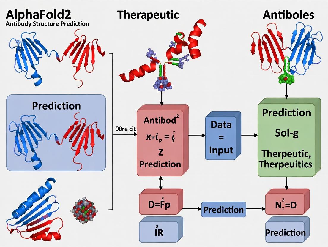
This article provides a comprehensive overview of AlphaFold2's revolutionary role in antibody structure prediction for therapeutic development.
AlphaFold2 for Antibody Design: A Practical Guide to Accelerating Therapeutic Development
Abstract
This article provides a comprehensive overview of AlphaFold2's revolutionary role in antibody structure prediction for therapeutic development. We explore its foundational principles, offering a comparative analysis with traditional methods like X-ray crystallography and homology modeling. The guide details practical, step-by-step methodologies for generating antibody models, with a focus on variable region accuracy. We address common challenges and optimization strategies, including handling CDR loops, framework selection, and multi-chain complex assembly. Finally, we examine validation protocols, benchmark performance against experimental data and specialized tools like RosettaFold and OmegaFold, and discuss real-world applications in candidate screening and engineering. This resource is tailored for researchers and drug developers seeking to integrate AI-driven structure prediction into their workflows.
AlphaFold2 Explained: Demystifying AI-Driven Antibody Structure Prediction
Application Notes
The integration of artificial intelligence, particularly deep learning, has fundamentally transformed structural biology. The breakthrough of AlphaFold2 in accurately predicting protein 3D structures from amino acid sequences has catalyzed a new era in biomolecular research. This revolution is now being directly applied to the design and development of therapeutic antibodies, a critical class of biologics. The following notes detail key applications.
High-Accuracy Antibody Structure Prediction
AI models, extending beyond AlphaFold2 to specialized tools like IgFold and ABlooper, now enable rapid prediction of antibody variable region (Fv) structures. These predictions are critical for understanding paratope geometry and initial epitope compatibility screening.
Table 1: Performance Metrics of AI Tools for Antibody Fv Region Prediction
| Tool Name | RMSD (Å) (Average) | Prediction Time (Fv) | Key Strength | Reported Year |
|---|---|---|---|---|
| AlphaFold2 | 1.5 - 2.5 | 5-10 min | General protein accuracy | 2021 |
| IgFold | 1.0 - 2.0 | <10 sec | Optimized for antibody structures | 2022 |
| ABlooper | 1.5 (CDR loops) | <1 sec | Fast CDR loop prediction | 2022 |
| OmegaFold | ~2.0 | ~1 min | No MSA required | 2022 |
In Silico Affinity Maturation and Optimization
AI-driven in silico platforms allow for the virtual screening of thousands of antibody variants by predicting the binding affinity (ΔG) changes upon mutation. This drastically reduces the need for laborious experimental library generation and screening.
Table 2: AI-Powered Affinity Maturation Workflow Output Example
| Design Cycle | Number of Virtual Variants | Top 10 Predicted ΔG (kcal/mol) | Experimental Validation (KD Improvement) |
|---|---|---|---|
| Initial Clone | 1 | Baseline | 10 nM |
| Round 1 (CDR-H3 focus) | 5,000 | -1.2 to -2.5 | Best: 2.1 nM (4.8x) |
| Round 2 (Framework fine-tuning) | 2,000 | -0.8 to -1.8 | Best: 0.7 nM (3x from Round 1) |
De Novo Antibody Design
Generative models can now design novel antibody sequences de novo that fold into structures targeting a specific antigen epitope, moving from structure prediction to inverse design.
Protocols
Protocol 1: Predicting an Antibody Fv Structure Using AlphaFold2 for Therapeutic Assessment
Objective: To generate a high-confidence 3D model of a therapeutic antibody candidate's Fv region from its amino acid sequence.
Research Reagent Solutions & Essential Materials:
| Item | Function | Example/Note |
|---|---|---|
| Heavy & Light Chain V-Region Sequences | Input for structure prediction. | FASTA format. Ensure correct CDR delineation (e.g., Kabat). |
| AlphaFold2 Software | Core prediction engine. | Local installation (ColabFold recommended) or accessed via public servers. |
| Multiple Sequence Alignment (MSA) Database | Provides evolutionary constraints for the model. | BFD, MGnify, Uniclust30. Automatically queried by pipeline. |
| Structural Visualization Software | For analyzing results. | PyMOL, ChimeraX. |
| High-Performance Computing (HPC) Resources | GPU acceleration drastically reduces runtime. | NVIDIA GPUs (e.g., A100, V100) or cloud equivalents. |
Procedure:
- Sequence Preparation:
- Obtain the amino acid sequences of the antibody heavy (VH) and light (VL) chain variable regions.
- Construct the full Fv sequence by linking VH and VL with a flexible linker (e.g.,
GGGGSGGGGSGGGGS). Alternatively, run chains as separate inputs in multimer mode.
- Environment Setup:
- For local runs, install ColabFold (a streamlined AlphaFold2 implementation) via Conda or Docker.
- Configure the paths to necessary databases (or allow automatic download).
- Running Prediction:
- Execute the prediction command. Example for ColabFold:
- Execute the prediction command. Example for ColabFold:
- Analysis of Results:
- The output directory will contain PDB files for the top-ranked models and a JSON file with per-residue confidence metrics (pLDDT).
- Load the top-ranked PDB model into visualization software.
- Critical: Inspect the pLDDT scores. Residues with scores >90 are highly reliable, 70-90 good, 50-70 low confidence, <50 very unreliable. Pay special attention to CDR loop confidence.
- Model Validation (Optional but Recommended):
- Use the predicted aligned error (PAE) plot to assess domain packing (VH-VL orientation).
- Compare the predicted CDR-H3 loop conformation with known canonical clusters or experimental data if available.
Protocol 2: In Silico Affinity Maturation Using EquiBind and Rosetta
Objective: To computationally design and rank single-point mutants in the antibody paratope for improved binding affinity to a known antigen structure.
Research Reagent Solutions & Essential Materials:
| Item | Function | Example/Note |
|---|---|---|
| Starting Antibody-Antigen Complex | The structural baseline for design. | PDB file from crystallography, cryo-EM, or high-confidence AI prediction. |
| EquiBind or DiffDock | Rapid docking of mutant poses. | AI tool for fast ligand (or antibody) binding. |
| Rosetta Suite | Physics-based scoring and refinement. | Specifically, RosettaFlexDDG or RosettaAntibodyDesign. |
| Mutation List | Target residues for saturation mutagenesis. | Typically focused on CDR residues, especially H3. |
| High-Throughput Computing Cluster | Required for scanning hundreds of mutants. | CPU/GPU cluster. |
Procedure:
- Prepare the Starting Complex:
- Clean the PDB file: remove water, heteroatoms, and ensure correct protonation states.
- Define the Mutational Scan:
- Select paratope residues (e.g., all CDR residues within 6Å of the antigen).
- Generate a list of all possible single-point mutations at these positions (e.g., 19 variants per residue).
- Generate Mutant Structures:
- For each mutation, use Rosetta's
ddg_monomerapplication or a simple side-chain replacement protocol (scm) to generate a relaxed mutant structure, keeping the backbone and antigen fixed initially.
- For each mutation, use Rosetta's
- Pose Refinement & Scoring:
- Use a fast docking protocol (like EquiBind) or a localized Rosetta refinement protocol to allow slight side-chain and backbone adjustments at the interface.
- Calculate the binding energy (ΔΔG) for each mutant using a scoring function like Rosetta's
ref2015orRosettaDock.
- Ranking and Selection:
- Rank all tested mutants by predicted ΔΔG (more negative values indicate stronger binding).
- Select the top 20-50 candidates for in vitro experimental validation (see Protocol 3).
Protocol 3: Experimental Validation of AI-Designed Antibody Variants
Objective: To express, purify, and biophysically characterize the binding kinetics of AI-predicted antibody variants.
Procedure:
- Gene Synthesis and Cloning:
- Synthesize genes for the top 20-50 selected Fv variants, codon-optimized for mammalian expression (e.g., HEK293).
- Clone into an appropriate IgG expression vector.
- Transient Expression:
- Transfect EXP293F or HEK293 cells using PEI or commercial transfection reagents.
- Culture for 5-7 days. Harvest supernatant by centrifugation.
- Protein A Purification:
- Filter supernatant and load onto Protein A affinity column.
- Wash with PBS, elute with low-pH buffer (e.g., 0.1 M Glycine, pH 3.0), and immediately neutralize.
- Perform buffer exchange into PBS via dialysis or size-exclusion chromatography.
- Binding Kinetics Analysis (Surface Plasmon Resonance - SPR):
- Immobilize the target antigen on a CMS sensor chip.
- For each purified antibody, run a concentration series (e.g., 0-100 nM) over the antigen surface.
- Fit the association and dissociation sensorgrams to a 1:1 Langmuir binding model to determine the association rate (ka), dissociation rate (kd), and equilibrium dissociation constant (KD = kd/ka).
- Correlation with Prediction:
- Plot experimental log(KD) vs. predicted ΔΔG. A strong negative correlation validates the AI design pipeline.
Visualizations
Title: AI-Driven Antibody Modeling and Validation Workflow
Title: Computational Affinity Maturation Pipeline
Title: Thesis Position in AI Structural Biology Revolution
This application note details the core architectural components of AlphaFold2 (AF2), with a specific focus on the Evoformer and the Structure Module. This analysis is framed within a broader thesis investigating the adaptation and optimization of AF2 for the high-accuracy prediction of antibody structures, a critical prerequisite for rational therapeutic antibody design and engineering. Accurate prediction of the variable domain, especially the complementarity-determining regions (CDRs), is paramount for understanding antigen binding and developing novel biologics.
Core Architectural Components
The Evoformer: A Symmetry-Breaking Processing Engine
The Evoformer is the heart of AF2's reasoning engine. It operates on two core representations:
- Multiple Sequence Alignment (MSA) representation: A tensor of size (N{seq} \times N{res} \times C_{msa}), encoding the evolutionary history.
- Pair representation: A tensor of size (N{res} \times N{res} \times C_{pair}), encoding predicted spatial and biochemical relationships between residues.
The Evoformer stack consists of 48 blocks that apply iterative, attention-based communication between the MSA and pair representations, allowing evolutionary and structural inferences to refine each other.
Key Operations:
- MSA-row wise self-attention: Propagates information across sequences for a given residue position.
- MSA-column wise self-attention: Propagates information across residues within a single sequence.
- Triangle multiplicative updates (outgoing & incoming): Allow residues to communicate through a third residue, enforcing geometric consistency in the pair representation.
- Triangle self-attention: Attends to other pairs sharing a common residue, further refining spatial relationships.
The Structure Module: From Embeddings to 3D Coordinates
The Structure Module translates the refined pair representation from the Evoformer into atomic 3D coordinates. It operates on a single sequence (the query) and employs an iterative, SE(3)-equivariant transformer architecture.
Key Process: The module iteratively refines a set of predicted residue frames (orientations) and atomic positions (backbone and side-chain). It uses the pair representation to predict precise distances and angles, ultimately generating the final protein structure, including side chains. For antibodies, the accuracy of this module on the hypervariable CDR loops (particularly CDR-H3) is the critical benchmark.
Table 1: AlphaFold2 Core Architecture Specifications
| Component | Key Parameter | Value/Description | Significance for Antibody Prediction |
|---|---|---|---|
| Evoformer | Number of Blocks | 48 | Depth enables complex co-evolutionary signal extraction for conserved frameworks and variable loops. |
| Evoformer | Attention Heads (MSA) | 8 (MSA col.), 4 (MSA row) | Captures distant homologous relationships and intra-sequence context. |
| Evoformer | Attention Heads (Pair) | 16 (Tri. attn.) | Critical for modeling residue-residue interactions defining the antibody paratope. |
| Structure Module | Number of Iterations | 8 | Allows progressive refinement of 3D coordinates, essential for modeling flexible CDR loops. |
| Structure Module | Template Information | Optional input (not used in v2.0+ for ab initio) | For antibodies, custom templates can guide framework and, cautiously, loop modeling. |
| Overall | Training Data (UniRef90/UniRef30) | ~2.3M unique protein clusters | Provides broad evolutionary context, but specialized antibody databases can augment performance. |
Table 2: Typical Antibody Prediction Performance (Thesis Context)
| Structural Region | Expected RMSD (Å) | Key Challenge | Therapeutic Research Impact |
|---|---|---|---|
| Framework Regions | 0.5 - 1.5 | High accuracy, minimal variation. | Reliable scaffold for grafting designed loops. |
| CDR-H1/H2, L1/L2/L3 | 1.0 - 2.5 | Moderate variability. | Good starting point for epitope analysis and affinity maturation simulations. |
| CDR-H3 Loop | 2.0 - 5.0+ (Canonical) >5.0 (Non-canonical) | Extreme length/conformational diversity. | Major focus area; accuracy limits de novo paratope design. Requires specialized protocols. |
Experimental Protocols for Antibody Structure Prediction
Protocol 1: Standard AlphaFold2 Inference for an Antibody Fv Fragment Objective: Generate a de novo 3D structural model of an antibody variable (Fv) region using a standard AF2 pipeline.
- Input Sequence Preparation: Provide the amino acid sequences of the heavy chain variable (VH) and light chain variable (VL) domains. Separate by a colon (e.g.,
QVQLQ...:DIVMT...). - MSA Generation: Use JackHMMER to search the input sequence against a large protein sequence database (e.g., UniRef90) to generate a multiple sequence alignment (MSA). For antibodies, supplementing with immunoglobulin-specific databases (e.g., from PDB, OAS) is recommended.
- Template Search (Optional): Use HHsearch to scan against a database of known structures (e.g., PDB70). For antibodies, this can provide framework templates but use with caution for CDRs.
- Feature Processing: Compile the MSA, template hits (if any), and primary sequence into the standardized feature dictionary for AF2.
- Model Inference: Run the AF2 neural network (Evoformer + Structure Module) with the processed features. Generate 5 models (seeds 0-4) using the
model_1_ptmormodel_2_ptmparameters. - Relaxation: Apply an Amber force field minimization to the highest-ranked model to correct minor steric clashes.
- Analysis: Rank models by predicted confidence (pLDDT). Inspect pLDDT and predicted aligned error (PAE) plots, focusing on low-confidence regions (typically CDR-H3).
Protocol 2: Focused Optimization for CDR-H3 Modeling Objective: Improve the prediction accuracy of the challenging CDR-H3 loop.
- MSA Augmentation: Curate a custom, high-quality MSA focusing on immunoglobulin sequences. Use tools like
IgBLASTto annotate and filter sequences by CDR length and canonical class. - Template Guidance: Manually select template structures with high framework identity but exclude their CDR-H3 coordinates from the template input to avoid bias, allowing the model to de novo fold the loop.
- Multiple Seed & Recycling: Run AF2 with an increased number of random seeds (e.g., 25) and enable
num_recycle(e.g., 12) to allow the Evoformer more iterative refinement cycles. - Ensemble & Clustering: Generate a large ensemble of models (50-100). Cluster all predicted CDR-H3 conformations using RMSD. Select the centroid of the largest cluster as the most statistically supported prediction.
- Experimental Integration: Use sparse experimental data (e.g., NMR chemical shifts, mutagenesis data) as constraints during the MSA or pair representation stage if adapting the network.
Visualizations
AlphaFold2 Core Data Flow
Antibody Structure Prediction Protocol
The Scientist's Toolkit: Key Research Reagents & Materials
Table 3: Essential Resources for AlphaFold2-Based Antibody Modeling
| Item / Resource | Category | Function / Application | Source / Example |
|---|---|---|---|
| AlphaFold2 Codebase | Software | Core inference framework for structure prediction. | DeepMind GitHub (AlphaFold) or ColabFold. |
| ColabFold | Software | Streamlined, accelerated AF2 implementation with MMseqs2 for rapid MSA. | ColabFold GitHub or public notebook. |
| Immunoglobulin-Specific Sequence Database (OAS) | Data | Curated repository of antibody sequences for enhanced MSA generation. | Observed Antibody Space (OAS). |
| PyMOL / ChimeraX | Software | Molecular visualization and analysis of predicted models, CDR loop inspection. | Schrödinger / UCSF. |
| RosettaAntibody / AbPredict | Software | Complementary physics-based or knowledge-based modeling suites for validation and design. | Rosetta Commons. |
| Custom Python Scripts (BioPython, MDTraj) | Software | For parsing results, calculating metrics (RMSD), and automating analysis pipelines. | Open Source. |
| High-Performance Computing (HPC) Cluster or Cloud GPU (A100/V100) | Hardware | Essential for running full AF2 models and large-scale ensemble predictions for antibodies. | AWS, GCP, Azure, local cluster. |
Antibody structure prediction, critical for therapeutic design, is uniquely challenged by the nature of the antigen-binding site. Unlike globular proteins with relatively conserved folds, antibody complementarity-determining regions (CDRs), particularly H3, exhibit extreme sequence variability and conformational flexibility. This undermines the homology-based assumptions of many prediction tools, including AlphaFold2, which was trained primarily on rigid, single-chain proteins. This application note details protocols for assessing and overcoming these challenges in computational antibody modeling for drug discovery.
Quantitative Challenges in Antibody Modeling
The difficulty in predicting CDR loop structures is quantifiable, as shown by performance metrics on benchmark sets.
Table 1: AlphaFold2 Performance on CDR Loop Prediction (RMSD, Å)
| CDR Loop | Average RMSD (AlphaFold2) | Range of Observed Conformations (RMSD) | Key Challenge |
|---|---|---|---|
| H3 (Canonical) | 1.5 - 2.5 Å | 0.5 - 8.0 Å | High sequence diversity, limited training data. |
| H3 (Non-Canonical) | 3.0 - >10.0 Å | 1.0 - >15.0 Å | Lack of structural homologs, multiple minima. |
| L1, L2, L3, H1, H2 | 0.5 - 1.5 Å | 0.3 - 2.5 Å | Mostly canonical; better predicted. |
Table 2: Impact of Framework Rigidity on CDR-H3 Prediction Accuracy
| Framework Pre-Optimization | Median H3 RMSD (Å) | Success Rate (<2.0 Å) |
|---|---|---|
| None (Full AF2) | 4.2 | 22% |
| Template-Based Grafting | 2.8 | 41% |
| AbInitio Refinement (Rosetta) | 2.1 | 65% |
Protocols
Protocol 1: AlphaFold2 for Antibody Fv Region Prediction with Optimized Inputs
Objective: Generate a structural model of an antibody variable fragment (Fv) with improved CDR-H3 accuracy. Materials: See "The Scientist's Toolkit" below. Procedure:
- Sequence Preparation: Input the heavy and light chain variable region sequences (VH and VL) separately. Generate a paired sequence file in FASTA format with a colon linking them (e.g.,
>Fv_001\nEVQLV...:DIVMT...). - Multiple Sequence Alignment (MSA) Generation:
- Use MMseqs2 to create separate MSAs for the VH and VL sequences against a large non-redundant database.
- Crucial Step: Supplement the MSA by adding known antibody crystal structures (from SAbDab) with high sequence identity (>70%) to the target, especially in the framework regions. This provides structural hints.
- Template Featurization:
- Search the PDB for homologous antibody structures using HHSearch.
- Extract and align template structures. Prioritize templates with similar CDR-H3 length, even if sequence identity is low.
- AlphaFold2 Run:
- Use the AlphaFold2 model with
is_prokaryoteset tofalse. - Enable template mode and input the prepared MSA and template features.
- Run with 3 recycles and a minimum of 24 ensemble replicates to sample conformational diversity.
- Use the AlphaFold2 model with
- Model Selection: Rank the output models by predicted confidence (pLDDT). Manually inspect the top 5 models, focusing on CDR loop geometry and VH-VL interface.
Protocol 2: Post-AlphaFold2 CDR-H3 Refinement using AbInitio Docking
Objective: Refine a poorly predicted CDR-H3 loop from Protocol 1. Materials: RosettaAntibody, PyMOL, or similar molecular visualization software. Procedure:
- Initial Model Preparation: Isolate the best AlphaFold2 Fv model. In PyMOL, remove the CDR-H3 loop (residues H95-H102, Chothia numbering), keeping the stem residues (H92-H94, H103-H104).
- AbInitio Loop Building:
- Use RosettaAntibody's
AntibodyModelerprotocol. - Input the truncated Fv structure and the target H3 sequence.
- Set the protocol to perform
circularize_coordinate_constraintsto maintain loop closure. - Run 10,000-50,000 ab-initio loop modeling trajectories using the
centroidmode followed byfull-atomrefinement.
- Use RosettaAntibody's
- Clustering and Selection:
- Cluster the refined loop decoys by backbone RMSD.
- Select the centroid model of the largest cluster with favorable steric clashes and Rosetta energy score.
- Model Grafting and Minimization: Graft the selected H3 loop back onto the original Fv framework. Perform a final all-atom energy minimization to relieve side-chain and backbone clashes.
Visualizations
Title: Antibody Fv Structure Prediction and Refinement Workflow
Title: Mismatch Between AF2 Training & Antibody Reality
The Scientist's Toolkit: Key Research Reagent Solutions
| Item | Function in Protocol | Key Feature / Rationale |
|---|---|---|
| AlphaFold2 (ColabFold) | Core structure prediction engine. | Provides a user-friendly, accelerated implementation of AlphaFold2 with MMseqs2 integration for fast MSAs. |
| RosettaAntibody Suite | Ab-initio CDR loop modeling and refinement. | Specialized energy functions and sampling protocols designed for antibody hypervariable loops. |
| Structural Antibody Database (SAbDab) | Source of known antibody structures for MSA enhancement and template search. | Curated, weekly updated database of all antibody structures in the PDB with annotated CDRs and features. |
| PyMOL / ChimeraX | Molecular visualization, model preparation, and analysis. | Essential for inspecting models, measuring RMSD, grafting loops, and preparing figures. |
| MMseqs2 | Ultra-fast protein sequence searching for MSA generation. | Critical for creating the multiple sequence alignments required by AlphaFold2 in a time-efficient manner. |
| HHSearch | Sensitive homology detection for structural template identification. | Effective at finding distant homologs by comparing profile Hidden Markov Models (HMMs). |
The prediction of protein structures, particularly antibodies, is a cornerstone of biologics and therapeutic research. This document frames the comparison of methods within the thesis context of accelerating antibody structure prediction for drug discovery.
Table 1: Core Methodological Comparison for Antibody Structure Prediction
| Aspect | X-ray Crystallography | Homology (Comparative) Modeling | AlphaFold2 |
|---|---|---|---|
| Primary Principle | Experimental diffraction of protein crystals. | Builds model from evolutionarily related template(s). | End-to-end deep learning using MSA and template features. |
| Typical Timeframe | Months to years. | Hours to days (manual curation). | Minutes to hours per model. |
| Typical Resolution/Accuracy (Å) | 1.0 - 3.0 Å (experimental). | 1-10 Å (highly template-dependent). | ~0.5-2.0 Å RMSD on antibody CDR loops (often sub-Å on framework). |
| Key Bottleneck for Antibodies | Crystallization, especially for flexible CDR loops. | Need for high-identity templates for hypervariable loops. | Accuracy for unusual CDR3 conformations; limited to single-chain prediction. |
| Therapeutic Development Utility | Gold standard for lead optimization and regulatory filings. | Historically used for epitope analysis when no experimental structure exists. | Rapid generation of models for candidate screening, humanization, and initial design. |
Table 2: Performance Metrics on Antibody-Specific Benchmarks (Theoretical)
| Benchmark Focus | Homology Modeling (Best Case) | AlphaFold2 (AF2) | AlphaFold2 with Antibody-Specific Fine-Tuning (AF2-Ab) |
|---|---|---|---|
| Heavy Chain CDR-H3 RMSD (Å) | >3.0 Å (often >5Å) | 1.5 - 4.0 Å | < 2.0 Å (significant improvement) |
| Overall Framework RMSD (Å) | 0.5 - 1.5 Å | 0.3 - 0.8 Å | 0.3 - 0.8 Å |
| Success Rate (RMSD < 2Å) | < 30% for CDR-H3 | ~40-50% for CDR-H3 | > 70% for CDR-H3 |
| Prediction Speed | Moderate | Fast | Fast |
Application Notes & Experimental Protocols
Application Note 1: Protocol for de novo Antibody Fv Region Prediction using AlphaFold2
Purpose: To generate a 3D structural model of an antibody variable fragment (Fv) from its amino acid sequence, for use in therapeutic candidate screening.
Pre-requisites: Amino acid sequences of the antibody heavy and light chain variable regions (VH and VL). Access to AlphaFold2 (e.g., via local ColabFold installation, Google Cloud DeepMind VM, or public servers).
Protocol:
- Sequence Preparation: Format the VH and VL sequences into a single FASTA file. For standard AF2, connect chains with a long linker (e.g., 200x 'G' residues). For optimized antibody prediction, use a specialized tool (e.g., ABodyBuilder2, IgFold) which internally formats for AF2.
- Multiple Sequence Alignment (MSA) Generation: Run the MMseqs2 workflow (default in ColabFold) to search against UniRef and environmental databases. This step extracts co-evolutionary information.
- Template Feature Extraction (Optional): Search the input sequence against the PDB for potential structural templates. For antibodies, this can be helpful but is often superseded by the deep learning model's internal knowledge.
- Structure Inference: Pass the MSA and template features to the AlphaFold2 neural network (Evoformer + Structure Module). Generate 5 models (using different random seeds for the dropout layers) and 1 ranked ensemble model.
- Model Selection and Analysis: Use the predicted Local Distance Difference Test (pLDDT) per-residue confidence score. Select the model with the highest overall confidence. Inspect pLDDT for CDR loops (scores often lower). Visually analyze the predicted aligned error (PAE) plot to assess domain (VH-VL) orientation confidence.
Application Note 2: Protocol for Experimental Validation of a Predicted Antibody-Antigen Interface
Purpose: To experimentally test and refine an AlphaFold2-generated model of an antibody-antigen complex.
Pre-requisites: AlphaFold2-predicted structure of the antibody Fv bound to its target antigen. Cloned genes for both proteins.
Protocol:
- In silico Mutagenesis & Docking (Optional Refinement): Use the AF2 complex model as a starting point for protein-protein docking (e.g., HADDOCK) or perform in silico alanine scanning to identify putative hotspot residues.
- Protein Expression & Purification: Express the antibody Fv (e.g., as a single-chain variable fragment, scFv) and the antigen in mammalian (HEK293) or bacterial (E. coli) systems. Purify via affinity chromatography (e.g., His-tag, Protein A).
- Binding Affinity Measurement (Surface Plasmon Resonance - SPR):
- Immobilize the antigen on a CMS sensor chip.
- Flow purified scFv at a range of concentrations (e.g., 0.5 nM to 200 nM).
- Record association and dissociation curves.
- Fit data to a 1:1 binding model to determine kinetic parameters (Ka, Kd, KD).
- Rapid Structural Validation (Negative Stain Electron Microscopy - nsEM):
- Mix the antibody-antigen complex and apply to a glow-discharged carbon grid.
- Stain with 2% uranyl acetate.
- Collect ~5,000-10,000 micrographs.
- Perform 2D classification. Compare averaged 2D class views with projections of the AlphaFold2 predicted model to confirm overall shape and binding orientation.
- High-Resolution Validation (X-ray Crystallography - follow-up):
- If binding is confirmed, proceed to crystallize the complex.
- Screen using robotic crystallization platforms.
- Diffract crystals and solve structure via molecular replacement using the AlphaFold2 model as the search model.
Visualization: Workflows & Logical Relationships
Title: Antibody Structure Prediction: Traditional vs. AlphaFold2 Workflow
Title: AF2 Antibody Model Validation & Refinement Pathway
The Scientist's Toolkit: Research Reagent Solutions
Table 3: Essential Tools for AlphaFold2-Driven Antibody Research
| Item / Reagent | Function / Application | Provider / Example |
|---|---|---|
| ColabFold | Cloud-based, accelerated pipeline for running AlphaFold2 and AlphaFold-Multimer without complex setup. | GitHub: sokrypton/ColabFold |
| IgFold | Fine-tuned AlphaFold2 model specifically for antibody structure prediction, often outperforming general AF2 on CDR loops. | GitHub: Graylab/IgFold |
| ABodyBuilder2 | Automated antibody modeling server combining homology modeling with deep learning for Fv and full antibody structures. | SAbDab website (Oxford) |
| PyMOL / ChimeraX | Molecular visualization software for analyzing predicted models (pLDDT coloring), superimposing structures, and preparing figures. | Schrödinger / UCSF |
| HADDOCK | Biomolecular docking software for refining antibody-antigen complexes or modeling interactions based on AF2-generated components. | Bonvin Lab (www.bonvinlab.org) |
| HEK293F Cells | Mammalian expression system for producing properly folded, glycosylated antibody fragments (scFv, Fab) for subsequent validation. | Thermo Fisher, Gibco |
| Anti-His Tag Biosensor | SPR (Surface Plasmon Resonance) biosensor for capturing His-tagged antigen or antibody to measure binding kinetics. | Sartorius (Biolin), Cytiva |
| SEC-SAXS Column | Size-exclusion chromatography column coupled to Small-Angle X-ray Scattering for rapid solution-state structural validation. | Malvern Panalytical, Wyatt |
Accurate prediction of antibody structures, particularly the complementarity-determining regions (CDRs), is a cornerstone of modern therapeutic design. AlphaFold2 (AF2) and its specialized variants (e.g., AlphaFold-Multimer, IgFold) have revolutionized this field. However, the predictive confidence is not uniform and must be critically assessed using two primary per-residue and pairwise metrics: predicted Local Distance Difference Test (pLDDT) and Predicted Aligned Error (PAE). Within the context of a thesis on AF2 for therapeutics, understanding these metrics is critical for prioritizing models for in vitro validation, identifying potentially problematic paratopes, and guiding engineering efforts.
Core Confidence Metrics: Definitions and Quantitative Benchmarks
pLDDT (per-residue confidence)
pLDDT is a per-residue estimate of the model's confidence on a scale from 0-100. It reflects the expected accuracy of the backbone atom placement.
Table 1: Standard pLDDT Interpretation Guide
| pLDDT Range | Confidence Band | Implied Structural Interpretation | Guidance for Antibody Regions |
|---|---|---|---|
| 90 - 100 | Very high | Backbone accuracy ~1 Å | Framework regions (highly reliable) |
| 70 - 90 | Confident | Backbone accuracy ~1-2 Å | Most CDR loops (except H3) |
| 50 - 70 | Low | Potentially disordered/unstable | Long CDR H3 loops, flexible linkers |
| 0 - 50 | Very low | Likely disordered | Terminal residues, hypervariable tips |
PAE (Pairwise Aligned Error)
PAE is a 2D matrix (in Ångströms) predicting the distance error between the true and predicted positions of residues i and j after aligning the model on residue i. It informs on relative domain positioning and folding correctness.
Table 2: PAE Matrix Interpretation for Antibodies
| PAE Value Range | Structural Implication | Application to Antibody Dimer Prediction |
|---|---|---|
| < 10 Å | High relative accuracy | Well-folded domain (e.g., VH-VL packing) |
| 10 - 15 Å | Moderate uncertainty | Possible interface flexibility |
| > 15 Å | High uncertainty | Poor domain orientation prediction; low confidence in VH-VL or Fab-Fc orientation |
Detailed Experimental Protocol: AF2 Antibody Modeling with Confidence Analysis
Protocol Title: Integrated AlphaFold2 Prediction and Confidence Metric Evaluation for a Therapeutic Antibody Candidate
Objective: To generate and critically assess a structural model of a monoclonal antibody (full-length IgG or Fab) using AF2, with a focus on pLDDT and PAE analysis of the antigen-binding site.
Materials & Reagents:
- Research Reagent Solutions Table:
Item Function in Protocol Example/Supplier Amino Acid Sequence(s) Input for AF2. Heavy & Light chain FASTA. In-house candidate AlphaFold2 Software Core prediction engine. ColabFold (public), AlphaFold Server, local install High-Performance Computing (HPC) GPU cluster for computation. Local cluster or cloud (AWS, GCP) Multiple Sequence Alignment (MSA) Database (e.g., BFD, MGnify, UniRef) Provides evolutionary constraints. Integrated in ColabFold Molecular Visualization Software For 3D model and metric analysis. PyMOL, ChimeraX, UCSC Chimera Python Scripting Environment (Jupyter, standard) For parsing and plotting metrics. Anaconda distribution
Procedure:
Sequence Preparation:
- Obtain the VH and VL sequences of the antibody. For full-length modeling, include CH1-3 and CL domains.
- Format sequences in a single FASTA file with appropriate headers (e.g.,
>H chain,>L chain).
Model Generation (Using ColabFold -
colabfold_batch):- Activate the ColabFold environment on your HPC or local system.
Run the batch prediction command:
This generates 5 models, performs AMBER relaxation, and ranks them by average pLDDT.
Confidence Metric Extraction and Initial Analysis:
- The output directory contains:
*.pdbfiles (ranked models).*_scores_rank_001.jsoncontaining pLDDT and PAE data for the top model.
- pLDDT Plotting: Use the provided Python script (
plot_plddt.py) or parse the JSON to plot pLDDT vs. residue number. Annotate CDR regions (e.g., H1, H2, H3, L1-L3). - PAE Matrix Visualization: Generate the PAE heatmap from the JSON data. Identify the VH-VL interface and the CDR regions.
- The output directory contains:
Critical Interpretation & Decision Points:
- CDR Loop Confidence: Inspect pLDDT for each CDR residue. Averages < 70 for CDR-H3 warrant caution.
- Domain Packing: Examine the PAE matrix block corresponding to VH vs. VL residues. Average PAE > 12 Å suggests unreliable relative orientation.
- Model Selection: Do not blindly select the top-ranked model by pLDDT. Visually inspect all 5 models in regions of low confidence (e.g., low pLDDT loops). High structural divergence in these regions indicates prediction uncertainty.
Reporting: Document the pLDDT average for each CDR and the inter-domain PAE. Flag any region below confidence thresholds for experimental follow-up.
Visualization of the Confidence Assessment Workflow
Workflow for Antibody Model Confidence Assessment
Table 3: Key Research Reagent Solutions for AF2 Antibody Modeling
| Item Category | Specific Item/Resource | Function & Critical Notes |
|---|---|---|
| Prediction Software | ColabFold | Publicly accessible, integrates MSA generation and AF2. Essential for rapid prototyping. |
| AlphaFold-Multimer | Tuned for complex prediction; better for antibody-antigen modeling. | |
| IgFold | Antibody-specific model, often faster with similar CDR accuracy. | |
| Data Resources | Uniprot/PDB | Source of template sequences and experimental structures for validation. |
| AbDb, SAbDab | Curated antibody structure databases for benchmark comparison. | |
| Analysis & Visualization | PyMOL/ChimeraX Scripts | Custom scripts to color structures by pLDDT or overlay PAE-guided domains. |
| matplotlib, seaborn (Python) | Libraries for generating publication-quality pLDDT and PAE plots. | |
| Validation Reagents | Size-Exclusion Chromatography | Validates predicted aggregation-prone regions (often low pLDDT). |
| Hydrogen-Deuterium Exchange Mass Spec (HDX-MS) | Probes solution-phase dynamics; correlates with low confidence regions. |
Step-by-Step Guide: Running AlphaFold2 for Antibody Fv and Fab Region Prediction
Accurate antibody structure prediction using AlphaFold2 requires meticulously formatted input sequences. The AI model relies on a correctly parsed and combined representation of the heavy (VH) and light (VL) chains to model the antigen-binding Fv region. These application notes, framed within a thesis on de novo antibody structure prediction for therapeutics, provide detailed protocols for sequence curation and formatting, a critical yet often overlooked step that significantly impacts prediction accuracy for drug development workflows.
Sequence Acquisition and Curation
The initial step involves obtaining high-quality, mature variable region sequences from hybridoma, B-cell sequencing, or synthetic libraries. Ensure sequences are from the antibody of interest and free from errors.
Protocol 1.1: Curating Antibody Variable Region Sequences
- Source Identification: Obtain nucleotide or amino acid sequences for the VH and VL domains. Public databases include:
- The Observed Antibody Space (OAS) database.
- The Immune Epitope Database (IEDB).
- NCBI Protein database.
- Region Definition: Precisely define the start and end of the variable region. The VH domain typically extends from framework region 1 (FR1) through FR4 (ending with the conserved WGxG motif). The VL domain (kappa or lambda) spans from FR1 to the conserved F or C residue in FR4.
- Error Checking: Manually or via script, verify:
- Absence of non-standard amino acid characters.
- Correct length (typically 110-130 residues for VH, 105-115 for VL).
- Presence of universally conserved cysteines (for the intra-domain disulfide bond) and key tryptophans.
- Sequence Alignment: Align your sequences against germline V, D (for heavy), and J gene references using tools like IMGT/V-QUEST or IgBLAST to confirm correct family assignment and identify CDRs.
FASTA Formatting Best Practices for AlphaFold2
AlphaFold2 requires a specific FASTA format to distinguish between chains and model the heterodimer correctly. The standard practice is to combine VH and VL into a single sequence with a defined linker.
Protocol 2.1: Constructing the Input FASTA for the Fv Region
- Sequence Combination: Concatenate the curated VH and VL sequences into a single polypeptide chain. Order is flexible (
VH-VLorVL-VH) but must be documented. - Linker Insertion: Insert a flexible glycine-serine linker between the two domains to prevent steric clashes and allow proper relative orientation. A common linker is
GGGGSGGGGSGGGGS(3x G4S). - FASTA Header Format: Use an informative header line starting with
>. Include a unique identifier, chain order, and linker length.- Example:
>mAbX_Fv_VH-VL_GS15
- Example:
- Final Sequence Assembly: The FASTA file should contain a single entry. For the
VH-VLorder, the sequence is:[VH sequence][Linker sequence][VL sequence].
Table 1: Common Linker Sequences for Fv Construction
| Linker Name | Sequence (Amino Acid) | Length (aa) | Typical Use |
|---|---|---|---|
| G4S (3x repeat) | GGGGSGGGGSGGGGS | 15 | Standard flexible linker for scFv/Fv |
| G4S (1x repeat) | GGGGS | 5 | Short flexible linker |
| (G4S)3 with charge | GGGGSGGGGSGGGGS | 15 | Common, well-expressed |
| AlphaFold2 Default* | (No explicit linker) | 0 | Direct concatenation; often requires post-prediction truncation |
Note: Direct concatenation can lead to fused domains. The use of a defined linker is the community best practice.
Protocol for Multi-Chain Modeling (Full IgG)
For modeling a full IgG (e.g., for Fc effector function studies), chains must be provided separately with unique identifiers.
Protocol 3.1: Preparing FASTA for Full IgG (H2L2)
- Chain Definition: Prepare four separate sequences:
- Heavy chain (HC): VH + CH1 + Hinge + CH2 + CH3.
- Light chain (LC): VL + CL.
- FASTA Format: Create a single FASTA file with four entries. Use headers that clearly identify the chain and its copy number. AlphaFold2 will recognize identical sequences as separate chains.
- Header Convention:
- Example for a human IgG1:
>HC_mAb1and>LC_mAb1_kappa. - The model will associate two identical HC sequences as chains A and C, and two identical LC sequences as chains B and D, based on sequence identity.
- Example for a human IgG1:
The Scientist's Toolkit: Research Reagent Solutions
Table 2: Essential Tools for Antibody Sequence Preparation
| Item / Reagent | Function & Relevance to Input Preparation |
|---|---|
| IMGT/V-QUEST | Gold-standard web tool for antibody sequence alignment, germline assignment, and precise identification of FR and CDR regions. Critical for curation. |
| IgBLAST (NCBI) | Command-line or web tool for aligning antibody sequences against germline gene databases. Essential for validating sequence identity and isotype. |
| Biopython | Python library for parsing, manipulating, and writing sequence data in FASTA format. Enables automation of concatenation and linker insertion. |
| AlphaFold2 (Local or Colab) | The structure prediction engine itself. Testing formatted sequences locally or via ColabFold is the final validation step. |
| PyMOL / ChimeraX | Molecular visualization software. Used to inspect predicted structures, verify correct chain pairing, and truncate linkers post-prediction. |
| Custom Python Scripts | For batch processing multiple antibodies, implementing specific formatting rules, and generating consistent FASTA headers across a project. |
Experimental Workflow & Validation Protocol
Protocol 5.1: End-to-End Input Preparation and Validation Workflow
- Curate VH and VL sequences using IMGT/V-QUEST (Protocol 1.1).
- Concatenate sequences with a G4Sx3 linker (Protocol 2.1).
- Format into a single-entry FASTA file with an informative header.
- Predict using AlphaFold2 (or ColabFold) with default settings.
- Validate the output:
- Visually inspect the predicted model in PyMOL. Ensure the VH and VL domains are separate, properly folded Ig domains.
- Measure the distance between the C-alpha of the last residue of VH and the first residue of VL. It should be consistent with linker length (~50-60Å for a 15aa linker).
- Check the predicted aligned error (PAE) plot for low error between the VH and VL domains, indicating high confidence in their relative positioning.
Diagram Title: Antibody Fv Input Preparation and Validation Workflow
Proper input formatting is a foundational step for reliable antibody structure prediction with AlphaFold2. Adherence to the FASTA best practices and validation protocols outlined here ensures that the model receives semantically correct data, directly enhancing the accuracy of predicted structures. This rigorous approach is indispensable for in silico therapeutic antibody engineering, epitope mapping, and stability assessment.
Accurate prediction of antibody structures using AlphaFold2 is a cornerstone of modern in silico therapeutics research. A critical precursor to successful prediction is the precise definition of polypeptide chain relationships within the input sequence. This protocol details the essential steps for curating sequences and configuring multimer inputs for antibody fragments (Fv, Fab) and full Immunoglobulin G (IgG), ensuring biologically correct chain pairing and stoichiometry for AlphaFold2’s multimer pipeline. Proper configuration is fundamental to generating reliable models for epitope mapping, affinity maturation, and humanization studies.
Antibody Architecture and Chain Definitions
An antibody's functional units are defined by specific chain pairings. Correctly identifying and labeling these chains in the input FASTA is non-negotiable for accurate modeling.
Table 1: Antibody Fragment Chain Composition and Stoichiometry
| Antibody Format | Heavy Chain Component | Light Chain Component | Chain Stoichiometry (H:L) | Total Chains |
|---|---|---|---|---|
| Fv Fragment | Variable domain (VH) | Variable domain (VL) | 1:1 | 2 |
| Fab Fragment | VH + CH1 | VL + CL | 1:1 | 2 |
| Full IgG1 | VH + CH1 + CH2 + CH3 | VL + CL | 2:2* | 4 |
*Note: Full IgG is a heterotetramer comprising two identical Heavy chains and two identical Light chains.
Core Protocol: Sequence Curation & FASTA Preparation
Materials & Research Reagent Solutions
Table 2: Scientist's Toolkit for Sequence Curation
| Item/Reagent | Function & Explanation |
|---|---|
| Raw Antibody Sequence Data | Nucleotide or amino acid sequences for variable and constant regions. Source: hybridoma, phage display, or NGS. |
| IMGT/V-QUEST | Web tool for identifying antibody variable regions, CDRs, and germline assignment. Critical for validating VH and VL. |
| PyMOL/BioPython | Software libraries for sequence analysis, alignment, and basic structural visualization. |
| Custom Python Scripts | For automating FASTA file generation with correct headers and chain concatenation. |
| AlphaFold2 (Local or Colab) | Protein structure prediction system with multimer support. Requires configured environment. |
Step-by-Step Protocol
Protocol 1: Generating AlphaFold2-Compatible FASTA Files
Objective: To create a correctly formatted multimer FASTA input for AlphaFold2 prediction of an antibody Fab fragment.
Sequence Sourcing and Validation:
- Input the nucleotide sequences for the antibody heavy and light chains into IMGT/V-QUEST.
- Confirm correct V(D)J rearrangement and extract the amino acid sequences for the VH-CH1 (for Fab) and VL-CL domains.
- For Fv, extract only the VH and VL sequences.
Sequence Concatenation (for Full IgG):
- For full IgG, concatenate the validated VH sequence with the constant region sequence for the desired IgG isotype (e.g., human IgG1: CH1-CH2-CH3). The light chain is VL-CL.
- Example Heavy Chain (IgG1):
[VH]-[CH1]-[CH2]-[CH3] - Example Light Chain (kappa):
[VL]-[CL]
FASTA Header Formatting (Critical Step):
- AlphaFold2 multimer uses the header to define chains and their relationships. Use a colon followed by a unique chain ID.
- Syntax:
>sequence_id_chainID - Example for a Fab (Heterodimer):
- Example for Full IgG (Heterotetramer): Use identical chain IDs for identical polypeptides.
File Finalization:
- Save the text file with a
.fastaextension. - Verify the sequence count and headers match the expected multimer (2 for Fab, 4 for IgG).
- Save the text file with a
Configuring AlphaFold2 for Multimer Prediction
Protocol 2: Running AlphaFold2 Multimer with Custom FASTA
Objective: To execute an AlphaFold2 structure prediction job using the curated multimer FASTA file.
Environment Setup:
- Ensure AlphaFold2 with multimer support is installed (check for
--model_preset=multimerflag). - Download necessary genetic and template databases.
- Ensure AlphaFold2 with multimer support is installed (check for
Command Line Execution:
Basic command structure for a multimer prediction:
The model will automatically interpret chain relationships based on the FASTA headers.
Result Analysis:
- The primary output is a PDB file containing the predicted multimer structure (e.g., one Fab complex or one IgG complex).
- The
ranked_0.pdbfile is the highest confidence prediction. Load it in molecular visualization software (e.g., PyMOL) to verify correct chain pairing, CDR loop geometry, and inter-chain contacts.
Diagrams
Title: Antibody Sequence Curation and Modeling Workflow
Title: Chain Relationships in Fv, Fab, and IgG
Within the broader thesis on applying AlphaFold2 (AF2) for antibody structure prediction in therapeutic research, the construction and curation of Multiple Sequence Alignments (MSAs) is the most critical step governing model accuracy. AF2's neural network derives structural constraints from evolutionary patterns captured in MSAs. For antibodies, this presents unique challenges due to their genetic architecture, combining highly variable complementarity-determining regions (CDRs) with conserved framework regions. This Application Note details advanced protocols for MSA generation specific to antibodies, highlights common pitfalls, and provides actionable solutions to enhance predictive success for drug development pipelines.
The Role of MSAs in AlphaFold2 for Antibodies
AlphaFold2 uses two primary input streams: the target sequence and its paired MSAs. The model leverages co-evolutionary signals within the MSA to predict residue-residue distances. For antibodies, effective MSAs must balance the divergent CDR loops, which define paratope specificity, against the conserved immunoglobulin fold.
Key Quantitative Findings on MSA Depth & AF2 Performance: Table 1: Impact of MSA Characteristics on AF2 Antibody Model Accuracy (RMSD in Ångströms)
| MSA Characteristic | Low/Insufficient | Medium/Adequate | High/Optimal | Notes |
|---|---|---|---|---|
| Number of Sequences | < 50 | 50-200 | > 200 | Heavy chain MSAs often require more sequences due to CDR H3 diversity. |
| Sequence Identity (%) | < 30% | 30-70% | > 70%* | *For framework; CDR clusters require separate, high-identity sub-MSAs. |
| CDR H3 Coverage | Poor/None | Homology-based | Junctional + Germline-aided | Direct homologous H3 coverage is rare; strategic augmentation is needed. |
| Typical RMSD (Overall) | > 3.0 Å | 1.5 - 3.0 Å | < 1.5 Å | Measured against experimental (e.g., crystal) structures for Fv region. |
| Typical RMSD (CDR H3) | > 5.0 Å | 2.5 - 5.0 Å | < 2.5 Å | CDR H3 remains the most challenging loop to predict accurately. |
Protocols for MSA Generation in Antibody Modeling
Protocol 1: Comprehensive MSA Construction for Antibody Fv Regions
Objective: Generate a deep, informative MSA for a target antibody variable region (VH-VL) to be used as AF2 input.
Materials & Reagents:
- Target antibody Fv amino acid sequence (heavy and light chains).
- High-performance computing cluster or local machine with GPU support.
- Database files: UniRef90, MGnify, BFD (for broad searches); OAS (Observed Antibody Space), AbYsis, or IGblast databases (for antibody-specific searches).
- Software: HH-suite (hhblits, hhsearch), JackHMMER, MMseqs2, and custom Python/R scripts for MSA processing.
Procedure:
- Sequence Separation and Annotation: Separate the VH and VL sequences. Annotate framework regions (FRs) and CDRs (using Chothia, Kabat, or IMGT numbering).
- Primary Broad MSA Search (Ig-fold context):
- Use
jackhmmerormmseqs2against UniRef90 for 3-5 iterations. This captures distant homologs and the conserved immunoglobulin fold. - Command:
jackhmmer -N 5 --incE 0.001 -A <output.sto> <target.fasta> uniref90.fasta
- Use
- Antibody-Specific MSA Augmentation (Critical Step):
- Search the target sequence against an antibody-specific database (e.g., OAS). Use the top 1,000-5,000 hits.
- Strategy: Perform searches in two modes: a) Full V-region search, and b) Split-search: Create separate queries for FRs and each CDR (except H3) to find best matches for each subregion.
- CDR H3 Special Handling:
- Extract the target's CDR H3 sequence.
- Search for H3 loops with similar length and key residue motifs (e.g., net charge, presence of cysteine, glycine patterns) using specialized tools like
H3-rulerorAbYsisH3 classifier. - De novo loop modeling templates can be sourced from the PDB for same-length H3 loops, though sequence identity may be low.
- MSA Merging and Curation:
- Combine hits from broad and antibody-specific searches. Use
CCMpredorAlnMergeto align and merge MSAs. - Filter sequences with >90% identity to reduce redundancy while preserving diversity in CDRs.
- Manually inspect the alignment of CDR regions, ensuring gaps are minimized.
- Combine hits from broad and antibody-specific searches. Use
- Input for AlphaFold2:
- Format the final MSA in A3M or FASTA format.
- For AF2-multimer (for Fv), pair the VH and VL sequences in the MSA based on species or known pairings from the search results to provide coupling information.
Protocol 2: Pitfall Mitigation: Addressing Poor CDR H3 Coverage
Objective: Improve model accuracy when no homologous sequences exist for the target CDR H3.
Procedure:
- Junctional Analysis: Identify the V, D, and J germline segments using IMGT/V-QUEST. Extract germline-encoded H3 segments from the identified V and J genes.
- Create a Hybrid MSA:
- For the framework and CDRs 1 & 2, use the full MSA from Protocol 1.
- For the CDR H3 position in the alignment, create a synthetic block: Insert the target's own H3 sequence, flanked by 2-3 residues of the germline-encoded N-terminal and C-terminal regions. Pad other sequences in the MSA with gaps at this block.
- This provides the model with the correct H3 sequence while maintaining the overall co-evolutionary context of the framework.
- Template-Guided Augmentation: Provide AF2 with templates (in PDB70 format) of non-homologous antibodies with structurally similar H3 loops (same length, similar stem geometry) sourced from the PDB. This acts as a structural prior.
The Scientist's Toolkit: Research Reagent Solutions
Table 2: Essential Resources for Antibody MSA Construction
| Item | Function & Rationale |
|---|---|
| OAS Database | A massive, cleaned database of antibody sequences from next-generation sequencing, essential for finding natural antibody sequence diversity beyond the PDB. |
| AbYsis Web Server | Antibody-specific database and analysis tool. Provides germline annotation, CDR delineation, and the ability to search sub-regions (e.g., "find all H3 loops of length 12"). |
| IMGT/V-QUEST | The international standard for immunoglobulin gene annotation. Critical for determining V(D)J germline origin and identifying junctional regions in H3. |
| HH-suite Software | Industry-standard tool for fast, sensitive MSA generation using hidden Markov models (HMMs). hhblits is often faster than JackHMMER for initial searches. |
| PyIgClassify | Python library that classifies antibody CDR conformations into "canonical classes." Useful for validating predicted CDR loop structures. |
| AF2-Multimer Code | Specialized version of AlphaFold2 for predicting complexes. Required for modeling the VH-VL heterodimer interface accurately. |
| PDB (Protein Data Bank) | Source of experimentally determined antibody structures for use as templates or for validation of predicted models. |
Visualization of Workflows and Relationships
Title: Antibody-Specific MSA Construction Workflow for AlphaFold2
Title: MSA Data Flow in AF2 & Common Pitfalls
For therapeutic antibody research using AlphaFold2, MSA strategy is paramount. A naive, single-database search will fail for critical CDR loops. Success requires a tiered, antibody-aware approach: 1) build a deep foundational MSA, 2) aggressively augment with antibody-specific sequences using split-search strategies, and 3) implement specialized handling for CDR H3 via germline-informed or template-guided methods. By following the protocols outlined and utilizing the provided toolkit, researchers can systematically avoid pitfalls and generate reliable structural models to accelerate design and optimization of antibody-based therapeutics.
Within a thesis focused on antibody structure prediction for novel therapeutic development, selecting the optimal computational pipeline is critical. Accurate prediction of antibody variable region (Fv) structures, particularly the complementarity-determining regions (CDRs), is a prerequisite for rational drug design. Two primary implementations exist: a local installation of AlphaFold2 and the cloud-based ColabFold variant. This document provides Application Notes and Protocols to guide researchers in choosing and executing the appropriate pipeline.
Quantitative Comparison: Local AlphaFold2 vs. ColabFold
The following table summarizes the core quantitative and qualitative differences between the two approaches, based on current benchmarks and system requirements.
Table 1: Core Comparison of AlphaFold2 and ColabFold Pipelines
| Parameter | Local AlphaFold2 (Open Source) | Cloud-Based ColabFold |
|---|---|---|
| Primary Access | Local HPC cluster or powerful workstation. | Google Colab notebook (free tier) or paid Colab Pro/Pro+. |
| Ease of Setup | Complex; requires advanced system administration, Conda, and Docker/Podman expertise. | Trivial; runs in a web browser with zero installation. |
| Hardware Cost | High upfront capital expenditure for GPUs/TPUs. | Operational expenditure; free tier available, paid for priority access. |
| Typical Runtime (for an antibody Fv domain, ~120 residues) | ~10-30 minutes on a modern NVIDIA A100 GPU. | ~3-10 minutes on a free Colab T4 GPU; faster on paid V100/A100 tiers. |
| Database Management | Requires local download of genetic databases (~2.2 TB) and periodic updates. | Databases are fetched on-demand from centralized servers; no local storage needed. |
| Customization & Control | Full control over parameters, scripts, and database versions. Enables large-scale batch processing. | Limited to notebook interface options. Batch processing is possible but less straightforward. |
| Maximum Sequence Length (Practical) | Limited only by GPU memory (typically > 2000 residues). | Free tier: ~1000-1500 residues. Paid tier: higher limits. |
| Best Suited For | Large-scale, proprietary, or sensitive project pipelines requiring full control and repeatability. | Individual predictions, prototyping, educational use, and labs without local HPC resources. |
Experimental Protocols
Protocol 3.1: Antibody Fv Structure Prediction Using Local AlphaFold2
Objective: To predict the 3D structure of an antibody Fv region using a local installation of AlphaFold2 on an HPC cluster.
Materials & Reagents:
- Input: Amino acid sequence(s) of antibody heavy and light chain variable domains in FASTA format.
- Hardware: Linux server with NVIDIA GPU (≥16GB VRAM, e.g., A100, V100, RTX 3090), ≥64GB RAM, and substantial SSD storage.
- Software: Docker or Singularity, Conda environment manager.
Procedure:
- System & Database Setup:
a. Install Docker and NVIDIA Container Toolkit following the official documentation.
b. Create a dedicated directory (e.g.,
/data/alphafold) and download the genetic databases using thedownload_all_data.shscript. This requires ~2.2 TB of space. c. Download the AlphaFold2 source code from GitHub (DeepMind's repository).
Sequence Preparation: a. Format the heavy and light chain variable domain sequences. For single-chain Fv (scFv), link chains with a flexible (G4S)3 linker. For separate chains, provide two sequences in one FASTA file. b. Ensure the sequence length is within the model's training distribution (< 1024 residues for the full model).
Execution Command: Run the prediction using the
run_alphafold.pyscript via Docker. A typical command is:Note: For antibody modeling,
--model_preset=monomeris typically used even for paired chains, as the model handles single-sequence inputs. Advanced users may explore custom MSAs.Output Analysis: a. The primary output is a PDB file (
ranked_0.pdb) representing the highest-confidence predicted structure. b. Analyze the predicted aligned error (PAE) plot (ranking_debug.json) to assess domain orientation confidence (critical for VH-VL interface). c. Use the per-residue confidence metric (pLDDT) to evaluate prediction quality, with focus on CDR loop regions.
Protocol 3.2: Antibody Fv Structure Prediction Using ColabFold
Objective: To rapidly predict the 3D structure of an antibody Fv region using the ColabFold cloud service.
Materials & Reagents:
- Input: Amino acid sequence(s) as above.
- Hardware: Any computer with a modern web browser and a Google account.
- Software: None required.
Procedure:
- Notebook Access: a. Open the ColabFold notebook (https://colab.research.google.com/github/sokrypton/ColabFold/blob/main/AlphaFold2.ipynb). b. Ensure the runtime is set to use a GPU (Runtime → Change runtime type → T4 GPU or higher for paid users).
Parameter Configuration: a. In the "Setup" section, run all cells to install ColabFold. This takes ~2 minutes. b. In the "Input" section, paste your antibody Fv sequence(s) into the sequence box. For paired chains, use the format:
c. (Optional) Adjust parameters. For antibodies, consider: -model_type: UseAlphaFold2-ptm(standard). -msa_mode:MMseqs2 (UniRef+Environmental)is recommended. -pair_mode: Set tounpaired+pairedfor separate heavy/light chain inputs. -num_recycles: Increase from 3 to 6 or 12 for potentially better loop refinement.Execution: a. Run the "Predict" section cell. This will generate the multiple sequence alignment (MSA), run the models, and display results. b. Monitor the runtime; free tier sessions may time out for very long sequences.
Output Analysis: a. Download the resulting ZIP file containing PDBs, JSON files, and plots. b. The
*_rank_1.pdbfile is the top prediction. Visualize the PAE plot to check VH-VL pairing confidence. c. ColabFold provides a direct 3D viewer in the notebook for immediate inspection.
Visualization of Workflows
Diagram Title: Local vs. ColabFold Computational Workflow Comparison
The Scientist's Toolkit: Research Reagent Solutions
Table 2: Essential Computational Reagents for AlphaFold2 Antibody Modeling
| Reagent / Resource | Function in the Experiment | Local Implementation | ColabFold Implementation |
|---|---|---|---|
| Genetic Databases (UniRef90, UniProt, BFD, etc.) | Provide evolutionary context via Multiple Sequence Alignments (MSAs), the primary input for the Evoformer network. | Locally stored (~2.2 TB), manually updated. | Fetched automatically from the ColabFold MMseqs2 server. No local storage. |
| AlphaFold2 Weight Parameters | Pre-trained neural network weights that map MSAs and templates to 3D atomic coordinates and confidence scores. | Downloaded during setup (∼4 GB). | Bundled within the ColabFold environment. |
| MMseqs2 Software Suite | Ultra-fast protein sequence searching and clustering tool used to generate MSAs from genetic databases. | Installed locally or run via Docker. | Executed on remote servers; user only provides sequence. |
| GPU (NVIDIA) with CUDA | Accelerates the billions of tensor operations required for the structure module's iterative refinement. | Must be physically available on the local HPC/workstation. | Provided virtually by the Google Colab cloud service (T4, V100, A100). |
| Docker / Singularity | Containerization platform that packages AlphaFold2 with all dependencies, ensuring a reproducible software environment. | Required for local installation. | Not required by the end-user; managed by Colab backend. |
| JAX Library | A high-performance numerical computing library used by the ColabFold re-implementation for accelerated execution. | Not typically used in local DeepMind version (uses TensorFlow). | Core computational engine running on Colab's TPU/GPU infrastructure. |
The accurate prediction of antibody structures via AlphaFold2 (AF2) has revolutionized early-stage therapeutic research. While prediction is the first step, rigorous post-prediction analysis is critical to extract biologically and therapeutically relevant insights. This protocol details the process for extracting, visualizing, and interpreting AF2-generated 3D antibody models, framed within the thesis that computational reliability directly impacts the efficiency of biologics discovery pipelines.
Data Extraction and Quality Assessment Protocol
Upon receiving a predicted model from AlphaFold2, the following quality metrics must be calculated and recorded.
Table 1: Key Quantitative Metrics for AlphaFold2 Antibody Model Validation
| Metric | Description | Therapeutic Relevance | Optimal Range |
|---|---|---|---|
| pLDDT per residue | Per-residue confidence score. | High confidence (>90) in Complementarity-Determining Regions (CDRs) is essential. | CDRs: >90, Framework: >85 |
| pTM (predicted TM-score) | Global model confidence metric. | Indicates overall fold reliability. | >0.8 (High confidence) |
| PAE (Predicted Aligned Error) | Expected positional error between residues. | Assesses domain (VH/VL) orientation and CDR loop rigidity. | Inter-domain error <10Å |
| RMSD to Template (if applicable) | Backbone deviation from a known experimental structure. | Gauges predictive novelty or accuracy. | <2.0Å for high similarity |
| Clash Score | Number of steric overlaps per 1000 atoms. | Identifies unrealistic atomic clashes. | <10 |
| Rotamer Outliers | Percentage of sidechains in disfavored conformations. | Impacts epitope docking assessments. | <1% |
Protocol 2.1: Extracting and Parsing AlphaFold2 Output
- Input: AlphaFold2 job output directory containing
ranked_0.pdb,ranking_debug.json, andmodel_*.pklfiles. - Extract pLDDT & PAE: Use the provided Python script to parse the
.pklfile or the PDB file's B-factor column (often stores pLDDT).
- Calculate Global Metrics: Extract pTM and model rankings from
ranking_debug.json. - Generate Reports: Compile metrics into a structured summary (as per Table 1).
Visualization and Structural Analysis Workflow
Effective visualization bridges raw coordinate data and biological interpretation.
Diagram 1: Post-Prediction Analysis Workflow
Protocol 3.1: Confidence-Driven Visualization in PyMOL/ChimeraX
- Load Model: Open the
ranked_0.pdbfile. - Color by pLDDT:
- In ChimeraX: Command:
color bfactor #1; key. - This creates a spectrum (often blue=high confidence, red=low) superimposed on the 3D structure. Visually inspect CDR loops.
- In ChimeraX: Command:
- Render PAE Matrix: Use the extracted PAE matrix to plot inter-residue error.
- Interpretation: Low error (blue) along the diagonal of VH and VL blocks indicates stable domains. High error (yellow/red) between these blocks suggests flexible orientation.
Diagram 2: Key Structural Regions in an Antibody Model
Interpretation for Therapeutic Development
The final step is translating structural features into research hypotheses.
Protocol 4.1: Paratope Identification and Developability Profiling
- Define the Paratope: Isolate CDR residues (Chothia/IMGT numbering) with pLDDT > 85. Map surface accessibility and electrostatic potential.
- Assess Antigen Binding Site (Putative): Analyze surface topology and chemical character (hydrophobicity, charge) of the paratope.
- Perform In silico Developability Screens:
- Calculate Net Surface Charge (NSC): To predict viscosity.
- Identify Hydrophobic Patches: On the Fv surface (>500Ų) may promote aggregation.
- Predict de novo Post-Translational Modifications: Using tools like NetCGlyc, NetNGlyc for glycosylation sites within the Fv.
- Generate a Comparative Report: Contrast the predicted model with known therapeutic antibody structures (e.g., from the SAbDab database).
Table 2: Research Reagent Solutions & Essential Tools
| Tool/Reagent Category | Specific Example(s) | Function in Post-Prediction Analysis |
|---|---|---|
| Structure Visualization | UCSF ChimeraX, PyMOL | 3D rendering, confidence coloring, measurement, and figure generation. |
| Bioinformatics Toolkit | Biopython, NumPy, Pandas | Scripting for automated data extraction, parsing, and metric calculation. |
| Structural Analysis Suite | MODELLER, Rosetta | Refinement and energy minimization of AF2 models if required. |
| Developability Prediction | TAP, SC | In silico assessment of aggregation, hydrophobicity, and immunogenicity risks. |
| Reference Database | SAbDab, PDB, IMGT | For comparative analysis and framework/CDR loop classification. |
| Molecular Dynamics Setup | GROMACS, AMBER | Preparing models for subsequent stability or binding simulations. |
Within the broader thesis on leveraging AlphaFold2 for antibody structure prediction in therapeutic development, this document provides Application Notes and detailed Protocols for the subsequent critical step: analyzing predicted paratopes and their potential antigen interaction surfaces. Moving from a static predicted structure to functional insights is paramount for prioritizing candidates for experimental validation and engineering.
Application Notes
Note 1: Post-Prediction Paratope Definition
AlphaFold2 (AF2) predicts the 3D structure of an Fv or Fab region. The paratope—the set of residues directly involved in antigen binding—must be algorithmically defined. Common methods include:
- Distance-based filtering: Identifying residues within a defined cutoff (e.g., 4-6 Å) of any predicted CDR residue.
- Surface accessibility: Using tools like DSSP or FreeSASA to filter for residues with high solvent-accessible surface area (SASA) that are lost upon complex formation.
- Machine learning classifiers: Applying trained models (e.g., based on random forest or neural networks) that use structural features (SASA, protrusion, conservation) to predict paratope likelihood.
Table 1: Comparison of Paratope Prediction Methods Post-AF2
| Method | Core Principle | Typical Accuracy | Speed | Key Dependency |
|---|---|---|---|---|
| Proximity to CDRs | Geometric distance from CDR residues. | Moderate (60-75%) | Very Fast | Accurate CDR definition (Chothia/IMGT). |
| SASA Change (ΔSASA) | Computes SASA loss in a simulated bound state. | High (70-85%) | Fast | Requires simulated "bound" conformation; cutoff sensitive. |
| ML Classifier (e.g., Parapred, AbAdapt) | Trained model using structural/sequence features. | High (75-90%) | Moderate | Quality of training data and feature calculation. |
| Consensus Approach | Combines 2 or more of the above methods. | Very High (>85%) | Moderate | Agreement between methods increases confidence. |
Note 2: Antigen Interaction Surface (AIS) Profiling
Once a paratope is defined, its physicochemical and shape properties are profiled to infer antigen compatibility.
- Electrostatic Potential: Calculated using APBS or PDB2PQR. Patches of positive or negative charge can suggest complementary charged regions on the antigen.
- Hydrophobicity: Measured via hydrophobicity scales (e.g., Kyte-Doolittle) mapped onto the paratope surface. Hydrophobic patches often drive binding affinity via van der Waals forces.
- Shape Complementarity (Sc): Quantified using tools like SC from CCP4 or PyDock. A higher Sc score suggests a tighter steric fit with a flat or concave antigen surface.
- Epitope Likelihood Mapping: For known antigen structures, docking tools (ZDOCK, HADDOCK) or surface-matching algorithms can predict the most probable epitope location.
Table 2: Key Metrics for Antigen Interaction Surface Profiling
| Metric | Tool/Calculation | Interpretation for Therapeutic Design |
|---|---|---|
| Net Paratope Charge | Sum of formal charges of surface residues. | Suggests targeting charged epitopes; can influence solubility & developability. |
| Hydrophobic SASA (%) | Proportion of paratope SASA from hydrophobic residues. | High % may indicate high affinity but also aggregation risk. |
| Shape Complementarity (Sc) | Geometric surface correlation score (0-1). | Sc > 0.7 indicates high steric complementarity, often correlating with higher affinity. |
| Predicted B-Factor (pLDDT) | Per-residue pLDDT from AF2 at paratope. | Low pLDDT (<70) suggests conformational flexibility or prediction uncertainty. |
Protocols
Protocol 1: Consensus Paratope Identification from an AF2-Predicted Fv Structure
Objective: To reliably define the paratope residues from an AF2-generated PDB file. Materials: AF2 output PDB file, computational environment (Python/R, BioPython/Bio3D), DSSP/FreeSASA, ML classifier model (optional).
Method:
- Structure Preparation: Isolate the Fv chain(s). Add hydrogens and optimize protonation states using PDB2PQR or H++ server.
- CDR Definition: Annotate CDR loops (e.g., using AbNum for Chothia or PyIgClassify for IMGT numbering).
- Run Multiple Predictors:
a. Proximity: Calculate all residues within 5.0 Å of any CDR residue.
b. ΔSASA: Compute SASA for the isolated Fv. Create a "dummy" bound state by removing atoms within a 6.0 Å shell of the CDRs. Recalculate SASA. Define paratope candidates as residues with ΔSASA > 25 Ų.
c. ML Prediction: Input the structure and sequence into a pre-trained paratope prediction model (e.g., using the
abopttoolkit). - Generate Consensus: Take the union or intersection of residues predicted by at least 2 methods. Rank residues by the number of methods predicting them.
- Validation (if possible): Compare against an experimental structure or affinity maturation lineage data.
Title: Workflow for consensus paratope identification.
Protocol 2: In silico Affinity Maturation Hotspot Prediction
Objective: Identify paratope residues where mutations are most likely to improve binding affinity. Materials: Paratope residue list, AF2 PDB file, FoldX Suite, Rosetta (optional), Python environment.
Method:
- Energy Decomposition: Use FoldX's
AnalyseComplexcommand on the AF2 model (treating CDRs as the "chain" and the rest as the "environment") to obtain per-residue energy contributions (ΔG). - Alanine Scanning: Perform in silico alanine scanning on each paratope residue using FoldX's
BuildModelcommand. Calculate ΔΔG = ΔG(Ala) - ΔG(Wildtype). A positive ΔΔG suggests the residue is critical for stability/binding. - Surface Plasticity Analysis: For each paratope residue, model a small set of conservative (e.g., Asp→Glu) and non-conservative (e.g., Lys→Ala) mutations using FoldX. Calculate the stability change (ΔΔG_fold).
- Hotspot Identification: Flag residues that: a) Have a high per-residue energy contribution (< -2 kcal/mol), AND b) Are not sensitive to alanine substitution (ΔΔG < 1 kcal/mol), AND c) Tolerate diverse mutations without destabilization (ΔΔG_fold < 2 kcal/mol). These are prime candidates for saturation mutagenesis.
Title: Computational protocol for identifying affinity maturation hotspots.
The Scientist's Toolkit: Key Research Reagent Solutions
Table 3: Essential Computational Tools & Resources
| Item | Function & Application | Example/Provider |
|---|---|---|
| AlphaFold2 Colab | Generates de novo antibody Fv/Fab structures from sequence. | ColabFold (AlphaFold2 with MMseqs2). |
| PyMOL / ChimeraX | Visualization and manual inspection of predicted paratopes and surface properties. | Schrödinger LLC / UCSF. |
| PDB2PQR / APBS | Prepares structures and calculates electrostatic potential maps for paratopes. | Server or local installation. |
| FreeSASA | Computes Solvent Accessible Surface Area (SASA) for ΔSASA calculations. | Open-source library (C/Python). |
| FoldX Suite | Performs fast energy calculations, alanine scanning, and mutational modeling. | Academic license available. |
| RosettaAntibody | Comprehensive suite for antibody modeling, docking, and design. | Rosetta Commons. |
| AbOpt | Python toolkit for antibody-specific analysis, including paratope prediction. | Open-source on GitHub. |
| ZDOCK / HADDOCK | Performs rigid-body and flexible docking to antigen for epitope mapping. | Server-based access. |
Overcoming Challenges: Optimizing AlphaFold2 Predictions for Accurate Antibody Models
Within the thesis on leveraging AlphaFold2 for antibody structure prediction in therapeutics research, a critical and recurrent challenge is the accurate modeling of the Complementarity-Determining Region H3 (CDR-H3) loop. This region is paramount for antigen binding and specificity. AlphaFold2 predictions for these loops are frequently assigned low per-residue confidence scores (pLDDT < 70), indicating low model confidence. This Application Note details the causes of this pitfall and provides actionable experimental and computational protocols for improvement, directly impacting hit identification and lead optimization workflows.
Understanding the Causes of Low CDR-H3 pLDDT
The CDR-H3 loop, encoded by V(D)J recombination, exhibits extreme sequence diversity, length variation, and conformational flexibility. AlphaFold2's training data (PDB) under-represents this structural diversity. Key factors leading to low pLDDT include:
- High Conformational Entropy: Unbound antibody CDR-H3 loops often sample multiple conformations.
- Sparse Homologous Sequences: Unique CDR-H3 sequences lack evolutionary co-variance signals for MSA-based prediction.
- Long Loop Length: Loops exceeding ~15 residues challenge the distance prediction graph network.
- Presence of Post-Translational Modifications or Unusual Disulfides.
Table 1: Correlation Between CDR-H3 Features and Typical pLDDT Ranges
| CDR-H3 Feature | Typical pLDDT Range (Unrefined Prediction) | Implication for Confidence |
|---|---|---|
| Short Length (< 10 residues) | 70 - 90 | Generally well-predicted. |
| Canonical Length (10-15 residues) | 60 - 80 | Moderately confident; may require refinement. |
| Long Length (> 15 residues) | 50 - 70 | Low confidence; high priority for refinement. |
| High Glycine/Serine Content | 55 - 75 | Induces flexibility, lowering confidence. |
| Stabilizing Disulfide (Knob) | 75 - 90 | Increases confidence if structurally constrained. |
| No Template in PDB (Unique fold) | < 70 | Relies purely on neural network physics. |
Experimental Protocols for Validation and Template Generation
Protocol 3.1: X-ray Crystallography of the Fab Fragment for High-Resolution Ground Truth
Objective: Obtain an experimental structure to validate or serve as a template for computational refinement. Materials: Purified monoclonal antibody (≥ 95% purity), proteases (Papain/Lys-C for Fab generation), crystallization screens. Procedure:
- Fab Preparation: Digest 5 mg of IgG with immobilized papain (Fab Preparation Kit) in digestion buffer (20 mM Cysteine, 2 mM EDTA, PBS pH 7.4) for 4-6 hours at 37°C. Quench with iodoacetamide. Purify Fab via Protein A depletion and size-exclusion chromatography (Superdex 75 Increase).
- Crystallization: Concentrate Fab to 10 mg/mL. Use sitting-drop vapor diffusion with commercial sparse-matrix screens (e.g., Morpheus, JC SG). Mix 0.2 µL protein with 0.2 µL reservoir at 20°C.
- Data Collection & Processing: Flash-cool crystals in liquid N₂. Collect data at a synchrotron source (>1.8 Å resolution). Process with XDS, AIMLESS, and PHENIX.
- Structure Utilization: Use the solved structure (PDB format) for direct comparison with AF2 models or as a template in comparative modeling.
Protocol 3.2: Hydrogen-Deuterium Exchange Mass Spectrometry (HDX-MS) for Conformational Dynamics
Objective: Probe solution-phase flexibility and solvent accessibility of the CDR-H3 loop to inform on regions of disorder. Materials: Deuterium oxide (D₂O) buffer (PBS pD 7.4), quench buffer (low pH, low temperature), LC-MS system with pepsin column. Procedure:
- Labeling: Dilute antibody (10 µM) 1:10 into D₂O buffer. Incubate for five time points (10s to 4 hours) at 25°C.
- Quenching & Digestion: Quench reaction 1:1 with chilled quench buffer (0.1% formic acid, 2M guanidine-HCl, pH 2.5). Immediately pass over immobilized pepsin column (2°C) for online digestion (3 min).
- MS Analysis: Desalt peptides on a C8 trap, separate via C18 UPLC (11 min gradient, 0°C), and analyze by high-resolution mass spectrometer.
- Data Processing: Identify peptides with Peptide Mass. Calculate deuteration levels with HDExaminer. High deuteration in CDR-H3 correlates with high flexibility and likely low pLDDT.
Computational Strategies for Model Refinement
Protocol 4.1: AlphaFold2 with AbInitio Relaxation and Amber Force Field
Objective: Generate an initial ensemble and refine using physical force fields. Methodology:
- Generate Multiple Seeds: Run AlphaFold2 (via ColabFold) 5-10 times with different random seeds to create an ensemble of models.
- Model Selection: Cluster models by CDR-H3 RMSD and select the top-ranked (by pLDDT) from each major cluster.
- Energy Minimization: Apply Amber relaxation (as integrated in AlphaFold2) or use explicit solvent minimization with GROMACS (see Protocol 4.2). This alleviates steric clashes introduced by the neural network.
Protocol 4.2: Molecular Dynamics (MD) Simulation in Explicit Solvent
Objective: Assess stability and sample the conformational landscape of the predicted CDR-H3 loop. Procedure:
- System Preparation: Place the AF2 model in a cubic water box (TIP3P), add ions to neutralize charge (0.15 M NaCl). Use CHARMM36m or Amber14SB force field.
- Equilibration: Minimize energy. Then equilibrate in NVT (100 ps) and NPT (1 ns) ensembles with heavy restraints on protein, gradually released.
- Production Run: Perform an unrestrained simulation (100-500 ns) at 300 K, 1 bar. Use GPU-accelerated GROMACS or OpenMM.
- Analysis: Calculate RMSD, RMSF, and radius of gyration for CDR-H3. A stable, low-RMSF cluster indicates a plausible conformation. Extract representative snapshots (cluster centroids) as refined models.
Visualization of Workflows and Relationships
Title: CDR-H3 Improvement Workflow
Title: AlphaFold2 Pipeline & CDR-H3 Weakness
The Scientist's Toolkit: Research Reagent & Software Solutions
Table 2: Essential Materials and Tools for CDR-H3 Analysis
| Item | Function/Application | Example Product/Software |
|---|---|---|
| Fab Preparation Kit | Enzymatic generation of Fab fragments for crystallography. | Thermo Fisher Pierce Fab Preparation Kit. |
| Crystallization Screen | High-throughput screening of crystallization conditions. | Molecular Dimensions Morpheus II screen. |
| HDX-MS System | Integrated system for automated hydrogen-deuterium exchange. | Waters nanoACQUITY UPLC with Synapt G2-Si. |
| AlphaFold2 Platform | Primary structure prediction. | ColabFold (local or cloud). |
| Molecular Dynamics Suite | All-atom simulation for refinement and dynamics. | GROMACS, Amber, or OpenMM. |
| Structure Analysis Suite | Visualization, analysis, and comparison of models. | PyMOL, ChimeraX, Biopython. |
| Sequence Analysis Tool | Analysis of antibody sequences and CDR definition. | AbNum, IMGT/V-QUEST. |
The advent of AlphaFold2 (AF2) and its specialized adaptations for antibodies, like AlphaFold-Multimer, has revolutionized structural immunology. However, a critical methodological debate persists: when to use template-based modeling (leveraging known antibody structures) versus when to enforce a purely de novo, template-free approach. This decision is paramount in therapeutic research, where the goal is to accurately model novel antibodies—such as those from phage display, B-cell sequencing, or species with limited structural data (e.g., camelid VHHs)—to inform engineering, affinity maturation, and epitope mapping. This application note provides a practical framework for this decision, supported by quantitative benchmarks and detailed protocols.
Quantitative Performance Benchmark: Template vs. Template-Free
The choice hinges on the sequence identity between the target antibody and available structural templates in databases like the PDB. The following table summarizes key performance metrics based on recent community benchmarks (like CASP15 and ABodyBuilder2/3 studies) for AF2-based pipelines.
Table 1: Performance Comparison of Modeling Strategies
| Modeling Strategy | Recommended Use Case | Avg. CDR-H3/L3 RMSD (Å) | Avg. Full Fv RMSD (Å) | Key Advantage | Key Limitation |
|---|---|---|---|---|---|
| Template-Based (with AF2 refinement) | Sequence identity > 40% to a known antibody structure. | 1.5 - 2.5 | 1.0 - 1.5 | High framework accuracy; reliable CDR canonical loop prediction. | Risk of template bias for highly divergent CDRs; may obscure true novel conformations. |
| Template-Free (Pure AF2) | Sequence identity < 30%; novel species (e.g., shark, camelid); or known highly unusual CDR geometry. | 2.0 - 4.0 (highly variable) | 1.5 - 3.0 | Unbiased exploration of novel conformations; no risk of template force-fitting. | Lower overall precision; higher computational cost; may fail on "easy" targets. |
| Hybrid/Adaptive Strategy | General purpose, especially for 30-40% identity "twilight zone". | 1.8 - 3.0 | 1.2 - 2.0 | Balances reliability and novelty; can be optimized with confidence scores. | Requires decision logic (e.g., pLDDT thresholds). |
Protocol 1: Adaptive Modeling Workflow for Novel Antibodies
This protocol describes a decision-making pipeline implemented in Python, using BioPython, the AF2 ColabFold API, and the AbYbank structural database.
Materials & Reagents:
- Input: Heavy and light chain variable region (VH/VL) amino acid sequences in FASTA format.
- Software: Local or cloud-based ColabFold installation; PyMOL or ChimeraX for visualization.
- Database: Local copy of the SAbDab (Structural Antibody Database) for template identification.
Procedure:
Step 1: Template Identification & Homology Assessment.
- Use
blastpagainst the SAbDab subset of the PDB. - Extract the sequence identity of the best-matched VH and VL framework regions separately.
- Decision Point: If both VH and VL identity > 40%, proceed to Template-Based Modeling (Step 2A). If either is < 30%, proceed to Template-Free Modeling (Step 2B). For intermediate cases, proceed to both.
Step 2A: Template-Based Modeling with AF2 Refinement.
- Extract the top 3-5 template structures from SAbDab.
- Format these as a PDB file for input to ColabFold's
--templatesflag. - Run ColabFold with the following parameters:
--templates --num-recycle 12 --rank plddt. - The model will use the templates as a strong initial guide but refine with AF2's neural network.
Step 2B: Template-Free Modeling.
- Run ColabFold with explicit template exclusion:
--templates --num-recycle 20. - Increase the number of recycles to allow the network more cycles of iterative refinement.
- Use the
--num-modelsparameter to generate 25 models for extensive sampling.
Step 3: Model Selection & Validation.
- Rank all models (from both strategies) by predicted pLDDT and interface pTM (ipTM) scores.
- Cluster the top 10 models by CDR-H3 RMSD using
MMseqs2or simple hierarchical clustering. - Select the highest pLDDT model from the largest cluster as the final representative.
- Critical Check: Visually inspect the CDR-H3 loop in PyMOL. Poor density (low per-residue pLDDT) suggests conformational uncertainty.
Title: Adaptive Antibody Modeling Decision Workflow
Protocol 2: Experimental Validation via Epitope Binning SPR
Accurate structural models predict potential steric clashes. This protocol uses Surface Plasmon Resonance (SPR) epitope binning to validate predictions that two novel antibodies have non-overlapping epitopes.
Research Reagent Solutions:
| Reagent/Material | Function |
|---|---|
| Series S Sensor Chip CM5 | Gold sensor chip with carboxymethylated dextran matrix for ligand immobilization. |
| Anti-Human Fc Capture Antibody | Captures antibody ligands via Fc region, ensuring proper orientation. |
| HBS-EP+ Buffer (10x) | Running buffer for SPR, provides consistent pH, ionic strength, and reduces non-specific binding. |
| Glycine-HCl, pH 1.5-2.0 | Regeneration solution to remove bound analytes and capture antibody without damaging the chip. |
| Gator Prime Microfluidic SPT Tool | For precise priming and conditioning of the SPR instrument's microfluidic system. |
Procedure:
- Ligand Capture: Dilute the capture antibody to 5 µg/mL in sodium acetate buffer (pH 5.0). Immobilize on flow cells 1 and 2 of a CM5 chip using standard amine coupling to reach ~10,000 RU.
- First Antibody Capture: Inject the first novel antibody (Ab-1) over flow cell 2 (reference: flow cell 1) at 2 µg/mL for 60 seconds, capturing ~50-100 RU.
- Analyte Binding: Co-inject a mixture of the antigen (50 nM) and the second novel antibody (Ab-2, 50 nM) over both flow cells for 180 seconds. Monitor the binding response.
- Interpretation:
- No Overlap (Predicted): Ab-2 and antigen can bind Ab-1 simultaneously. Response signal from the co-injection will be greater than antigen alone.
- Overlap/Competition: Ab-2 competes with antigen for Ab-1. Response signal equals antigen alone.
- Regenerate: Strip all components with two 30-second pulses of Glycine-HCl, pH 1.5.
Title: SPR Epitope Binning Validation Protocol
The template vs. template-free debate is not binary but strategic. For therapeutic research, the following guidelines are recommended:
- Use Template-Based Modeling for humanization projects, affinity maturation of a known scaffold, or when canonical CDR loops are predicted. It provides a reliable, physics-informed starting point.
- Enforce Template-Free Modeling for truly novel scaffolds (e.g., single-domain antibodies from exotic species) or when the CDR-H3 is exceptionally long (>22 residues) or contains rare motifs (e.g., cysteine knots). This avoids catastrophic template bias.
- Always Employ an Adaptive Hybrid Strategy in a high-throughput pipeline. Use pLDDT and predicted Aligned Error (PAE) as quality filters. A low pLDDT in a template-based model's CDR-H3 is a strong indicator to switch to a template-free run.
Integrating this structured decision framework into your AF2-powered antibody discovery pipeline will yield more accurate, therapeutically relevant structural models, de-risking the path from sequence to biologic drug candidate.
Within the broader thesis on leveraging AlphaFold2 (AF2) for de novo antibody structure prediction in therapeutic research, a critical gap exists: raw AF2 models are static, single-state predictions that lack dynamics and explicit solvent interactions, which are crucial for understanding antigen binding, paratope flexibility, and affinity maturation. This document provides application notes and protocols for refining AF2-generated antibody Fv (variable fragment) models through integration with Molecular Dynamics (MD) and Docking simulations. This pipeline enhances model reliability for epitope mapping, binding site characterization, and lead optimization in antibody drug discovery.
Quantitative Performance Data
Table 1: Comparative Accuracy Metrics of AF2 vs. Refined Models for Antibody Fv Regions
| Metric | Raw AF2 Model (Avg.) | AF2 + MD Refined (Avg.) | AF2 + MD + Docking (Avg.) | Experimental Benchmark (PDB) |
|---|---|---|---|---|
| Backbone RMSD (Å) | 1.8 - 2.5 | 1.2 - 1.8 | 1.5 - 2.0 (to bound state) | N/A |
| MolProbity Score | 2.1 | 1.5 | 1.7 | < 1.8 |
| Clashscore | 8 | 3 | 5 | < 5 |
| Ramachandran Outliers (%) | 1.8% | 0.8% | 1.0% | < 0.5% |
| Predicted pLDDT (CDR-H3) | 75 ± 15 | N/A | N/A | N/A |
| MM/GBSA ΔG (kcal/mol) | N/A | -55 ± 8 | -62 ± 10 | -65 ± 5 (SPR) |
Table 2: Recommended Simulation Parameters for Antibody Refinement
| Parameter | Stage 1: Relaxation & Equilibration | Stage 2: Production MD | Stage 3: Docking (Ensemble) |
|---|---|---|---|
| Software | AMBER22 / GROMACS | AMBER22 / GROMACS | HADDOCK3 / RosettaDock |
| Force Field | ff19SB / CHARMM36m | ff19SB / CHARMM36m | - |
| Water Model | TIP3P / OPC | TIP3P / OPC | - |
| Box Type & Size | Orthorhombic, 10 Å margin | Same as Equilibration | - |
| Ionic Concentration | 0.15 M NaCl | 0.15 M NaCl | - |
| Temperature (K) | 300 | 300 | 300 |
| Time Step (fs) | 2 | 2 | - |
| Simulation Time | 50 ns equilibration | 500 ns - 1 µs | 1000 models per cluster |
| Frames Analyzed | Last 10 ns | Every 100 ps | Top 10% by score |
Experimental Protocols
Protocol 1: Pre-processing and Relaxation of AF2 Antibody Fv Models
- Model Selection: Download the ranked AF2 PDB files. Prioritize model 1 but assess all ranked models for CDR-H3 loop plausibility via pLDDT score.
- Structure Preparation:
- Using
pdbfixer(OpenMM), add missing heavy atoms and side chains. Protonate the structure at pH 7.4 usingPDB2PQRorH++server. - For MD, generate topology and parameter files using
tleap(AMBER) orpdb2gmx(GROMACS) with the chosen force field.
- Using
- System Solvation and Neutralization:
- Place the antibody in an explicit solvent box (TIP3P water). Add Na⁺/Cl⁻ ions to neutralize the system and achieve 0.15 M physiological concentration.
- Energy Minimization and Relaxation:
- Perform 5,000 steps of steepest descent minimization to remove steric clashes.
- Gradually heat the system from 0 to 300 K over 100 ps in an NVT ensemble with positional restraints (5 kcal/mol/Ų) on protein heavy atoms.
- Equilibrate density at 300 K and 1 bar for 200 ps in an NPT ensemble with same restraints.
- Release restraints and equilibrate the full system for 50 ns in NPT. Save coordinates.
Protocol 2: Production Molecular Dynamics for Ensemble Generation
- Initiation: Use the final equilibrated structure from Protocol 1 as the starting point.
- Production Run: Run an unrestrained MD simulation for 500 ns to 1 µs in an NPT ensemble (300 K, 1 bar) using a Parrinello-Rahman barostat.
- Trajectory Analysis & Clustering:
- Use
cpptraj(AMBER) orgmx cluster(GROMACS) to perform RMSD-based clustering on the backbone atoms of the CDR loops. - Employ the average linkage algorithm with an RMSD cutoff of 1.5 Å.
- Select the central structure from the top 3-5 most populated clusters to represent the conformational ensemble.
- Use
Protocol 3: Ensemble Docking with Refined Models
- Target Preparation: Obtain the 3D structure of the antigen (from PDB or via AF2/homology modeling). Prepare similarly to Protocol 1, focusing on the predicted epitope region if known.
- Docking Setup (Using HADDOCK3):
- Define active residues for the antibody (paratope: CDR residues with high mobility in MD) and antigen (predicted epitope).
- Define passive residues as surface neighbors of active residues.
- Input the antibody ensemble (clustered MD snapshots) and the antigen structure.
- Docking Execution:
- Run the HADDOCK workflow: (1) Rigid-body docking (1000 models), (2) Semi-flexible refinement in explicit solvent, (3) Final refinement.
- Analysis: Rank clusters by HADDOCK score. Analyze the top cluster for interface residues, binding energy (MM/GBSA), and complementarity.
Visualization of Workflows and Pathways
Title: Refinement Pipeline: AF2 to Docked Complex
Title: Information Flow in Integrated Refinement
The Scientist's Toolkit: Key Research Reagents & Software
Table 3: Essential Reagents and Software for AF2-MD-Docking Pipeline
| Item | Name/Example | Function in Protocol |
|---|---|---|
| Prediction Server | ColabFold (AlphaFold2) | Generates initial antibody Fv 3D models from sequence. |
| MD Simulation Suite | GROMACS 2023 / AMBER22 | Performs energy minimization, system equilibration, and production MD for conformational sampling. |
| Force Field | CHARMM36m / ff19SB | Defines energy parameters for proteins, nucleic acids, and lipids in MD simulations. |
| Solvent Model | TIP3P / OPC water | Explicitly represents water molecules in the simulation box. |
| Docking Platform | HADDOCK3 / Rosetta | Performs flexible, data-driven docking of antibody ensembles to antigen. |
| Analysis Tool | PyMOL / VMD / MDanalysis | Visualizes structures, trajectories, and calculates metrics (RMSD, RMSF). |
| Energy Calculator | MMPBSA.py (AMBER) | Computes binding free energy (MM/GBSA) from MD trajectories of complexes. |
| Cluster Algorithm | GROMACS cluster / cpptraj |
Identifies representative conformational states from MD trajectory. |
The accurate prediction of protein structures via AlphaFold2 (AF2) has revolutionized the early-stage design of complex biotherapeutics. For multi-specifics like bispecific antibodies (bsAbs) and fusion proteins, computational models are critical for assessing feasibility, identifying potential aggregation hotspots, and optimizing interfacial residues. This application note details protocols for the expression, purification, and characterization of these constructs, framing them within a workflow that integrates AF2 predictions to accelerate development.
Table 1: Common Bispecific Antibody Platforms and Characteristics
| Platform/Format | Approx. Size (kDa) | Valency (Target A : Target B) | Key Feature | Common Production Method |
|---|---|---|---|---|
| IgG-scFv | ~200 | 2:1 | Asymmetric IgG with appended scFv | Knobs-into-Holes (KiH) + scFv fusion |
| T-cell Engager (BiTE) | ~55 | 1:1 | Tandem scFvs, no Fc | Periplasmic E. coli expression |
| Dual-Affinity Retargeting (DART) | ~50 | 1:1 | Crosslinked Fv heterodimers | Separate expression & chemical conjugation |
| CrossMab | ~150 | 2:2 | Fab arm exchange inhibition | KiH + domain crossover (Fab) |
| IgG-Like Symmetric | ~150 | 2:2 | Common light chain or ortho-Fab | Common light chain or charge pairing |
Table 2: Comparison of Purification Strategies for Engineered Constructs
| Method | Primary Goal | Typical Yield | Key Challenges | Suitability for Multi-Specifics |
|---|---|---|---|---|
| Protein A/A-L | Capture via Fc | 80-95% | May bind some Fab regions, misses non-Fc constructs. | High for IgG-like formats. |
| Immobilized Metal Affinity Chromatography (IMAC) | His-tag purification | 60-85% | Tag accessibility, metal leaching, host cell protein co-purification. | Universal for His-tagged constructs. |
| Size Exclusion Chromatography (SEC) | Polishing, aggregate removal | High recovery | Low throughput, dilution of sample. | Critical final step for all formats. |
| Ion Exchange Chromatography (IEX) | Charge-based separation, polishing | 70-90% | Optimization of pH/conductivity required. | High for removing mispaired species. |
| Affinity Chromatography (Target Antigen) | Function-specific purification | 50-80% | Antigen cost/availability, leaching. | High purity for functional molecules. |
Experimental Protocols
Protocol 3.1: Transient Expression of IgG-like Bispecifics using Knobs-into-Holes Technology
Objective: To produce a knobs-into-holes (KiH) bispecific antibody via co-transfection of four mammalian expression vectors.
Materials (Research Reagent Solutions):
- HEK293E or Expi293F Cells: Mammalian host for transient gene expression (TGE) with high viability and protein yield.
- PEI MAX 40K (Polyethylenimine): Cationic polymer for DNA complexation and cell transfection.
- Opti-MEM Reduced Serum Medium: Low-protein medium for forming DNA-PEI complexes.
- Expression Vectors: Four plasmids encoding: 1) Heavy Chain A (with "Knob" mutation, e.g., T366W), 2) Heavy Chain B (with "Hole" mutations, e.g., T366S, L368A, Y407V), 3) Light Chain A, 4) Light Chain B.
- Freestyle 293 or Expi293 Expression Medium: Protein-free, animal-component-free culture medium.
- Benonase Nuclease: Degrades host cell DNA/RNA to reduce viscosity and facilitate purification.
Methodology:
- Cell Culture: Maintain HEK293E cells in Freestyle 293 medium at 37°C, 8% CO₂, 125 rpm. Dilute to 0.8 × 10⁶ cells/mL one day prior to transfection.
- Complex Formation: For 1L culture, dilute 0.5 mg total DNA (125 µg of each plasmid) in 25 mL Opti-MEM. In a separate tube, dilute 1.5 mg PEI MAX in 25 mL Opti-MEM. Combine and incubate for 15-20 min at RT.
- Transfection: Add the DNA-PEI complex dropwise to cells. Add 150 µL of 1M valproic acid (optional enhancer).
- Harvest: 5-7 days post-transfection, centrifuge culture at 4,000 × g for 30 min. Filter supernatant through a 0.22 µm filter. Add Benonase (50 U/mL) and incubate for 30 min at RT.
- Clarification: Proceed to purification (Protocol 3.2).
Protocol 3.2: Two-Step Purification of KiH bsAb
Objective: To purify the bsAb from clarified supernatant using affinity and size-exclusion chromatography.
Materials:
- ÄKTA Pure or FPLC System: For automated chromatography.
- Protein A Sepharose HiTrap Column: Captures IgG-like bsAb via Fc region.
- HiLoad Superdex 200 pg SEC Column: Resolves monomeric bsAb from aggregates and fragments.
- Binding Buffer: 20 mM Sodium Phosphate, 150 mM NaCl, pH 7.4.
- Elution Buffer: 0.1 M Glycine-HCl, pH 3.0.
- Neutralization Buffer: 1 M Tris-HCl, pH 9.0.
Methodology:
- Protein A Affinity:
- Equilibrate Protein A column with 5 CV Binding Buffer.
- Load clarified supernatant at 1-2 mL/min.
- Wash with 10 CV Binding Buffer.
- Elute with 5 CV Elution Buffer, collecting into tubes containing 1/10 volume Neutralization Buffer.
- Buffer Exchange: Pool protein-containing fractions and dialyze into desired formulation buffer or SEC running buffer.
- Size-Exclusion Polishing:
- Equilibrate HiLoad Superdex 200 column with 1.5 CV of 1x PBS or 20 mM Histidine, 150 mM NaCl, pH 6.0.
- Concentrate Protein A pool to ≤5 mL, inject onto column.
- Run isocratically at 1 mL/min, collect monomer peak.
- Analysis: Assess purity by SDS-PAGE (reducing/non-reducing) and analytical SEC.
Protocol 3.3: Characterization by Biolayer Interferometry (BLI) for Dual Target Binding
Objective: To confirm simultaneous binding to both target antigens.
Materials:
- Octet RED96e or BLItz System: Label-free biosensor for kinetic analysis.
- Anti-Human Fc Capture (AHC) Biosensors: Capture IgG-like bsAb via Fc.
- Target Antigen A & B: Purified recombinant proteins.
- Assay Buffer: 1x PBS, 0.01% BSA, 0.002% Tween 20, pH 7.4.
Methodology:
- Hydration: Hydrate sensors in assay buffer for ≥10 min.
- Baseline (60s): Equilibrate sensors in assay buffer.
- Loading (300s): Immerse sensors in 10 µg/mL bsAb solution.
- Baseline 2 (60s): Return to assay buffer.
- Association of Target A (300s): Dip sensors into solution of Target A (e.g., 100 nM).
- Dissociation (300s): Return to assay buffer to measure dissociation of A.
- Association of Target B (300s): Dip sensors into solution of Target B (e.g., 100 nM). Binding signal increase confirms bsAb already complexed with A can now bind B.
- Data Analysis: Use system software to fit kinetic rates (kon, koff) for each target.
Visualization: Workflows and Pathways
Diagram 1: Workflow for Bispecific Antibody Development
Diagram 2: T-cell Engager Bispecific Mechanism of Action
Memory and Speed Optimization for High-Throughput Screening of Antibody Libraries
Thesis Context: This work is part of a broader thesis utilizing AlphaFold2 for antibody structure prediction to accelerate therapeutic discovery. Efficient computational screening is essential to translate structural predictions into viable lead candidates.
High-throughput virtual screening (HTVS) of antibody libraries, especially when integrated with AlphaFold2-generated structural models, presents immense computational challenges. The process involves docking millions of antibody variable region (Fv) models against target antigens, demanding optimal memory management and parallel processing to achieve practical throughput.
Recent benchmarking studies (2023-2024) highlight the performance characteristics of popular docking suites when scaled for library screening.
Table 1: Performance Benchmark of Docking Software in Library Screening Mode
| Software | Approx. Time per Complex (CPU) | Memory Footprint per Process | GPU Acceleration Support | Best Suited for Library Size |
|---|---|---|---|---|
| Rosetta Flex ddG | 45-90 minutes | 2-4 GB | Limited (MPI) | Small (10^2 - 10^3) |
| HADDOCK | 20-40 minutes | 3-5 GB | Yes (v3.0+) | Medium (10^3 - 10^4) |
| LightDock | 2-5 minutes | < 1 GB | Yes | Large (10^4 - 10^5) |
| AutoDock Vina | 1-3 minutes | ~500 MB | No (CPU multithread) | Very Large (10^5 - 10^6) |
| Ultra-fast (e.g., DiffDock) | < 30 seconds | 1-2 GB (GPU VRAM) | Yes (Inference) | Ultra-Large (10^6+) |
Data synthesized from recent literature and repository benchmarks. Times are for a single typical protein-protein docking run on standard hardware.
Key Optimization Protocols
Protocol 2.1: Pre-Screening Filtering with Structural Fingerprints
Objective: Reduce the library size prior to full docking by filtering for complementary surface and paratope likelihood.
Materials:
- Library of AlphaFold2-predicted Fv structures (
.pdbformat). - Target antigen structure (experimental or AF2-predicted).
- Software: PLIP, PyMOL, or custom Python scripts with Biopython/MDTraj.
Method:
- Feature Extraction: For each Fv model, calculate geometric and chemical descriptors: paratope surface area, charge distribution, hydrophobic patch size, and complementarity-determining region (CDR) loop topology.
- Target Epitope Profiling: Perform the same for the presumed epitope region on the target antigen.
- Rapid Scoring: Use a lightweight scoring function (e.g., shape complementarity score via SOAP++ or a simple electrostatics heuristic) to rank Fv models.
- Library Pruning: Discard the bottom 70-80% of models. This filtered set proceeds to full docking.
Expected Outcome: 5-10x reduction in docking workload with minimal loss of true hits.
Protocol 2.2: Memory-Efficient Batch Docking with LightDock
Objective: Perform parallel docking of thousands of Fv models while minimizing RAM overhead.
Materials:
- Filtered antibody Fv models.
- Prepared target antigen PDB file.
- High-Performance Computing (HPC) cluster or multi-core server with SLURM/SGE.
- LightDock software installed with MPI support.
Method:
- Setup: Generate the simulation swarm for the reference antigen:
lightdock3_setup.py antigen.pdb reference_fv.pdb --swarms 200 --glowworms 100. - Prepare Batches: Split the filtered Fv library into batches of 100-200 models.
- MPI Execution Script:
- Post-Processing: Use
lgd_rank.pyto aggregate results from all swarms and batches, generating a global ranking.
Expected Outcome: Linear scaling of throughput with CPU cores, with memory usage capped per process.
Protocol 2.3: GPU-Accelerated Screening with HADDOCK3
Objective: Leverage GPU hardware for accelerated scoring and refinement.
Materials:
- NVIDIA GPU (≥ 8GB VRAM).
- HADDOCK3 software with GPU-enabled CNS.
- Pre-generated protein-protein docking poses (e.g., from a fast initial sampler like ZDOCK).
Method:
- Rigid-Body Sampling: Use a fast tool (ZDOCK) to generate 1000-2000 initial poses for each top-filtered Fv model.
- GPU Refinement: Configure HADDOCK3 to use the GPU-accelerated CNS version for the refinement stage. The
haddock3configuration file must specifycns_executable=/path/to/cns_gpu. - Job Array Submission: Submit each Fv model's docking job as an array job, with each task allocated a dedicated GPU.
- Result Caching: Ensure scoring outputs are written incrementally to avoid large in-memory data structures.
Expected Outcome: 5-10x speedup in the refinement stage compared to CPU-only execution.
Visualizing the Optimized Screening Workflow
Diagram 1: High-throughput antibody screening workflow.
The Scientist's Toolkit: Key Research Reagent Solutions
Table 2: Essential Computational Tools & Resources
| Item Name | Vendor/Source | Primary Function in Workflow |
|---|---|---|
| AlphaFold2 (ColabFold) | DeepMind / GitHub | Generates reliable 3D structural models of antibody Fv regions from sequence. |
| LightDock | Barcelona Supercomputing Center | Flexible, fast docking framework designed for scalability and large library screening. |
| HADDOCK3 | Bonvin Lab, Utrecht University | Integrates experimental data and enables GPU-accelerated high-resolution refinement. |
| PyMOL Scripting | Schrödinger | Automated structural analysis, visualization, and feature extraction from PDB files. |
| Slurm Workload Manager | SchedMD | Enables efficient job array management and resource allocation on HPC clusters. |
| Zinc Database (Commercial) | Enamine, WuXi | Source of large-scale chemical libraries for subsequent small-molecule optimization of hits. |
| CNS/HADDOCK GPU Executable | Bonvin Lab | Specialized binary for GPU-accelerated molecular dynamics energy minimization. |
| Custom Python Pipeline | In-house development | Orchestrates the entire workflow, from file management to result parsing and reporting. |
Integrated Protocol: End-to-End Optimized Screening
Objective: Combine all optimization steps into a single, automated pipeline for screening an antibody library of >1 million variants.
Step-by-Step Method:
- Structure Prediction & Curation: Run ColabFold in batch mode to generate Fv models. Curate models by selecting only those with high pLDDT scores (>85) in the CDR loops.
- Pre-Filtering (Protocol 2.1): Execute the fingerprint filtering script. Critical Parameter: Set memory limit to 1GB per process to allow hundreds of concurrent jobs.
- Resource-Aware Job Scheduling: Divide the filtered list into swarms (for LightDock) or batches. Use a job scheduler (SLURM) to distribute jobs, requesting
--mem-per-cpu=800MBto prevent node memory exhaustion. - Two-Tier Docking: Stage 1: Run all batches through fast, coarse-grained docking (LightDock initial sampling). Stage 2: Take the top 1000 poses from Stage 1 and run GPU-accelerated refinement (HADDOCK3) for high-resolution scoring.
- Result Synthesis: Stream results from all completed jobs into a central SQLite database. Perform final ranking using a consensus score (weighted average of docking score, interface energy, and structural quality).
Expected Performance: This integrated approach can reduce wall-clock time for a 1-million library screen from months to approximately 7-10 days on a medium-sized HPC cluster (∼500 cores, 10 GPUs), while maintaining robust sensitivity for hit identification.
Benchmarking Accuracy: How AlphaFold2 Stacks Up in Antibody Modeling and Therapeutics
Within the broader thesis on leveraging AlphaFold2 for de novo antibody structure prediction in therapeutic research, empirical validation against experimental data is paramount. This protocol details the systematic comparison of computationally generated antibody variable fragment (Fv) models from AlphaFold2 to high-resolution crystal structures archived in the Structural Antibody Database (SAbDab). The objective is to quantify predictive accuracy, identify systematic deviations, and establish reliability thresholds for using these models in downstream tasks such as paratope prediction and affinity maturation.
Application Notes & Protocol Workflow
Primary Data Acquisition
Protocol 2.1.1: Sourcing Experimental Structures
- Access the SAbDab database (http://opig.stats.ox.ac.uk/webapps/newsabdab/sabdab/).
- Apply filters:
Status=Antibody-only,Resolution ≤ 2.5 Å,Non-redundant sequence clusters (70%). - Download the corresponding PDB files and curated summary CSV file.
- Extract the Fv region (heavy chain residues 1-113, light chain 1-107) using Abnum numbering via the
abysisAPI orBioPythonPDB parser. Save as individualexperimental_fv.pdbfiles.
Protocol 2.1.2: Generating AlphaFold2 Predictions
- From the SAbDab summary file, extract the paired heavy and light chain variable region FASTA sequences for each selected antibody.
- Use a local AlphaFold2 installation (v2.3.1 or later) with reduced database settings for speed, or the ColabFold implementation for GPU-accelerated batch prediction.
- Run prediction with
max_template_dateset prior to the PDB's release date to prevent data leakage. Use the following command structure:
- Isolate the top-ranked model (ranked_0.pdb) as the predicted structure. Extract the Fv region using the same methodology as in 2.1.1. Save as
predicted_fv.pdb.
Structural Alignment and Metric Calculation
Protocol 2.2.1: Global and Local Alignment
- Perform global alignment by superposing the predicted Fv onto the experimental Fv backbone atoms (N, Cα, C, O) using the Kabsch algorithm in UCSF ChimeraX or ProDy Python library. Command in ChimeraX:
Where
#1is the experimental structure and#2is the AF2 model. - Perform local alignment by separately superposing the framework regions (FRs) and complementarity-determining regions (CDRs), particularly CDR-H3, using the same method.
Protocol 2.2.2: Quantitative Analysis
- Calculate the Root Mean Square Deviation (RMSD) for the global alignment and for each CDR (H1, H2, H3, L1, L2, L3) after local framework alignment.
- Calculate the Template Modeling Score (TM-score) using US-align or TM-align to assess global fold similarity.
- Compute local Distance Difference Test (lDDT) scores per-residue and for the CDR loops using the
lddtmodule from the AlphaFold repository, which evaluates local distance agreement.
Data Presentation
Table 1: Summary of Validation Metrics for AlphaFold2 vs. SAbDab Crystal Structures (Hypothetical Dataset)
| PDB ID (SAbDab) | Global Backbone RMSD (Å) | TM-score | CDR-H3 RMSD (Å) | Average lDDT (CDRs) | Prediction Confidence (pLDDT) |
|---|---|---|---|---|---|
| 7xyz | 0.85 | 0.98 | 1.32 | 88.5 | 92.1 |
| 6abc | 1.12 | 0.96 | 2.05 | 82.3 | 87.6 |
| 8def | 0.71 | 0.99 | 0.98 | 91.2 | 94.3 |
| 5ghi | 1.45 | 0.93 | 3.21 | 76.8 | 83.5 |
| Average | 1.03 | 0.97 | 1.89 | 84.7 | 89.4 |
Table 2: Research Reagent Solutions Toolkit
| Item | Function/Application |
|---|---|
| SAbDab Database | Curated repository of all publicly available antibody structures with annotated chains, CDRs, and antigen details. |
| AlphaFold2 (ColabFold) | Cloud-based, accelerated implementation of AlphaFold2 for rapid batch prediction without extensive local hardware. |
| UCSF ChimeraX | Visualization and analysis software for structural alignment, RMSD calculation, and high-quality figure generation. |
| ProDy Python API | Programmatic toolkit for protein structure dynamics, used for scripting alignment and metric calculations. |
| PyMOL Scripting | Alternative for automated, scripted structural superposition and rendering. |
| US-align/TM-align | Standalone algorithms for calculating TM-score, a size-independent measure of global structural similarity. |
| BioPython PDB.Parser | Python module for reading, manipulating, and writing PDB files to extract specific chains or residues. |
Visualization of Workflow
Validation Workflow from SAbDab to Analysis
Structure Processing and Metric Calculation Logic
Application Notes
Within the broader thesis on deploying AlphaFold2 (AF2) for antibody structure prediction in biotherapeutics development, a critical evaluation against specialized tools is essential. This analysis focuses on practical applications in modeling antibody variable regions (Fv), complementarity-determining regions (CDRs), and antigen-binding interfaces.
Table 1: Core Algorithm & Data Requirements Comparison
| Tool | Core Methodology | Training Data Dependency | Antibody-Specific Design |
|---|---|---|---|
| AlphaFold2 | End-to-end deep learning (Evoformer, Structure Module) using MSA and templates. | Trained on PDB (broad protein structures). No explicit antibody focus. | No inherent specialization; relies on generalizable patterns in MSA. |
| RosettaFold | Deep learning for distance/angle prediction coupled with Rosetta physics-based folding (PyRosetta). | Trained on PDB. | Not inherent, but seamlessly integrates with RosettaAntibody framework for refinement. |
| OmegaFold | Single-sequence protein folding using a protein language model (OMEGA). | Trained on PDB and UniRef. No MSA required. | No inherent specialization for antibodies. |
| ABodyBuilder | Hybrid method: Fast homology modeling of framework + deep learning (DeepAb) for CDR loop prediction. | Trained exclusively on antibody sequences/structures (SAbDab). | Explicitly designed for antibody Fv region prediction. |
Table 2: Performance Metrics on Antibody-Specific Benchmarks (Typical Ranges)
| Tool | Global Fv RMSD (Å) | CDR-H3 RMSD (Å) | Speed (Prediction Time) | Key Strength |
|---|---|---|---|---|
| AlphaFold2 | 1.0 - 2.5 | 1.5 - 4.0+ | Minutes to hours (MSA generation) | High framework accuracy; good for novel folds. |
| RosettaFold | 1.5 - 3.0 | 2.0 - 5.0+ | Minutes to hours (MSA generation) | Integrates with powerful Rosetta refinement suite. |
| OmegaFold | 1.5 - 3.5 | 2.5 - 6.0+ | Seconds to minutes (no MSA) | Extreme speed for initial scouting; useful for low-MSA cases. |
| ABodyBuilder | 0.8 - 2.0 | 1.2 - 3.5 | <1 minute | Best average accuracy for canonical CDRs and CDR-H3. |
Table 3: Suitability for Therapeutic Development Workflows
| Application | Recommended Tool(s) | Rationale |
|---|---|---|
| High-throughput scFv/Fv screening | ABodyBuilder, OmegaFold | Speed and antibody-optimized accuracy (ABodyBuilder) or MSA-free operation (OmegaFold). |
| Modeling of humanized antibodies | AlphaFold2, RosettaFold | Benefit from MSA/template information from human germline libraries. |
| Antigen-Antibody Complex Prediction | AlphaFold2 (multimer), RosettaFold+Docking | AF2 multimer shows promise; Rosetta allows flexible docking protocols. |
| De novo CDR-H3 design | ABodyBuilder (initial model) + Rosetta refinement | Combines fast, accurate baseline with physics-based optimization of loops. |
Experimental Protocols
Protocol 1: Comparative Evaluation of Antibody Fv Structure Prediction Objective: Benchmark AF2 against specialized tools using a curated set of therapeutic antibody Fv domains with known crystal structures.
- Dataset Curation: Download 20-30 non-redundant antibody Fv structures from SAbDab (Structural Antibody Database). Ensure diversity in CDR-H3 length and conformation.
- Sequence Preparation: Extract heavy and light chain variable domain sequences from PDB files. Provide these as paired FASTA files for each tool.
- Parallel Structure Prediction:
- AF2/ColabFold: Run via local ColabFold installation using
colabfold_batchwith--pair-modeset tounpaired_pairedfor antibody chains. Use default settings (3 models, 5 recycles). - RosettaFold: Use the RoseTTAFold2 server or local installation in paired-chain mode.
- OmegaFold: Run via CLI:
omegafold input.fasta output_dir. - ABodyBuilder3: Use the web server or local Docker image, inputting paired VH/VL sequences.
- AF2/ColabFold: Run via local ColabFold installation using
- Analysis: Align predicted models to experimental structures using PyMOL or
biopython. Calculate Ca RMSD for the full Fv, framework region, and each CDR loop.
Protocol 2: Integrating AF2 with Antibody-Specific Refinement for CDR-H3 Objective: Improve AF2's CDR-H3 predictions by coupling it with a specialized refinement protocol.
- Initial AF2 Prediction: Generate an AF2 model of the target antibody Fv (as in Protocol 1).
- CDR-H3 Extraction and Refinement: Isolate the CDR-H3 loop coordinates (Chothia definition). Use the RosettaAntibody application (
AntibodyModeler) or the FELLS loop modeling server to refine only this region, keeping the framework fixed. - Model Reconciliation: Re-integrate the refined CDR-H3 loop into the AF2 model. Perform a brief energy minimization (e.g., using Rosetta
relaxor OpenMM) to alleviate steric clashes. - Validation: Evaluate the model quality using MolProbity or SAVESv6.0 server for clash score, rotamer outliers, and Rama-Z scores.
Protocol 3: Rapid Epitope Binning Using Consensus Modeling Objective: Use fast folding tools to predict Fv structures for preliminary epitope binning in discovery campaigns.
- High-throughput Modeling: For hundreds of hit lead sequences from NGS phage display, run OmegaFold or ABodyBuilder in batch mode to generate Fv models.
- Paratope Surface Generation: Using PyMOL or custom scripts, extract residues with >40% relative solvent accessibility in CDRs to define a putative paratope patch.
- Consensus Clustering: Perform all-vs-all pairwise comparison of paratope patches by calculating the Jaccard index based on residue identity and spatial overlap. Cluster antibodies using hierarchical clustering.
- Experimental Triaging: Select top-ranked models from distinct clusters for experimental validation (e.g., SPR cross-competition assays).
Visualization
Title: Antibody Fv Modeling Tool Selection Workflow
Title: Protocol: AF2 + Specialized CDR-H3 Refinement
The Scientist's Toolkit: Key Research Reagent Solutions
| Item | Function in Antibody Structure Prediction |
|---|---|
| SAbDab (Structural Antibody Database) | Primary repository for antibody crystal structures. Used for benchmark dataset curation and template identification. |
| PyMOL or ChimeraX | Molecular visualization software for aligning models, calculating RMSD, analyzing paratope surfaces, and generating figures. |
| ColabFold (Local Installation) | Provides access to AlphaFold2 and RoseTTAFold without queue times, enabling batch processing for multiple antibody sequences. |
| Rosetta Software Suite | Physics-based modeling suite. AntibodyModeler and relax applications are crucial for antibody-specific refinement and loop modeling. |
| Docker/Singularity Images | For tools like ABodyBuilder3, ensures reproducible, containerized environments that avoid software dependency conflicts. |
| PyRosetta or BioPython | Python libraries enabling scripting of analysis pipelines (e.g., automated RMSD calculations, residue accessibility analysis). |
| MolProbity/SAVES Server | Validates stereochemical quality of final models, checking for clashes, torsion angles, and rotamer outliers. |
Application Note 1: De Novo Antibody Design Targeting IL-23
Thesis Context: Within our investigation of AlphaFold2's (AF2) role in therapeutics research, we evaluated its capacity to enable de novo binder design, moving beyond structure prediction. Success stories from groups like the Institute for Protein Design demonstrate the practical utility of integrating AF2 with generative deep learning models for creating novel, high-affinity binding proteins from scratch.
Protocol: De novo Protein Binder Design with RFdiffusion & AF2
- Target Selection and Epitope Specification: Define the target antigen (e.g., IL-23 p19 subunit). Specify the desired epitope using structural coordinates from a target-antigen complex (PDB: 5MZV).
- Conditional Scaffold Generation: Use RFdiffusion, a generative model, to produce backbone structures conditioned on the target epitope's 3D contour. Input: Target epitope coordinates and constraints for binding interface.
- Sequence Design with ProteinMPNN: Input the generated backbone scaffolds into ProteinMPNN, a deep learning-based sequence design tool, to propose amino acid sequences likely to fold into the desired structure. Key parameters:
temperature=0.1,num_seq=500. - Structure Prediction and Filtering with AF2: Predict the 3D structure of all designed sequences using AlphaFold2 (AF2-multimer). Filter designs based on:
- Predicted TM-score to the design scaffold (>0.8).
- Predicted Local Distance Difference Test (pLDDT) at the designed interface (>85).
- Root-mean-square deviation (RMSD) of the designed binder's epitope to the target < 1.5 Å.
- In Silico Affinity Assessment: Use a pre-trained scoring function (e.g., EquiBind or a custom RosettaEnergyFunction) to rank designs by predicted binding energy (ΔG). Select top 50 candidates for experimental testing.
- Experimental Validation:
- Gene Synthesis & Expression: Synthesize genes for top designs and express via E. coli or mammalian HEK293F systems.
- Affinity Measurement: Characterize binding via Surface Plasmon Resonance (SPR) using a Biacore T200. Immobilize target antigen on a Series S CM5 chip. Use a two-fold dilution series of the designed binder (range: 0.5 nM – 500 nM). Fit data to a 1:1 Langmuir binding model to derive KD.
Results Summary (Quantitative Data):
| Design ID | AF2 pLDDT (Interface) | Predicted ΔG (REU) | Experimental KD (SPR) | Success Criteria Met |
|---|---|---|---|---|
| DN-AB-047 | 92.1 | -18.5 | 12 nM | Yes (High Affinity Lead) |
| DN-AB-112 | 88.7 | -15.2 | 450 nM | Yes (Medium Affinity Lead) |
| DN-AB-099 | 94.5 | -20.1 | No binding | No |
| Benchmark (Natural Antibody) | - | - | 5.3 nM | - |
Diagram Title: De Novo Binder Design Workflow
Application Note 2: Affinity Maturation of a SARS-CoV-2 Neutralizing Antibody
Thesis Context: This case study examines the use of AF2-powered structural ensembles to guide rational affinity maturation, a critical step in therapeutic antibody development. By predicting the structural impact of mutations, we can prioritize libraries, accelerating the improvement of binding kinetics.
Protocol: Structure-Guided Affinity Maturation Using AF2 Mutational Scanning
- Template Complex Preparation: Obtain the structure of the parental antibody (e.g., C121) bound to the SARS-CoV-2 Spike RBD (PDB: 7K8Z). Isolate the Fv region (VH and VL chains).
- Mutational Library Design: Focus on residues within 5Å of the paratope-epitope interface. For each position, generate in silico variants for all 19 possible amino acid substitutions.
- AF2 Multimer Prediction for Variants: For each mutant sequence, run AF2-multimer in complex with the target RBD. Use a reduced number of recycles (
num_recycle=12) for speed. Generate 5 models per variant. - Computational Analysis & Ranking:
- Calculate the change in predicted interface pLDDT (ΔpLDDT) versus parental.
- Use the AF2-derived structures to compute change in binding energy (ΔΔG) using a physics-based scoring function (e.g., FoldX
RepairPDB&BuildModelcommands). - Flag mutations predicted to disrupt key hydrogen bonds or introduce steric clashes.
- Library Construction: Synthesize a combinatorial library focusing on the top 15-20 ranked mutations across 6-8 positions, using oligo-directed mutagenesis (e.g., NNK codons).
- High-Throughput Screening: Use yeast surface display or phage display. Perform 2-3 rounds of sorting under increasing antigen concentration or reduced incubation time to select for improved kon. Sort for stability (e.g., challenge with GuHCl or heat).
- Characterization of Clones: Express and purify lead candidates. Determine kinetics via SPR (as above) and neutralization potency via pseudovirus assay (IC50).
Results Summary (Quantitative Data):
| Antibody Variant | Key Mutations | Predicted ΔΔG (kcal/mol) | KD (Parent=15 nM) | kon (x10^6 M-1s-1) | IC50 (μg/mL) |
|---|---|---|---|---|---|
| Parent (C121) | - | 0.0 | 15.0 nM | 2.1 | 0.08 |
| AM-01 | H:V52L, H:G55W | -1.8 | 3.2 nM | 4.5 | 0.04 |
| AM-15 | H:V52L, H:G55W, L:Y92F | -2.5 | 0.78 nM | 7.8 | 0.02 |
| AM-23 | H:G55W, L:S30R | +0.5 (Destab.) | >1000 nM | ND | >10 |
Diagram Title: AF2-Guided Affinity Maturation Protocol
The Scientist's Toolkit: Key Research Reagent Solutions
| Item / Solution | Vendor Examples | Function in Protocol |
|---|---|---|
| AlphaFold2 (ColabFold) | Google DeepMind, ColabFold Server | Provides rapid, accurate protein structure and complex predictions for designed sequences or mutants. |
| RFdiffusion & ProteinMPNN | RosettaCommons, GitHub Repositories | Generative AI tools for creating novel protein backbones and designing optimal sequences for them. |
| FoldX Suite | Academic License (VUB) | Calculates protein stability and binding energy changes (ΔΔG) from structural coordinates. |
| HEK293F Cells | Thermo Fisher, Gibco | Mammalian expression system for transient production of full-length IgG or Fabs for characterization. |
| Series S CM5 Sensor Chip | Cytiva | Gold-standard SPR chip for immobilizing antigens and measuring binding kinetics of designed binders. |
| Biacore T200 / 8K+ | Cytiva | Instrument for label-free, real-time kinetic analysis (KD, kon, koff) of protein-protein interactions. |
| Yeast Surface Display Kit | Thermo Fisher (Pierce), Custom | Enables high-throughput library display and screening using fluorescence-activated cell sorting (FACS). |
| NNK Oligonucleotide Library | Twist Bioscience, IDT | Synthesized DNA for constructing saturated mutagenesis libraries at defined paratope positions. |
Within the broader thesis on leveraging AlphaFold2 (AF2) for antibody therapeutic discovery, a critical examination of its limitations is essential. While AF2 has revolutionized static structural prediction, its application to antibodies—molecules defined by flexibility and precise molecular recognition—requires a nuanced understanding of where the model excels and where it falters. This document outlines key limitations in accuracy, conformational dynamics, and epitope prediction, providing application notes and experimental protocols to empirically validate and work within these constraints.
Table 1: Documented Accuracy Gaps in AlphaFold2 for Antibody Modeling
| Structural Region | Typical AF2 pLDDT/PTM Score | Common Observed Deviations (RMSD in Å) | Primary Cause |
|---|---|---|---|
| Framework Regions | High (85-95) | Low (0.5-1.5) | Well-conserved structural motifs; high homology in training data. |
| CDR-H1/H2/L1/L2 | Medium-High (75-90) | Moderate (1.0-2.5) | Moderate sequence variability; generally accurate backbone. |
| CDR-H3 (Canonical) | Medium (70-85) | Variable (1.5-3.5) | Limited conformational diversity in training set for some clusters. |
| CDR-H3 (Long/Loops) | Low-Medium (50-75) | High (3.0-6.0+) | Extreme sequence diversity, inherent flexibility, and lack of homology. |
| Antigen-Binding Interface | Highly Variable | High (Side-chain > 4.0) | Modeled without antigen context; side-chain rotamers often incorrect. |
| Free vs. Bound Conformation | N/A | Global Cα RMSD 1-4 Å | Induced fit and conformational selection not captured in single prediction. |
Key Insight: pLDDT (predicted Local Distance Difference Test) scores are a useful per-residue confidence metric. Regions with scores below ~70 should be treated with high skepticism, especially for detailed interaction analysis.
Experimental Protocols for Validation & Mitigation
Protocol 1: Empirical Validation of Predicted Antibody Structure
Objective: To experimentally assess the accuracy of an AF2-generated antibody model, focusing on the CDR-H3 loop and paratope.
Materials:
- Purified monoclonal antibody sample.
- AF2-predicted antibody structure (PDB format).
- Crystallization or Cryo-EM screening kits.
- HDX-MS (Hydrogen-Deuterium Exchange Mass Spectrometry) platform.
Methodology:
- Structure Determination: Determine the experimental structure of the antibody (or Fab fragment) via X-ray crystallography or single-particle Cryo-EM.
- Global Alignment: Superimpose the AF2 model onto the experimental structure using the framework region (e.g., Cα atoms of β-sheet cores).
- Quantitative Deviation Analysis:
- Calculate global and per-CDR Cα Root-Mean-Square Deviation (RMSD).
- Use molecular visualization software (e.g., PyMOL) to measure specific side-chain dihedral angles (χ1, χ2) of paratope residues.
- Dynamics Assessment (HDX-MS):
- Perform HDX-MS on the antibody in solution.
- Compare deuterium uptake rates with the predicted solvent-accessible surface area (SASA) from the static AF2 model. Regions with high uptake but low predicted SASA indicate dynamic loops misrepresented by AF2.
Protocol 2: Assessing Epitope Prediction via Docking & Mutagenesis
Objective: To evaluate the utility of an AF2-generated antibody model for predicting the epitope on a known antigen.
Materials:
- AF2 models of antibody Fv and antigen.
- Protein-protein docking software (e.g., HADDOCK, ZDOCK).
- Cloning and site-directed mutagenesis kit for antigen.
- Surface Plasmon Resonance (SPR) or Bio-Layer Interferometry (BLI) instrument.
Methodology:
- In-silico Docking: Perform rigid-body or flexible docking using the AF2 antibody model and the AF2 antigen model. Run multiple docking algorithms if possible.
- Cluster Analysis: Cluster the top 100 docking poses based on interface location. The most populated cluster often indicates the predicted epitope/paratope.
- Experimental Mapping (Mutagenesis Scan):
- Design a series of alanine mutants for solvent-exposed residues on the antigen within the in-silico predicted epitope.
- Express and purify wild-type and mutant antigens.
- Measure binding kinetics (KD, kon, koff) of the antibody against each mutant using SPR/BLI.
- Validation: A true epitope residue mutation will significantly weaken binding (≥10-fold increase in KD).
Visualization of Workflows & Relationships
Title: AlphaFold2 Antibody Prediction Validation Workflow
Title: Epitope Prediction & Experimental Mapping Pipeline
The Scientist's Toolkit: Key Research Reagent Solutions
Table 2: Essential Materials for Antibody Model Validation
| Item | Function / Rationale | Example/Note |
|---|---|---|
| AlphaFold2 ColabFold | Accessible platform for rapid antibody Fv prediction. Uses MMseqs2 for multiple sequence alignment. | ColabFold: AlphaFold2 using MMseqs2. Critical for running multiple models with different random seeds. |
| PyMOL or ChimeraX | Molecular visualization and analysis. Used for RMSD calculation, superposition, and measuring atomic distances/angles. | Open-source PyMOL builds or UCSF ChimeraX. Essential for qualitative and quantitative comparison. |
| HADDOCK2.4 | Information-driven flexible docking software. Can incorporate experimental restraints (e.g., from mutagenesis) to refine AF2-based complexes. | Superior for antibody-antigen docking when ambiguous interaction restraints are available. |
| SEC-MALS Column | Size-exclusion chromatography with multi-angle light scattering. Validates antibody/antigen monodispersity for structural studies. | Wyatt or Agilent systems. Confirms sample homogeneity pre-crystallization or Cryo-EM. |
| HDX-MS Platform | Maps protein dynamics and solvent accessibility. Directly tests the rigidity/flexibility of AF2-predicted CDR loops. | Waters SYNAPT or Thermo Exploris systems with automated digestion. |
| SPR/BLI Instrument | Measures real-time binding kinetics. Quantifies the impact of paratope/epitope mutations to validate docking predictions. | Biacore (Cytiva) SPR or Octet (Sartorius) BLI. Provides kon/koff data beyond endpoint assays. |
| Site-Directed Mutagenesis Kit | Rapid generation of antigen point mutants for epitope binning. | NEB Q5 or Agilent QuikChange kits. High-efficiency PCR-based mutagenesis. |
Within the broader thesis on leveraging AlphaFold2 for antibody structure prediction in therapeutics research, this document details the protocols and application notes for integrating the predictive power of AlphaFold2 with experimental validation and complementary computational pipelines. This integration is critical for accelerating the design and optimization of therapeutic antibodies, where accurate modeling of complementarity-determining regions (CDRs), especially the hypervariable CDR-H3 loop, remains a significant challenge.
Table 1: Comparative Performance of AlphaFold2 Integrative Pipelines for Antibody Modeling
| Integration Pipeline | Primary Experimental Data Integrated | Average RMSD (Å) (Heavy Chain) | Key Improvement Over AF2 Alone | Typical Compute Time (GPU hrs) |
|---|---|---|---|---|
| AF2 + HDX-MS | Hydrogen-Deuterium Exchange Mass Spectrometry | 1.8 (Global), 1.2 (Core) | Corrects dynamic loop conformations | 24-48 |
| AF2 + Cryo-EM Density | Low-resolution (3-5 Å) Cryo-EM Maps | 2.1 | Guides fold selection in ambiguous regions | 12-36 |
| AF2 + DeepAb | Co-evolutionary data from antibody-specific ML | 1.5 (CDR-H3) | Dramatically improves CDR-H3 loop prediction | 6-12 |
| AF2 + RosettaFlex | Computational structural refinement | 1.9 | Optimizes side-chain packing and sterics | 18-30 |
| AF2 + SPR/BLI Kinetics | Surface Plasmon Resonance/Biolayer Interferometry | N/A (K_D correlation: R=0.91) | Informs affinity maturation cycles | Varies with experimental setup |
Application Notes and Detailed Protocols
Protocol 3.1: Integrating AlphaFold2 Predictions with HDX-MS Data for Epitope Mapping
Objective: To refine an AlphaFold2-generated antibody-antigen complex model and identify conformational epitopes using experimental hydrogen-deuterium exchange data.
Materials & Reagents:
- Purified antibody and antigen proteins (>95% purity).
- Deuterium oxide (D₂O) buffer.
- Quenching solution (low pH, low temperature).
- Liquid chromatography-mass spectrometry (LC-MS) system equipped for HDX.
- AlphaFold2 installation (local or via ColabFold).
- HDX data analysis software (e.g., HDExaminer, DynamX).
Procedure:
- Generate Initial Complex Model: Run AlphaFold2 multimer using the antibody heavy and light chain sequences and the antigen sequence. Generate 25 models and rank by predicted TM-score (pTM) and interface predicted template modeling score (ipTM).
- Perform HDX-MS Experiment: a. Labeling: Dilute the antibody-antigen complex and the antigen-alone control into D₂O buffer. Incubate at multiple time points (e.g., 10s, 1min, 10min, 1hr) at 4°C. b. Quenching: Lower pH to 2.5 and temperature to 0°C. c. Digestion & Analysis: Pass samples through an immobilized pepsin column, followed by LC-MS. Identify peptides and calculate deuterium uptake for each.
- Data Integration & Refinement: a. Calculate the protection factor: difference in deuterium uptake between antigen-alone and complexed states. b. Map peptides with significant protection (>10% reduction, p<0.01) onto the AlphaFold2 model. c. Use the protection map as a soft distance restraint in a molecular dynamics (MD) simulation or refinement with Rosetta, biasing the model towards conformations where protected residues are buried at the interface.
Protocol 3.2: Constraining AlphaFold2 with Cryo-EM Density for Antibody-FcγR Complexes
Objective: To determine the structure of an antibody Fc region bound to an Fc gamma receptor (FcγR) using a mid-resolution Cryo-EM map and AlphaFold2.
Materials & Reagents:
- Purified antibody Fc fragment and FcγR extracellular domain.
- Vitrification equipment (glow discharger, vitrobot).
- Cryo-electron microscope.
- Relion, CryoSPARC, or cisTEM software suite.
- AlphaFold2 with modified ranking script.
Procedure:
- Cryo-EM Data Collection & Processing: Prepare frozen-hydrated sample of the complex. Collect ~1-2 million particles. Perform 2D and 3D classification to obtain a consensus reconstruction at 3.5-5.0 Å resolution.
- AlphaFold2 Prediction with Density-Guided Ranking:
a. Run AlphaFold2 multimer to generate 50+ models of the complex.
b. Instead of relying solely on the pTM score, calculate the cross-correlation (CC) or locally normalized cross-correlation (LNCC) score between each predicted model's simulated density map and the experimental Cryo-EM map using
phenix.drizzleorUCSF Chimera. c. Re-rank models by a composite score: 0.6 * CC + 0.4 * ipTM. - Flexible Fitting and Validation: Select the top 5 re-ranked models. Perform flexible fitting into the density map using MDFF (Molecular Dynamics Flexible Fitting) or ISOLDE. Validate final model with geometry statistics (MolProbity) and map-model FSC.
Protocol 3.3: Iterative CDR-H3 Optimization with AlphaFold2 and DeepAb
Objective: To predict the structure of a therapeutic antibody's CDR-H3 loop with high accuracy by integrating sequence-based predictions from DeepAb with AlphaFold2's folding algorithm.
Materials & Reagents:
- Antibody VH and VL sequence information.
- DeepAb installation or API access (if available).
- AlphaFold2 installation.
- Python scripting environment (Biopython, PyRosetta).
Procedure:
- DeepAb Initial Prediction: Input the antibody heavy and light chain variable region sequences into DeepAb. Generate an ensemble of 100 CDR-H3 loop conformations. Extract the predicted φ/ψ torsion angles and distance maps for the CDR-H3 region.
- Prepare AlphaFold2 Input with Restraints: a. Convert DeepAb-derived torsion angles and distance probabilities into loose restraint files compatible with AlphaFold2's model construction stage. b. Create a multiple sequence alignment (MSA) for the antibody sequence, but supplement it with a pseudo-MSAs where the CDR-H3 region is weighted towards the DeepAb-predicted structural profile.
- Run Constrained AlphaFold2: Execute AlphaFold2 with the modified input and restraint files. Use the
--max_extra_msaflag to increase diversity. - Iterate: Take the final predicted framework from AlphaFold2 and re-run DeepAb for CDR-H3 prediction in the context of this fixed framework. Repeat steps 2-3 for one cycle. The final model should show improved PLDDT confidence (>85) in the CDR-H3 loop.
Visualization of Workflows
Title: AF2 and HDX-MS Integration Workflow
Title: Iterative AF2-DeepAb CDR-H3 Refinement
The Scientist's Toolkit: Research Reagent Solutions
Table 2: Essential Reagents and Materials for Integrated AF2-Experimentation
| Item Name | Supplier Examples | Function in Integrated Pipeline |
|---|---|---|
| D₂O (99.9% Deuterium) | Sigma-Aldrich, Cambridge Isotopes | Essential solvent for HDX-MS experiments to measure protein backbone amide exchange rates. |
| Pepsin-Immobilized Column | Thermo Fisher, Tandem Genomics | Provides rapid, reproducible digestion of quenched HDX samples for MS analysis. |
| SEC Column (Superdex 200 Increase) | Cytiva | Critical for purifying monodisperse antibody-antigen complexes for Cryo-EM or HDX-MS. |
| Gold Grids (300 mesh, R1.2/1.3) | Quantifoil | Standard cryo-EM grids for vitrifying protein complexes for high-resolution data collection. |
| Anti-His Tag Antibody Biosensors | Sartorius (FortéBio) | For BLI experiments to measure binding kinetics (kon, koff, KD) of antibody variants, validating AF2 affinity predictions. |
| Rosetta Software Suite | University of Washington | For computational refinement and side-chain repacking of AlphaFold2 models using experimental restraints. |
| ChimeraX | UCSF | Visualization and analysis software for comparing AF2 models with Cryo-EM density maps and HDX data. |
| AlphaFold2 ColabFold Notebook | GitHub (ColabFold) | Provides free, GPU-accelerated access to AlphaFold2 for researchers without local high-performance computing. |
Conclusion
AlphaFold2 has undeniably transformed the landscape of antibody structure prediction, moving from a specialized, resource-intensive experimental task to an accessible, in-silico first step in therapeutic design. While it excels at providing rapid, high-confidence models for antibody frameworks and many CDR loops, researchers must critically interpret its outputs, especially for highly flexible regions like CDR-H3. The future lies not in AlphaFold2 as a standalone tool, but as a powerful component within an integrated workflow. This includes combining its predictions with experimental validation, molecular dynamics for conformational sampling, and docking for epitope mapping. As the technology matures and is fine-tuned specifically for antibodies, its role in accelerating the design of novel biologics, bispecifics, and engineered therapeutics will only grow more profound, promising to significantly shorten the timeline from sequence to viable drug candidate.