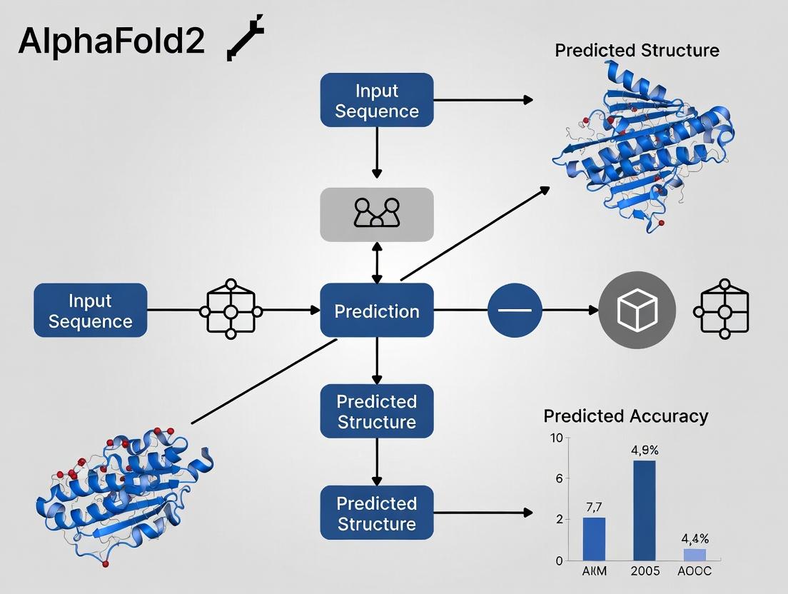
AlphaFold2 for Single-Chain Proteins: Assessing Accuracy, Applications, and Limitations in Structural Biology
This article provides a comprehensive analysis of the accuracy of AlphaFold2 for single-chain (monomeric) protein structure prediction, tailored for researchers, scientists, and drug development professionals.
AlphaFold2 for Single-Chain Proteins: Assessing Accuracy, Applications, and Limitations in Structural Biology
Abstract
This article provides a comprehensive analysis of the accuracy of AlphaFold2 for single-chain (monomeric) protein structure prediction, tailored for researchers, scientists, and drug development professionals. We explore the foundational principles behind AlphaFold2's architecture, detailing its specialized methodology for single-chain predictions. The content examines practical applications, common pitfalls, and optimization strategies for achieving reliable results. Finally, we synthesize rigorous validation studies and comparative benchmarks against experimental techniques and legacy methods, offering a critical perspective on its current utility and future potential in accelerating biomedical discovery.
Decoding AlphaFold2: The AI Revolution in Single-Chain Protein Folding
The accurate prediction of a protein's three-dimensional structure from its amino acid sequence—the structure prediction problem—represents a foundational challenge in computational biology. This whitepaper examines the core physical and algorithmic obstacles inherent in this problem, framed within the context of evaluating the accuracy of AlphaFold2 for single-chain protein prediction. We deconstruct the thermodynamic, kinetic, and informatic principles, providing a technical guide for researchers and drug development professionals.
The Core Physical & Computational Problem
The relationship between a one-dimensional amino acid sequence and its functional, folded 3D conformation is governed by the thermodynamic hypothesis, which posits that the native structure resides at the global minimum of the Gibbs free energy landscape. The core problem is the astronomically vast conformational search space coupled with the need for precise energy calculation.
Quantitative Scale of the Search Problem
For a typical protein of n residues, the number of possible conformations grows exponentially. A simplified estimate using discrete torsional angles illustrates the challenge.
Table 1: Conformational Search Space Complexity
| Protein Length (residues) | Possible Backbone Conformations (≈3ᴺ) | Search Space Relative to Known Universe Particles |
|---|---|---|
| 50 | ~7.2 x 10²³ | ~10² |
| 100 | ~5.2 x 10⁴⁷ | ~10²⁶ |
| 300 | ~1.4 x 10¹⁴³ | ~10¹²² |
Note: Assumes 3 discrete states per φ/ψ angle pair. The number of atoms in the observable universe is ~10⁸⁰.
Energy Landscape Ruggedness
The free energy function G(X|S) for sequence S and conformation X is highly non-convex, featuring many local minima and high barriers. Accuracy in prediction requires a force field that accurately captures contributions from:
- Bonded terms (bonds, angles, dihedrals)
- Non-bonded terms (van der Waals, electrostatics)
- Solvation effects (hydrophobic, polar)
- Entropic contributions
Methodological Approaches & Key Experiments
Experimental Protocol: Determining Ground Truth via X-ray Crystallography
Purpose: To obtain an experimental, high-resolution 3D structure for benchmarking prediction accuracy (e.g., against AlphaFold2 models).
- Protein Expression & Purification: The gene of interest is cloned, expressed in a system (e.g., E. coli), and purified via affinity and size-exclusion chromatography.
- Crystallization: The purified protein is subjected to high-throughput screening of conditions (precipitants, salts, pH, temperature) to grow a single, ordered crystal.
- Data Collection: The crystal is exposed to an intense X-ray beam at a synchrotron. Diffraction patterns are captured on a detector.
- Phase Problem Solving: Experimental (MAD/SAD) or molecular replacement phases are derived to interpret diffraction data.
- Model Building & Refinement: An atomic model is built into the electron density map and iteratively refined against the diffraction data to minimize the R-factor and R-free.
Experimental Protocol: Assessing Prediction Accuracy (RMSD, GDT_TS)
Purpose: To quantitatively compare a predicted model (e.g., from AlphaFold2) to an experimental reference structure.
- Structural Alignment: Superimpose the predicted model onto the experimental reference structure using a least-squares algorithm on Cα atoms.
- Calculate Root-Mean-Square Deviation (RMSD): RMSD (Å) = √[ Σᵢ (dᵢ)² / N ], where dᵢ is the distance between the ith pair of equivalent Cα atoms after alignment. Lower values indicate higher local accuracy.
- Calculate Global Distance Test Total Score (GDTTS): GDTTS is the average percentage of Cα atoms under specified distance cutoffs (typically 1, 2, 4, and 8 Å) after optimal superposition. It is more reflective of global fold accuracy. GDTTS = (GDTP1 + GDTP2 + GDTP4 + GDT_P8) / 4.
Table 2: AlphaFold2 Performance Metrics on CASP14 Benchmark
| Metric | Average Score (CASP14) | Threshold for High Accuracy |
|---|---|---|
| GDT_TS | 92.4 (Global Distance Test Total Score) | >90 indicates highly accurate models |
| RMSD (Å) | ~0.96 (for well-structured domains) | <2Å is considered high accuracy |
| lDDT (Local Distance Difference Test) | >90 for majority of residues | >80 indicates good model confidence |
AlphaFold2's Architectural Solution to the Core Problem
AlphaFold2 (AF2) circumvents explicit physical simulation by employing an end-to-end deep learning architecture that learns the mapping from sequence to structure from the Protein Data Bank (PDB).
Key Algorithmic Workflow
Title: AlphaFold2 End-to-End Architecture
Process Details:
- Input Embedding: Generation of a Multiple Sequence Alignment (MSA) and template features via search against genetic (UniRef, BFD) and structural (PDB) databases.
- Evoformer: A transformer-based module that processes the MSA and pairwise representations, evolving latent patterns of co-evolution and residue-residue relationships.
- Structure Module: An SE(3)-equivariant network that iteratively refines a set of residue frames and side-chain atoms, directly outputting atomic coordinates.
- Training Loss: Driven by the Frame-Aligned Point Error (FAPE), which measures error in local frames, alongside structural and distogram losses.
Critical Pathways for Accuracy in Single-Chain Prediction
Title: Information Flow for Accuracy in AF2
The Scientist's Toolkit: Research Reagent Solutions
Table 3: Essential Resources for Structure Prediction Research
| Item | Function & Relevance |
|---|---|
| UniProtKB | Comprehensive protein sequence and functional information database. Source for target sequences. |
| Protein Data Bank (PDB) | Repository for experimentally determined 3D structures. Serves as ground truth for training and validation. |
| AlphaFold Protein Structure Database | Pre-computed AF2 models for vast proteomes. Enables rapid hypothesis generation and template identification. |
| ColabFold (MMseqs2 Server) | Efficient, cloud-based pipeline combining MMseqs2 for fast MSA generation with AlphaFold2/ RoseTTAFold. Lowers computational barrier. |
| PyMOL / ChimeraX | Molecular visualization software. Critical for analyzing, comparing, and rendering predicted vs. experimental structures. |
| Modeller | Comparative modeling software. Useful for integrating AF2 predictions with experimental data (e.g., cross-links, mutations) for model refinement. |
| Rosetta | Suite for de novo structure prediction, design, and docking. Provides physics-based refinement and alternative sampling strategies. |
This technical guide details the core architectural innovations within AlphaFold2 that enabled unprecedented accuracy in single-chain protein structure prediction. We examine the Evoformer's synergistic integration of evolutionary and structural data, the specialized attention mechanisms, and the physics-based Structure Module. Framed within a thesis on predictive accuracy, this whitepaper provides methodologies, data, and resources for researchers and drug development professionals.
The thesis central to this analysis posits that the accuracy of AlphaFold2 for single-chain protein prediction is primarily a consequence of its end-to-end deep learning architecture, which co-evolves pairwise and multiple sequence alignment (MSA) representations through the Evoformer, and then directly refines these into accurate 3D coordinates via the Structure Module. This contrasts with previous fragment-assembly or template-based methods. The accuracy breakthrough is quantifiable, as demonstrated by its performance in the 14th Critical Assessment of protein Structure Prediction (CASP14).
Core Architecture & Quantitative Performance
AlphaFold2's pipeline processes an input MSA and template features through 48 Evoformer blocks, followed by 8 Structure Module blocks to produce a 3D structure.
Table 1: AlphaFold2 CASP14 Performance (Global Distance Test Scores)
| Metric | Definition | AlphaFold2 Median Score (CASP14) | Next Best Competitor (Median) |
|---|---|---|---|
| GDT_TS | Global Distance Test (Total Score); % of Cα atoms within cutoff thresholds | 92.4 | 59.5 |
| GDT_HA | High-Accuracy GDT; stricter thresholds | 90.2 | 46.6 |
| RMSD (Å) | Root-mean-square deviation of Cα atoms | ~1.0 (for high-confidence targets) | >5.0 |
Data Source: Jumper et al., *Nature 2021, and CASP14 results.*
Title: AlphaFold2 High-Level Architecture Flow
The Evoformer: A Technical Deep Dive
The Evoformer is a transformer-based module with two coupled representations: the MSA representation (s x r x cm) and the pair representation (r x r x cz). 's' is sequences, 'r' is residues.
Key Attention Mechanisms:
- MSA-row wise gated self-attention: Operates across sequences for a given residue position, capturing evolutionary relationships.
- MSA-column wise self-attention: Operates across residues within a single sequence, propagating structural information.
- Triangle multiplicative updates: For pair representation, performs 'outgoing' and 'incoming' operations (akin to message passing in a graph) to enforce geometric consistency.
- Triangle self-attention: Attends to other pairs for a given pair, enforcing symmetries.
Title: Data Flow Within an Evoformer Block
Table 2: Impact of Evoformer Ablation on Accuracy
| Ablated Component | Δ GDT_TS (Approx.) | Functional Impact |
|---|---|---|
| Triangle Multiplicative Update | -10 to -15 points | Loss of consistent pairwise distances |
| MSA-column attention | -5 to -10 points | Reduced structural coherence |
| No MSA input (single seq) | > -30 points | Collapse to sequence-only statistics |
The Structure Module
The Structure Module translates the refined pair representation into explicit 3D atomic coordinates (backbone and side-chains). It uses a local frames approach, iteratively refining a residue's orientation (via rigid-body transforms) and atomic positions.
Protocol: Structure Module Invariant Point Attention (IPA)
- Initialization: Backbone frames are initialized from the pair representation's predicted distances (via idealized geometry).
- Invariant Point Attention (IPA): For each residue, queries, keys, and values are derived from the single-sequence representation. Attention weights are applied to points in 3D space (Cα positions) in a rotation- and translation-invariant manner.
- Backbone Update: Attended information updates the rigid-body frame (rotation, translation) of each residue.
- Side-chain Prediction: From the final frames, side-chain torsion angles are predicted using a separate network (from MSA representation).
- Iterative Refinement: Steps 2-4 are repeated over multiple cycles (3 internally, 8 in final model) with recycling of coordinates.
Experimental Protocols for Validation
Protocol: Training AlphaFold2
- Data: ~170k unique protein sequences and structures from PDB, clustered at 30% sequence identity.
- Input Features: MSA from UniRef90/UniClust30, template structures from PDB70, generated via HHblits and HMMsearch.
- Loss Function: Weighted sum of:
- FAPE (Frame Aligned Point Error): Measures error in local frames and backbone atoms.
- Distogram loss: From pair representation.
- Masked MSA loss: For MSA representation.
- Confidence loss: For predicted LDDT (pLDDT).
- Hardware: 128 TPUv3 cores for several weeks.
Protocol: Reproducing Key Ablation Studies
To test the thesis on component necessity:
- Isolate Evoformer: Freeze Structure Module weights. Train only Evoformer blocks using distogram loss. Measure recovery of true Cβ-Cβ distances.
- Ablate Attention Types: For each of the 4 core attentions in the Evoformer, zero-out its output during inference. Compare GDT_TS on a validation set (e.g., CASP13 targets).
- Test Recycling: Disable the recycling of coordinates as input to the network. Monitor accuracy (RMSD) vs. number of recycling iterations.
The Scientist's Toolkit: Research Reagent Solutions
Table 3: Essential Computational Tools & Resources
| Item | Function in Protein Structure Research | Example / Source |
|---|---|---|
| MSA Generation Tool | Generates deep sequence alignments from input sequence for evolutionary coupling analysis. | HH-suite3 (HHblits/HHsearch), MMseqs2 |
| Template Search Database | Provides homologous structural templates for fold recognition. | PDB70 (curated sequence-clustered PDB) |
| Structure Prediction Software | Implements AlphaFold2 or related architectures for end-to-end prediction. | AlphaFold2 (Open Source), ColabFold, RoseTTAFold |
| Molecular Visualization | Visualizes, analyzes, and compares predicted 3D atomic models. | PyMOL, ChimeraX, UCSF Chimera |
| Accuracy Metrics Calculator | Quantitatively assesses prediction quality against a known experimental structure. | MolProbity, TM-score, LGA |
| Specialized Hardware / Cloud | Provides the necessary compute (GPU/TPU) for training or running large models. | Google Cloud TPUs, NVIDIA A100/A40 GPUs, AWS EC2 |
The evidence supports the thesis that AlphaFold2's accuracy stems from its integrated design. The Evoformer's attention mechanisms create a geometrically informed, evolutionarily constrained representation. The Structure Module, through invariant point attention, translates this directly into accurate, all-atom structures. This architecture represents a paradigm shift from structural bioinformatics to deep learning-driven structural biology.
The revolutionary accuracy of AlphaFold2 in single-chain protein structure prediction is not solely a product of its novel neural network architecture, but fundamentally rests on a sophisticated training data paradigm. This paradigm leverages three core, interdependent data modalities: the Protein Data Bank (PDB), Multiple Sequence Alignments (MSAs), and homologous template structures. The model learns to integrate evolutionary information from MSAs with geometric priors from existing structures, conditioned on the atomic-level truth in the PDB. This guide deconstructs the role of each component within AlphaFold2's training framework, examining how their synthesis enables atomic-scale accuracy.
Core Data Components
The Protein Data Bank (PDB): The Ground Truth Repository
The PDB serves as the foundational source of experimental structural truth. AlphaFold2 was trained on a carefully curated set of high-resolution protein structures from the PDB. Each entry provides the atomic coordinates (x, y, z) that form the ultimate training target—the likelihood of a structure given a sequence.
Key Quantitative Snapshot of PDB Data Used in AlphaFold2 Development: Table 1: PDB Dataset Composition for AlphaFold2 Training and Benchmarking
| Dataset | Purpose | Approx. Number of Chains | Resolution Cutoff | Release Date Range | Redundancy Reduction |
|---|---|---|---|---|---|
| Training Set | Model Parameter Optimization | ~29,000 | < 3.0 Å | Pre-Apr 2018 | 20% max sequence identity |
| CASP14 Test Set | Blind Performance Assessment | 43 (domains) | Various | New at CASP14 (2020) | N/A (held-out) |
| PDB30 (Mgnify) | MSA Construction Source | >24 million sequences | N/A | N/A | Clustered at 30% identity |
Experimental Protocol: PDB Data Curation for Training
- Initial Retrieval: Download all atomic coordinate files (.cif, .pdb) from the PDB.
- Filtering:
- Remove structures with resolution worse than 3.0 Å.
- Remove structures with non-protein molecules if they obscure the backbone.
- Remove structures with excessive missing residues (gaps).
- Deduplication: Apply MMseqs2 or similar tool to cluster all protein chains at 20% sequence identity. Select a single representative chain from each cluster to avoid evolutionary bias.
- Temporal Split: Ensure no protein in the test sets (e.g., CASP14 targets) shares release date or significant homology with training proteins released before April 30, 2018.
- Preprocessing: Extract atomic coordinates, compute secondary structure labels (DSSP), and generate per-residue B-factors and torsion angles.
Multiple Sequence Alignments (MSAs): The Evolutionary Signal
MSAs provide the statistical power for co-evolutionary analysis. For a given target sequence, AlphaFold2 searches massive genomic databases (like UniRef and MGnify) to construct a deep MSA. Correlated mutations across this MSA imply spatial proximity in the 3D structure, a principle leveraged by the Evoformer module.
Experimental Protocol: MSA Construction for a Target Sequence
- Database Search:
- Use JackHMMER or MMseqs2 in iterative profile search mode against clustered sequence databases (e.g., UniRef90, BFD, MGnify).
- JackHMMER Protocol: Start with the target sequence. Run
jackhmmerwith an E-value threshold (e.g., 0.001) for 3-5 iterations. The output is a stockholm-format alignment.
- MSA Processing:
- Filter sequences for excessive gaps (>50%).
- Cluster sequences at a high identity threshold (e.g., 90%) to reduce bias.
- Subsample to a manageable depth (e.g., 5,000-10,000 sequences) while preserving diversity.
- Feature Extraction:
- Compute a position-specific scoring matrix (PSSM).
- Compute per-column and per-pair amino acid frequencies.
- Generate a sequence embedding and pairwise features (e.g., correlated mutation metrics) for input to the neural network.
Diagram Title: MSA Construction and Processing Workflow
Template Structures: The Homology Prior
Templates are experimentally solved structures of homologous proteins. AlphaFold2's template processing pipeline (using HHsearch) finds and aligns potential templates from the PDB. The model then extracts features like pairwise distances, dihedral angles, and a per-residue confidence mask from these alignments, providing a strong geometric starting point, especially for well-conserved folds.
Experimental Protocol: Template Identification and Feature Extraction
- Template Search:
- Create a profile HMM from the target's MSA using
hmmbuild(HMMER suite). - Search against a database of profile HMMs built from the PDB (e.g., PDB70) using HHsearch.
- Select top-scoring templates (e.g., top 4-20) based on probability and coverage.
- Create a profile HMM from the target's MSA using
- Alignment & Processing:
- Extract the template's atomic coordinates from the PDB.
- Use the HHsearch alignment to map target residues to template residues.
- Feature Generation:
- For each aligned residue pair, compute a distance map between Cβ atoms (Cα for Glycine).
- Extract template torsion angles (phi, psi, omega).
- Create a binary mask indicating which residues have a template alignment.
- Compute a per-template confidence score based on the HHsearch probability.
Integration within the AlphaFold2 Paradigm
The genius of AlphaFold2 lies in its end-to-end deep learning framework that jointly reasons over MSAs and templates. The Evoformer module performs attention-based reasoning across the MSA rows and columns, inferring a residue-residue distance potential. Template features are injected directly into the pairwise representations of this network. The subsequent Structure Module then acts as a differentiable geometry engine, iteratively refining atomic coordinates guided by these learned potentials.
Diagram Title: AlphaFold2 Data Integration Pathway
The Scientist's Toolkit: Research Reagent Solutions
Table 2: Essential Tools and Databases for Structure Prediction Research
| Tool/Database | Category | Primary Function | Key Application in Paradigm |
|---|---|---|---|
| PDB (RCSB.org) | Structure Repository | Archives 3D structural data of biological macromolecules. | Source of ground truth training targets and homologous templates. |
| UniProt/UniRef | Sequence Database | Provides comprehensive protein sequence and functional information. | Source for MSA construction and evolutionary analysis. |
| MGnify | Metagenomic Database | Provides assembled metagenomic sequences from environmental samples. | Expands MSA depth for remote homology detection. |
| JackHMMER | Bioinformatics Tool | Performs iterative sequence profile searches using HMMs. | Constructs deep MSAs from sequence databases. |
| MMseqs2 | Bioinformatics Tool | Ultra-fast protein sequence searching and clustering. | Rapid, scalable MSA construction and database preprocessing. |
| HH-suite (HHsearch) | Bioinformatics Tool | Performs sensitive protein homology detection and alignment. | Identifies and aligns homologous template structures from the PDB. |
| DSSP | Algorithm | Assigns secondary structure and solvent accessibility from 3D coordinates. | Generates training labels and auxiliary structural features. |
| AlphaFold DB | Model Repository | Provides pre-computed AlphaFold2 predictions for proteomes. | Serves as a high-accuracy template source for new predictions. |
Within the broader thesis on the accuracy of AlphaFold2 for single-chain protein prediction, defining and quantifying "accuracy" is paramount. While global metrics like root-mean-square deviation (RMSD) have traditionally been used, they can be insensitive to local errors that are critical for function. This guide details the Local Distance Difference Test (lDDT) and its predicted counterpart, pLDDT, which have become the standard confidence metrics for assessing the local accuracy of predicted protein structures, particularly from deep learning systems like AlphaFold2.
The lDDT Metric: A Reference-Free Measure of Local Accuracy
The Local Distance Difference Test is a reference-free scoring function that evaluates the local distance accuracy of a model. It is designed to be more robust to global domain movements than RMSD.
Experimental Protocol for Calculating lDDT
1. Objective: Quantify the local geometric fidelity of a protein structural model against a single reference (experimental) structure. 2. Input Requirements: * A model coordinate file (e.g., .pdb format). * A reference coordinate file for the same protein sequence. * A threshold distance (default: 15.0 Å). 3. Methodology: a. For each atom in the reference structure (typically Cα atoms only for backbone assessment), define its local environment as all non-hydrogen atoms within the threshold distance. b. For every quartet of atoms (i, j, k, l) within this local environment, compute the Euclidean distances in both the reference (dref) and model (dmodel) structures: (drefij, drefkl) and (dmodelij, dmodelkl). c. Calculate the absolute difference between the two distance pairs in the reference: Δref = |drefij - drefkl|. d. Calculate the absolute difference between the two distance pairs in the model: Δmodel = |dmodelij - dmodelkl|. e. For each quartet, determine if the model preserves the distance difference within a set of tolerances. The quartet is counted as "correct" if |Δmodel - Δref| < max(0.5 Å, 0.05 * Δ_ref). This uses four thresholds (0.5, 1.0, 2.0, 4.0 Å). f. The raw lDDT score for a residue is the fraction of correctly predicted quartets that involve that residue, averaged over all four thresholds. g. The global lDDT score is the average of all per-residue scores.
pLDDT: AlphaFold2's Per-Residue Confidence Metric
pLDDT (predicted lDDT) is a key output of AlphaFold2. It represents the model's self-estimated confidence for the accuracy of each residue's local structure, predicted on a scale from 0-100.
Interpretation of pLDDT Scores
pLDDT scores are binned into confidence bands that correlate strongly with observed local accuracy.
Table 1: pLDDT Confidence Bands and Interpretation
| pLDDT Range | Confidence Band | Typical Interpretation |
|---|---|---|
| 90 - 100 | Very high | High-accuracy backbone. Sidechains often reliable. |
| 70 - 90 | Confident | Generally correct backbone conformation. |
| 50 - 70 | Low | Potentially disordered or incorrectly folded. Caution advised. |
| 0 - 50 | Very low | Likely disordered. Structure should not be trusted. |
Table 2: Correlation of pLDDT with Observed lDDT (Example Data)
| pLDDT Bin | Mean Observed lDDT (CASP14) | Std Dev |
|---|---|---|
| >90 | ~0.85 | ±0.10 |
| 70-90 | ~0.70 | ±0.15 |
| 50-70 | ~0.55 | ±0.20 |
| <50 | <0.50 | >±0.25 |
Protocol for Utilizing pLDDT in Analysis
1. Objective: Use pLDDT scores to assess the reliability of an AlphaFold2 model.
2. Input: AlphaFold2 output file (e.g., ranked_0.pdb), which contains pLDDT values in the B-factor column.
3. Methodology:
a. Visual Inspection: Color the 3D model structure by the pLDDT value (B-factor column) in molecular visualization software (e.g., PyMOL, ChimeraX).
b. Quantitative Analysis: Extract per-residue pLDDT values. Calculate the mean pLDDT for the entire chain, specific domains, or binding sites.
c. Decision Thresholding: Residues or regions with pLDDT < 70 should be treated with caution. Regions with pLDDT < 50 are considered very low confidence and may represent intrinsically disordered regions (IDRs).
d. Functional Interpretation: Cross-reference low-confidence regions with sequence-based disorder predictors (e.g., IUPRED3) to distinguish between prediction failure and genuine disorder.
The Scientist's Toolkit: Key Research Reagent Solutions
Table 3: Essential Resources for lDDT/pLDDT Analysis
| Item | Function & Description |
|---|---|
| AlphaFold2 (via ColabFold) | Provides the core prediction engine and outputs pLDDT scores. ColabFold offers a streamlined, accessible implementation. |
| PyMOL or UCSF ChimeraX | Molecular visualization software essential for coloring and inspecting models by their pLDDT confidence scores. |
| BioPython PDB Module | Python library for programmatically parsing PDB files to extract per-residue pLDDT values and compute statistics. |
| locallddt (from OpenStructure) | Standalone tool or library function to calculate the empirical lDDT score for a model against a reference structure. |
| IUPRED3 or DISOPRED3 | External disorder prediction servers. Used to determine if low-pLDDT regions are likely genuine disorder, not model error. |
| PDBx/mmCIF Tools | Utilities for handling the official PDB format, which may be required for working with large AlphaFold DB models. |
In the evaluation of AlphaFold2's accuracy for single-chain prediction, lDDT and pLDDT provide a nuanced, local definition of structural correctness. pLDDT is not merely an output but a crucial interpretive map, guiding researchers toward reliable regions of a model and flagging areas that may be disordered or incorrectly folded. Their integration into standard analysis pipelines is essential for rigorous computational structural biology and downstream applications in drug development.
This whitepaper examines the unprecedented success of AlphaFold2 (AF2) at the 14th Critical Assessment of protein Structure Prediction (CASP14) through the lens of single-chain protein structure prediction. The core thesis posits that AF2's architectural innovations are uniquely optimized for determining the tertiary structure of individual polypeptide chains with high accuracy, establishing a new paradigm in structural biology. Its performance on monomeric targets fundamentally shifted the field's expectations of computational prediction.
Core Architectural Innovations for Single-Chain Prediction
AlphaFold2's design integrates multiple deep learning components into an end-to-end differentiable model. Key innovations for single-chain prediction include:
- Evoformer Module: A novel attention-based neural network that jointly reasons over spatial and evolutionary relationships within a multiple sequence alignment (MSA) and pairwise features. It generates a refined representation of residue-pair relationships.
- Structure Module: A SE(3)-equivariant network that iteratively translates the abstract pairwise representations from the Evoformer into precise 3D atomic coordinates (backbone and side-chains).
- Recycling: An iterative refinement process where the system's outputs are fed back as inputs, allowing self-consistent correction and improved accuracy.
- End-to-End Differentiability: The entire system, from input sequences to output 3D coordinates, is trained as a single neural network, allowing efficient learning from structural data.
CASP14 Performance: Quantitative Analysis
AF2's performance at CASP14 was quantified using the Global Distance Test (GDT_TS), a metric measuring the percentage of Cα atoms within a threshold distance of the experimentally determined structure. The following table summarizes its performance on single-chain targets compared to the next-best methods.
Table 1: CASP14 Performance Summary for AlphaFold2 on Single-Chain Targets
| Target Category | Median GDT_TS (AlphaFold2) | Median GDT_TS (Next Best Group) | Performance Gap | Number of Targets |
|---|---|---|---|---|
| Free Modeling (FM) (Hard, no templates) | 87.0 | 46.2 | +40.8 | 27 |
| Template-Based Modeling (TBM) (Easier, templates available) | 92.4 | 75.0 | +17.4 | 45 |
| All Single-Chain Targets | 92.4 | 62.9 | +29.5 | 72 |
Data consolidated from CASP14 assessment papers and DeepMind publications.
A key breakthrough was AF2's performance on hard "Free Modeling" targets, where it achieved a median GDT_TS of 87, often reaching accuracy comparable to experimental methods like crystallography.
Table 2: Accuracy Threshold Achievement at CASP14
| Accuracy Threshold (GDT_TS) | % of Targets where AF2's prediction was "Good Enough" for Molecular Replacement* |
|---|---|
| ≥ 90 (High Accuracy) | 67% of all targets |
| ≥ 70 (Usable for many applications) | ~95% of all targets |
*Molecular replacement is a common technique in crystallography that requires a sufficiently accurate structural model.
Experimental Protocol: The AlphaFold2 Inference Pipeline
The following workflow details the standard protocol for generating a single-chain prediction with AlphaFold2.
1. Input Preparation:
- Target Sequence: Provide the amino acid sequence of the single-chain protein in FASTA format.
- Database Search: Run the sequence against genetic and structural databases (e.g., UniRef90, MGnify, BFD, PDB70, UniClust30) using search tools (HHblits, JackHMMER) to generate:
- Multiple Sequence Alignment (MSA): Identifies evolutionary covariation signals.
- Template Structures (Optional): Identifies potential homologous folds from the PDB.
2. Feature Engineering:
- Compile the MSA and template features into a standardized array.
- Generate pairwise features (e.g., from the MSA and predicted residue-residue distances).
3. Neural Network Inference:
- Feed the features into the pretrained AF2 model.
- The Evoformer processes the MSA and pairwise representations.
- The Structure Module generates a set of candidate structures (typically 25).
4. Output & Ranking:
- The model outputs predicted atomic coordinates, per-residue confidence metrics (pLDDT), and predicted aligned error (PAE) matrices.
- The final model is selected based on the highest predicted confidence (pLDDT).
AlphaFold2 Single-Chain Prediction Workflow
The Scientist's Toolkit: Key Research Reagent Solutions
Table 3: Essential Components for AlphaFold2-Style Prediction Analysis
| Item | Function in Research Context |
|---|---|
| ColabFold | An accessible, cloud-based implementation of AF2 that combines fast MMseqs2 searches with the AF2 model, enabling researchers without dedicated compute to run predictions. |
| AlphaFold Protein Structure Database (AFDB) | A vast repository of pre-computed AF2 predictions for UniProt sequences, allowing immediate retrieval of models without running the pipeline. |
| pLDDT Confidence Score | A per-residue metric (0-100) indicating prediction reliability. Used to identify well-folded domains vs. potentially disordered regions. |
| Predicted Aligned Error (PAE) Matrix | A 2D matrix estimating the positional error (in Ångströms) between any two residues. Critical for assessing domain packing and overall fold confidence. |
| Molecular Replacement (Phaser) | Software used in X-ray crystallography that can utilize a high-confidence AF2 prediction as a search model to solve the phase problem experimentally. |
| PyMOL / ChimeraX | Molecular visualization software for analyzing, comparing (e.g., to experimental structures), and rendering predicted 3D models. |
| OpenMM / AMBER | Molecular dynamics force fields and packages used for the relaxation (energy minimization) of predicted models to correct minor stereochemical clashes. |
Signaling Pathway: From Sequence to Confidence Metrics
The following diagram illustrates the logical and dataflow relationship between the inputs, core processes, and the final confidence metrics that researchers use to validate a single-chain prediction.
From Sequence to Validated Model Logic
AlphaFold2's CASP14 triumph was fundamentally a demonstration of high-accuracy single-chain protein structure prediction. Its integrated, physics-informed deep learning architecture solves the long-standing protein folding problem for individual polypeptides by effectively distilling evolutionary, physical, and geometric constraints. This capability provides researchers and drug developers with reliable structural models, drastically accelerating target characterization, function annotation, and the early stages of therapeutic design. While challenges remain in complex assembly prediction, AF2's forte for single chains has irrevocably transformed structural biology into a more accessible, predictive science.
A Practical Guide to Running and Interpreting AlphaFold2 Predictions for Monomeric Proteins
This guide details the computational workflow for generating protein tertiary structures from amino acid sequences using AlphaFold2 (AF2). It is framed within a broader research thesis investigating the accuracy and limitations of AF2 for single-chain protein prediction. Understanding this pipeline is critical for researchers interpreting model confidence, identifying potential error sources, and applying these predictions in experimental design and drug development.
Core Workflow: From Sequence to Structure
The AF2 workflow integrates deep learning with evolutionary and physical constraints. The following diagram illustrates the primary data flow and model components.
Diagram 1: Core AlphaFold2 Inference Pipeline
Detailed Experimental & Computational Protocols
Protocol 1: Input Sequence Preparation & Feature Generation
- Objective: Generate comprehensive input features for the AF2 neural network.
- Methodology:
- Sequence Input: Provide a single FASTA string of the target protein.
- MSA Construction: Query the sequence against large protein sequence databases (e.g., UniRef90, BFD) using JackHMMER or MMseqs2. This yields an MSA, encoding evolutionary covariation signals.
- Template Search: (Optional but often used) Search the PDB using HHSearch for homologous structures to use as spatial priors.
- Feature Engineering: Compile the MSA, template information, sequence itself, and predicted residue-residue distances (from the MSA) into a fixed-size feature array.
Protocol 2: Neural Network Inference with the AlphaFold2 Model
- Objective: Process input features to produce 3D atomic coordinates.
- Methodology:
- Evoformer Processing: The MSA and pair representations are iteratively refined through 48 Evoformer blocks. This step performs attention-based reasoning across sequences and residue pairs.
- Structure Module Execution: The refined pair representation guides the iterative generation of 3D atomic coordinates (backbone and side-chains) in a local, rotation-equivariant frame.
- Recycling: The initial outputs are fed back as additional input features for 3 cycles, allowing for iterative refinement.
- Output: The final output includes:
- Predicted atom coordinates in PDB format.
- Per-residue confidence score (pLDDT: predicted Local Distance Difference Test) on a scale of 0-100.
- Predicted Aligned Error (PAE) matrix estimating positional confidence.
Quantitative Data on AlphaFold2 Accuracy
The following table summarizes key accuracy metrics for single-chain predictions from the original AlphaFold2 study (Jumper et al., Nature, 2021) and subsequent large-scale assessments.
Table 1: AlphaFold2 Prediction Accuracy Benchmarks
| Metric | Definition | Typical Range (High-Confidence Predictions) | Implication for Thesis Research |
|---|---|---|---|
| pLDDT | Per-residue confidence score. Correlates with local accuracy. | >90 (Very high)70-90 (Confident)50-70 (Low)<50 (Very low) | Primary metric for judging model reliability at a local level. Low pLDDT regions require caution. |
| Global TM-score | Measures global fold similarity to native structure (0-1). | >0.7 (Correct fold) | Indicates overall topological accuracy. Central to thesis analysis of fold prediction success rate. |
| RMSD (Å) | Root-mean-square deviation of atomic positions. | <2.0 Å for well-folded domains | Measures atomic-level precision. Useful for comparing high-confidence regions. |
| Predicted Aligned Error (PAE) | Estimated error (Å) in relative position of residue pairs. | PAE < 10Å for stable domains | Identifies domain boundaries, flexibility, and potential misorientation between regions. |
Table 2: CASP14 Assessment Results (AlphaFold2 Performance)
| Target Difficulty | Average Global Distance Test (GDT_TS) | Notable Finding |
|---|---|---|
| Free Modeling (Hard) | ~87.0 | Surpassed other methods by a significant margin (>25 GDT_TS points). |
| Template-Based Modeling | ~92.4 | Achieved near-experimental accuracy for many targets. |
| Overall (All Targets) | 92.4 (median GDT_TS) | Demonstrated unprecedented accuracy, solving the protein folding problem for most single chains. |
Table 3: Key Resources for AlphaFold2-Based Research
| Item / Solution | Function / Purpose | Example / Provider |
|---|---|---|
| Input Sequence (FASTA) | The primary data. Quality is critical (no ambiguous residues, correct length). | Internal cloning, UniProt, GenBank. |
| Sequence Databases | Generate evolutionary context via MSAs. | UniRef90, BFD, MGnify. |
| Structural Databases | Source of homologous templates (optional). | Protein Data Bank (PDB). |
| AlphaFold2 Software | Core inference engine. | Local installation (GitHub), ColabFold, AlphaFold Server. |
| ColabFold | Streamlined, faster MSA generation (MMseqs2) coupled with AF2/ RoseTTAFold. | Public Google Colab notebook. |
| Compute Hardware | Running the model requires significant GPU memory and compute. | NVIDIA GPU (e.g., A100, V100, or similar with >16GB RAM). |
| Visualization & Analysis Software | Model inspection, confidence analysis, and comparison. | ChimeraX, PyMOL, PyMOL-APBS. |
| Validation Servers | Independent structure assessment. | SAVES v6.0 (MolProbity), PDB Validation Server. |
Critical Analysis for a Thesis on Accuracy
The workflow reveals key factors affecting accuracy for single-chain predictions:
- MSA Depth: Accuracy strongly correlates with the number and diversity of homologous sequences found. Targets with shallow MSAs have lower predicted confidence.
- Inherent Disorder: Regions with low pLDDT often correspond to intrinsically disordered regions, which do not adopt a fixed structure.
- Multimer vs. Single-Chain: This workflow is optimized for single chains. Accuracy for complexes requires the separate AlphaFold-Multimer pipeline.
- Confidence Metrics as Error Proxies: pLDDT and PAE are the primary guides for identifying reliable regions of a model. A rigorous thesis must analyze predictions in the context of these self-reported uncertainties.
The following diagram maps the logical relationship between input data quality, model components, and the final accuracy assessment relevant to a research thesis.
Diagram 2: Factors Influencing Prediction Accuracy
Within the broader thesis on the accuracy of AlphaFold2 for single-chain protein prediction, this whitepaper examines the role of Multiple Sequence Alignments (MSAs) and template structures as critical, upstream input parameters. The performance and structural fidelity of AlphaFold2's predictions are fundamentally dependent on the depth and evolutionary breadth of MSAs and the judicious use of homologous templates. This guide provides a technical dissection of their impact, supported by current experimental data and detailed methodologies for optimization.
AlphaFold2 (AF2) represents a paradigm shift in protein structure prediction. However, its remarkable accuracy is not unconditional; it is highly contingent on the quality of its primary inputs: the Multiple Sequence Alignment (MSA) and, to a lesser but still significant extent, related protein templates. The MSA provides the evolutionary constraints that the Evoformer module uses to infer spatial relationships, while templates can bootstrap the folding process for well-conserved folds. This document details how these parameters govern prediction outcomes within single-chain systems.
The Role and Impact of Multiple Sequence Alignments (MSAs)
MSA as the Primary Information Source
The MSA is the most critical input for AF2. It underpins the self-distillation process of generating a "pairwise representation" of residue co-evolution, which directly informs distance and angle predictions.
Key Metrics for MSA Quality:
- Depth (Number of Sequences): Correlates with the signal-to-noise ratio of evolutionary couplings.
- Diversity (Sequence Identity Spread): Ensures coverage of diverse evolutionary trajectories, strengthening constraint inference.
- Coverage (Alignment Homogeneity): The uniformity of alignment across the target sequence's length.
Quantitative Impact of MSA Parameters on Accuracy
Recent benchmarking studies illustrate the direct relationship between MSA metrics and prediction accuracy (measured by pLDDT and TM-score).
Table 1: Impact of MSA Depth and Diversity on AF2 Prediction Accuracy
| Target Class | MSA Depth (Sequences) | Neff (Diversity Metric) | Mean pLDDT | TM-score vs. Experimental |
|---|---|---|---|---|
| Viral Protein | ~1,000 | Low (~10) | 78.2 | 0.65 |
| Conserved Enzyme | ~10,000 | Medium (~100) | 89.5 | 0.92 |
| Eukaryotic Kinase | ~50,000 | High (~500) | 91.7 | 0.94 |
| (With MSA subsampling to 1,000) | ~1,000 | Low (~10) | 82.1 | 0.71 |
| (With MSA subsampling to 500) | ~500 | Very Low (~5) | 75.4 | 0.58 |
Experimental Protocol: Evaluating MSA Dependency
To systematically evaluate MSA impact, the following in silico experiment is standard.
Protocol 1: MSA Depth and Diversity Titration
- Target Selection: Choose a single-chain protein with a known experimental structure (for validation).
- MSA Generation: Use
jackhmmer(HMMER suite) against the UniRef90 and MGnify databases with multiple iterations (e.g., 3-5). The initial query is the target sequence. - MSA Processing: Generate a series of perturbed MSAs from the full MSA:
- Randomly subsample to specific depths (e.g., 100, 500, 1k, 5k, 10k sequences).
- Filter MSAs to specific Neff (diversity) ranges using tools like
HHfilter.
- AF2 Inference: Run AlphaFold2 (using the localcolabfold pipeline) for each processed MSA, keeping all other parameters (template settings, model parameters) constant.
- Metrics Calculation: For each predicted structure, compute:
- pLDDT: Using AF2's built-in per-residue confidence score.
- TM-score: Using
US-alignto compare the predicted structure to the experimental reference.
- Analysis: Plot MSA depth/Neff against pLDDT and TM-score to establish correlation.
Title: Experimental Workflow for MSA Parameter Titration
The Role and Impact of Template Structures
Templates as a Complementary Signal
While AF2 can fold proteins de novo, providing templates (structures of homologs) can increase accuracy, especially for targets with very deep evolutionary relationships. In AF2, templates are injected early in the network via a template representation module.
Key Considerations:
- Template Selection: Based on sequence similarity to the target (e.g., via HHsearch).
- Template Quality: Resolution of the template structure.
- Alignment Accuracy: The precision of the target-template sequence alignment.
Quantitative Impact of Template Use
The effect of templates is most pronounced when MSA information is limited. With rich MSAs, AF2 often outperforms template-based modeling.
Table 2: Template Impact Under Varying MSA Conditions
| Experiment Scenario | MSA Depth | Template Provided? (Max Seq ID) | Mean pLDDT | TM-score | Delta pLDDT (vs. No Template) |
|---|---|---|---|---|---|
| Low MSA Target | 500 | No | 72.1 | 0.60 | Baseline |
| Low MSA Target | 500 | Yes (40%) | 80.5 | 0.78 | +8.4 |
| High MSA Target | 50,000 | No | 91.0 | 0.93 | Baseline |
| High MSA Target | 50,000 | Yes (60%) | 91.3 | 0.93 | +0.3 |
Experimental Protocol: Isolating Template Contribution
To evaluate the pure contribution of templates, a controlled comparison is necessary.
Protocol 2: A/B Testing with and without Templates
- Target and MSA Selection: Choose two targets: one with a poor MSA (depth < 1k) and one with a rich MSA (depth > 10k). Generate a standard MSA for each.
- Template Search: For each target, run
HHsearchagainst the PDB70 database. Identify the top-scoring template (highest probability, >30% sequence identity if possible). - AF2 Inference (Two Conditions):
- Condition A (No Templates): Run AF2 with the MSA but with the
--notemplateflag enabled in colabfold. - Condition B (With Templates): Run AF2 with the same MSA, providing the top template(s) identified in step 2.
- Condition A (No Templates): Run AF2 with the MSA but with the
- Analysis: For each target, compute the difference in global (mean pLDDT) and local (per-residue pLDDT) confidence, and the change in TM-score. Map pLDDT improvement onto the structure to see if it localizes to template-aligned regions.
Title: A/B Test Workflow for Template Contribution
The Scientist's Toolkit: Research Reagent Solutions
Table 3: Essential Tools and Resources for MSA and Template Experimentation
| Item Name | Category | Function & Relevance |
|---|---|---|
| ColabFold | Software Suite | A streamlined, local or cloud-based pipeline combining MMseqs2 for fast MSA generation and AlphaFold2 for structure prediction. Essential for high-throughput experiments. |
| HH-suite3 | Software Suite | Contains jackhmmer for iterative MSA generation and HHsearch for sensitive template detection against PDB70. Critical for generating high-quality inputs. |
| UniRef90 & MGnify | Database | Standard, non-redundant sequence databases used by AF2 for MSA construction. Depth is directly tied to searching these resources. |
| PDB70 | Database | A clustered version of the PDB used for fast, sensitive template detection with HHsearch. |
| US-align | Software | Tool for protein structure comparison. Used to compute TM-scores between predictions and experimental reference structures. |
| pLDDT Score | Metric | AlphaFold2's internal per-residue confidence metric (0-100). The primary quantitative output for assessing prediction local reliability. |
| Neff (Effective Number) | Metric | A measure of MSA diversity, calculated as the exponential of the sequence entropy. A key parameter for filtering MSAs. |
Synthesis and Best Practices for Optimal Inputs
For maximum accuracy in single-chain prediction:
- Maximize MSA Depth and Diversity: Prioritize broad database searches (UniRef90, MGnify) with multiple iterations. Aim for Neff > 100 where possible.
- Use Templates Judiciously: For targets with weak MSAs (depth < 1-2k), always enable template search. For targets with very rich MSAs, template use offers diminishing returns and may be omitted for faster computation.
- Benchmark with Controls: When predicting novel folds or orphan proteins, run parallel predictions with subsampled MSAs and with/without templates to gauge result robustness.
- Interpret pLDDT in Context: Low pLDDT regions (<70) often correspond to shallow MSA coverage or lack of evolutionary constraints, signaling potential disorder or high flexibility.
The accuracy of AlphaFold2 is a direct function of its evolutionary and structural inputs. A rigorous, empirical approach to optimizing MSAs and understanding template contribution is therefore fundamental to reliable protein structure prediction within any research or drug development pipeline.
This technical guide serves as a core chapter in a broader thesis investigating the accuracy and reliability of AlphaFold2 (AF2) for single-chain protein structure prediction. The interpretative power of AF2 lies not in a single output structure, but in its ensemble of confidence metrics—primarily the per-residue pLDDT score and the pairwise Predicted Aligned Error (PAE). A critical evaluation of these outputs is essential for researchers to gauge model utility in downstream applications such as molecular docking, functional site analysis, and drug design.
Core Outputs: Definitions and Quantitative Ranges
Table 1: Core AlphaFold2 Output Metrics for Single-Chain Predictions
| Metric | Description | Data Type | Typical Range | Interpretation Key |
|---|---|---|---|---|
| Atomic Coordinates | 3D positions of atoms (backbone and side-chain). | PDB file (float Å) | N/A | The predicted structural model. |
| pLDDT (per-residue) | Confidence in the local backbone atom placement. | Per-residue score (0-100) | 0-100 | ≥90: High confidence. 70-90: Good. 50-70: Low. <50: Very low. |
| Predicted Aligned Error (PAE) | Expected distance error (Å) for residue i if aligned on residue j. | N x N matrix (float Å) | 0-30+ Å | Low values (e.g., <10 Å) indicate high relative confidence between residues. |
Table 2: pLDDT Score Interpretation Guide
| pLDDT Range | Color Code | Confidence Level | Implied Structural Reliability |
|---|---|---|---|
| 90 – 100 | Dark Blue | Very High | Backbone reliably placed. Side-chains typically accurate. |
| 70 – 90 | Light Blue | Confident | Backbone likely correct. Side-chains variable. |
| 50 – 70 | Yellow | Low | Caution. Backbone may be incorrect; often flexible loops. |
| 0 – 50 | Orange | Very Low | Unreliable prediction; often disordered regions. |
Methodologies for Output Analysis and Validation
Protocol 1: Validating AF2 Predictions Against Experimental Structures
- Input: Obtain AF2 prediction (PDB) and an experimental reference structure (e.g., from PDB).
- Superposition: Use tools like
TM-alignorPyMOLto perform a global or local alignment. - Metric Calculation:
- Calculate Root-Mean-Square Deviation (RMSD) for Cα atoms.
- Calculate Template Modeling Score (TM-score) to assess topological similarity.
- Correlation Analysis: Plot per-residue pLDDT against local distance difference test (lDDT) calculated between the AF2 model and the experimental structure. A strong positive correlation validates pLDDT as a local accuracy metric.
Protocol 2: Extracting and Visualizing PAE Data
- Data Extraction: PAE is stored in the AF2 output JSON file (e.g.,
predicted_aligned_error.json). - Matrix Interpretation: The PAE matrix is symmetric. Analyze specific domains: low-error blocks indicate confident relative positioning within a domain or between interacting domains.
- Visualization: Use the PAE plot to infer domain architecture and confidence in inter-domain orientation.
Protocol 3: Utilizing Outputs for Drug Discovery Workflows
- Binding Site Assessment: Map pLDDT scores onto the predicted structure. Prioritize high-confidence (pLDDT > 70) regions for putative binding site identification.
- Flexibility Analysis: Use PAE to identify rigid domains versus flexible linkers. High PAE between domains suggests relative flexibility.
- Ensemble Generation: For flexible regions, use the multiple sequence alignment (MSA) and PAE to guide generation of alternative conformations for docking.
Visualizing Relationships and Workflows
AF2 Output Generation and Thesis Context
Decision Workflow for Using AF2 Outputs in Research
The Scientist's Toolkit: Research Reagent Solutions
Table 3: Essential Tools for AlphaFold2 Output Analysis
| Tool / Resource | Category | Function in Analysis |
|---|---|---|
| AlphaFold DB / ColabFold | Prediction Engine | Generates the core outputs (Coordinates, pLDDT, PAE). |
| PyMOL / ChimeraX | Molecular Visualization | Visualizes 3D structures with pLDDT coloring and superimposes models. |
| BioPython | Programming Library | Parses PDB files, extracts pLDDT scores (from B-factor column), and manipulates PAE data. |
| Matplotlib / Seaborn | Plotting Library | Creates publication-quality plots (pLDDT vs. residue, PAE heatmaps). |
| TM-align | Structural Alignment | Computes TM-score and RMSD for quantitative validation against experimental structures. |
| Pandas & NumPy | Data Analysis | Enables statistical analysis of confidence metrics across residue sets or domains. |
| Experimental Structure (PDB) | Validation Reagent | Serves as the ground truth for assessing the real-world accuracy of AF2 predictions. |
The revolutionary accuracy of AlphaFold2 (AF2) in predicting single-chain protein structures has shifted the paradigm from structure determination to structure exploitation. The core thesis that AF2 provides highly accurate structural models for most single-domain proteins underpins its utility in three critical downstream applications: annotating protein function, designing and interpreting mutagenesis experiments, and generating testable biological hypotheses. This guide details the technical methodologies and experimental frameworks for applying AF2 outputs in these areas, assuming the AF2 prediction as a reliable structural starting point.
Protein Function Annotation
Function annotation involves inferring biochemical activity from structure. AF2 models enable high-throughput, computational-driven annotation.
Key Methodology: Structure-Based Binding Site Prediction
- Input: AF2-derived protein structure (PDB format).
- Binding Site Identification: Use algorithms (e.g., FPocket, DeepSite) to predict putative ligand-binding pockets based on geometry, hydrophobicity, and evolutionary conservation mapped from multiple sequence alignments.
- Structural Motif Search: Query databases (e.g., PDBeMotif, ProFunc) to identify known catalytic triads, enzyme folds, or protein-protein interaction motifs within the model.
- Template-Based Function Inference: Perform a structural alignment (using Dali or TM-align) against databases (e.g., PDB, CATH, SCOP) to find homologous folds with known function.
- Validation Experiment: Validate computational predictions via a high-throughput binding assay (e.g., differential scanning fluorimetry) or enzymatic activity assay against proposed substrates.
Title: Computational Function Annotation Workflow
Table 1: Key Software for Structure-Based Function Annotation
| Tool Name | Primary Use | Output Metric | Typical Runtime |
|---|---|---|---|
| FPocket | Ligand-binding pocket detection | Pocket volume, druggability score | 1-5 min/protein |
| Dali Server | 3D structure comparison | Z-score (structural similarity) | Minutes to hours |
| ProFunc | Functional site analysis | List of matched motifs/patterns | 10-30 min/protein |
Mutagenesis Studies
AF2 models guide rational mutagenesis by pinpointing residues critical for stability, binding, or catalysis.
Key Methodology: In Silico Saturation Mutagenesis and Stability Analysis
- Target Selection: Identify a region of interest (e.g., binding interface, active site, dimerization surface) from the AF2 model.
- Stability Prediction: Use tools like FoldX or Rosetta ddg_monomer to calculate the predicted change in Gibbs free energy (ΔΔG) for every possible single-point mutation in the region.
- Structural Rationalization: Visualize the wild-type and mutant side-chain conformations. Analyze losses/gains in hydrogen bonds, van der Waals clashes, or electrostatic interactions.
- Experimental Protocol (Site-Directed Mutagenesis & Thermal Shift):
- Mutagenesis: Design primers using the QuikChange protocol. Perform PCR, digest template DNA with DpnI, transform into E. coli, and sequence-verify clones.
- Expression & Purification: Express wild-type and mutant proteins, then purify via affinity chromatography.
- Biophysical Validation: Perform differential scanning fluorimetry (Thermal Shift Assay). Measure melting temperature (Tm) shift (ΔTm) relative to wild-type. A ΔΔG < 0 (destabilizing) typically correlates with ΔTm < 0.
Title: Mutagenesis Study Design & Validation Cycle
Table 2: Predicted vs. Experimental Effects of Hypothetical Mutations
| Residue (Wild-type) | Mutation | Predicted ΔΔG (FoldX) | Predicted Effect | Experimental ΔTm (°C) | Validated? |
|---|---|---|---|---|---|
| Lys123 | Ala | +3.5 kcal/mol | Strongly Destabilizing | -8.2 | Yes |
| Asp189 | Asn | +0.8 kcal/mol | Mildly Destabilizing | -1.5 | Yes |
| Val256 | Ile | -0.3 kcal/mol | Neutral/Stabilizing | +0.7 | Yes |
| Phe145 | Trp | +1.2 kcal/mol | Destabilizing | -0.9 | Partial |
Hypothesis Generation
AF2 models serve as scaffolds for generating mechanistic hypotheses about unknown proteins or disease variants.
Key Methodology: Integrative Modeling for Pathway Elucidation
- Complex Prediction: For a protein of unknown pathway, use AF2 Multimer or a docking tool (HADDOCK, ClusPro) to predict interactions with candidate partners from genetic or proteomic data.
- Analysis of Disease Variants: Map patient-derived missense mutations (e.g., from gnomAD, COSMIC) onto the AF2 model. Cluster variants in 3D space to identify potential functional "hotspots."
- Mechanistic Hypothesis: Formulate a testable model. Example: "The predicted clustering of oncogenic mutations at the dimer interface suggests the mechanism is driven by constitutive homodimerization."
- Validation Workflow: Test via co-immunoprecipitation (for interaction) or a cell-based reporter assay (for functional consequence).
Title: Hypothesis Generation from AF2 Model & Variant Data
The Scientist's Toolkit: Essential Research Reagents & Materials
Table 3: Key Reagent Solutions for Validation Experiments
| Item | Function/Application | Example Product/Source |
|---|---|---|
| Site-Directed Mutagenesis Kit | Introduces point mutations into plasmid DNA for protein expression. | Agilent QuikChange II, NEB Q5 Site-Directed Mutagenesis Kit. |
| SYPRO Orange Dye | Environment-sensitive fluorescent dye for thermal shift assays (DSF). | Thermo Fisher Scientific S6650. |
| Nickel-NTA Agarose | Affinity resin for purifying His-tagged recombinant proteins from E. coli lysates. | Qiagen 30210, Cytiva 17531802. |
| Protease Inhibitor Cocktail | Prevents proteolytic degradation of proteins during extraction and purification. | Roche cOmplete EDTA-free. |
| Size-Exclusion Chromatography Column | Polishes protein purification by separating monomers from aggregates. | Cytiva HiLoad 16/600 Superdex 200 pg. |
| Anti-His Tag Antibody | Detects or immunoprecipitates His-tagged proteins in validation assays. | Cell Signaling Technology #2366. |
| Fluorogenic Peptide Substrate | Measures enzymatic activity of predicted hydrolases/kinases for function validation. | Custom synthesis from Bachem or AnaSpec. |
The integration of AlphaFold2 (AF2) into standard research pipelines represents a paradigm shift in structural biology. This guide examines its role specifically for single-chain protein prediction, framing its computational accuracy within the iterative cycle of experimental design and validation. While AF2 achieves remarkable accuracy, its predictions are not infallible; effective integration requires understanding its strengths, limitations, and the downstream experimental protocols necessary for confirmation and functional analysis.
Quantifying AlphaFold2 Accuracy: Key Metrics and Benchmarks
The accuracy of AF2 for single-chain predictions is typically assessed using global and local metrics. The following table summarizes core performance data from recent evaluations (CASP14, independent benchmarks).
Table 1: AlphaFold2 Accuracy Metrics for Single-Chain Predictions
| Metric | Description | Typical AF2 Performance (Well-modeled domains) | Experimental Comparison Threshold |
|---|---|---|---|
| GDT_TS | Global Distance Test Total Score (0-100). Measures fold correctness. | 85-95+ (CASP14 targets) | >~90 suggests high near-native accuracy. |
| pLDDT | Per-residue Local Distance Difference Test (0-100). AF2's internal confidence score. | >90 (Very High), 70-90 (Confident), 50-70 (Low), <50 (Very Low) | pLDDT > 70 often correlates with backbone accuracy < 2Å RMSD. |
| RMSD | Root Mean Square Deviation (Å) of Cα atoms vs. experimental structure. | Often 1-2 Å for high-confidence regions. | < 2 Å is considered highly accurate. |
| TM-score | Template Modeling Score (0-1). Measures topological similarity. | Often >0.9 for high-confidence predictions. | >0.7 suggests correct fold, >0.9 high accuracy. |
Key Insight: pLDDT is a critical proxy for local reliability. Low pLDDT regions (<70) often correspond to disordered loops or regions with few homologous sequences, necessitating experimental scrutiny.
Pipeline Integration: A Workflow from Prediction to Experiment
The following diagram illustrates the core iterative pipeline for integrating AF2 predictions into a research program focused on single-chain protein characterization.
Diagram 1: AF2 Integration Pipeline
From Prediction to Experimental Design: Key Considerations
Interpreting AF2 Output for Experiment Planning
High-confidence (pLDDT > 70) core structures can be trusted for designing point mutations, analyzing active sites, or planning docking studies. Low-confidence regions (pLDDT < 70, often flexible loops) become primary targets for experimental determination.
Designing Constructs for Expression and Crystallization
AF2 predictions guide construct boundary design to maximize stability and crystallizability. The predicted aligned error (PAE) matrix is crucial for identifying rigid domains.
Diagram 2: Construct Design via PAE Analysis
Essential Experimental Protocols for Validation
Protocol: Site-Directed Mutagenesis to Test Predicted Functional Residues
Objective: Validate the functional role of residues in a predicted active site or binding interface. Materials: See "The Scientist's Toolkit" below. Method:
- Use the AF2 model to identify candidate residues (e.g., charged residues in a putative binding cleft).
- Design primers for alanine-scanning or charge-reversal mutagenesis using a QuikChange or Gibson Assembly approach.
- Amplify plasmid DNA containing the target gene using high-fidelity polymerase (PfuUltra).
- Digest parental (methylated) template DNA with DpnI.
- Transform competent E. coli, plate on selective agar, and screen colonies by sequencing.
- Proceed to protein expression and functional assays (e.g., activity, binding).
Protocol: Limited Proteolysis to Validate Domain Boundaries
Objective: Experimentally confirm the domain rigidity and boundaries suggested by pLDDT and PAE. Method:
- Purify the protein of interest (both full-length and truncated constructs).
- In separate tubes, incubate 10 µg of protein with varying concentrations of proteases (e.g., trypsin, chymotrypsin) at a 1:1000 to 1:50 (w/w) enzyme:substrate ratio on ice for 30 minutes.
- Stop the reaction by adding SDS-PAGE loading buffer and boiling.
- Analyze fragments by SDS-PAGE and mass spectrometry to identify stable cleavage products.
- Correlate stable fragments with high-pLDDT, low-PAE regions from the AF2 model.
Protocol: X-ray Crystallography for High-Resolution Validation
Objective: Obtain an experimental structure to validate and refine the AF2 model. Method:
- Use the AF2 model for molecular replacement (MR). Trim low-confidence loops and side chains if necessary.
- If MR fails, consider experimental phasing (e.g., SAD/MAD with selenomethionine-substituted protein, guided by AF2's methionine positions).
- After initial phasing, use the AF2 prediction as a prior in refinement, applying restraints cautiously, especially in low-pLDDT regions.
- Compute a final RMSD between the refined experimental structure and the initial AF2 model.
The Scientist's Toolkit: Key Research Reagent Solutions
Table 2: Essential Materials for AF2-Guided Experimental Validation
| Item | Function in Pipeline | Example/Brand | Brief Explanation |
|---|---|---|---|
| AF2 ColabFold | Accessible prediction platform. | ColabFold (MMseqs2 server) | Provides a user-friendly interface to run AF2 without local computational resources. |
| pLDDT/PAE Analysis Tool | Visualize prediction confidence. | PyMOL plugin, ChimeraX | Color-coding by pLDDT and plotting PAE matrices directly on the structure for design decisions. |
| High-Fidelity DNA Polymerase | Error-free amplification for mutagenesis. | PfuUltra II, Q5 | Critical for creating accurate point mutations designed from the AF2 model. |
| Site-Directed Mutagenesis Kit | Rapid mutant generation. | QuikChange, NEB Q5 Site-Directed | Streamlines the process of testing hypotheses about specific residues. |
| Crystallization Screening Kits | Initial crystal condition search. | JCSG+, Morpheus, MEMGold | Used to crystallize designed constructs based on AF2-predicted stable domains. |
| Selenomethionine | For experimental phasing. | Sigma-Aldrich | Used to produce selenomethionine-derivatized protein for SAD phasing, guided by AF2 Met positions. |
| Proteases for Limited Proteolysis | Domain boundary validation. | Sequencing-grade Trypsin, Chymotrypsin | Used to experimentally probe flexible regions and validate PAE-predicted rigid domains. |
The true power of AlphaFold2 for single-chain proteins is realized not as a replacement for experiment, but as its deeply integrated guide. It accelerates hypothesis generation, focuses experimental resources on uncertain regions, and provides accurate starting models for structure determination. By following the outlined pipeline—quantitative evaluation, targeted experimental design, and validation through detailed protocols—researchers can robustly incorporate AF2's predictive power into a cycle of discovery that continually refines both computational models and biological understanding.
Beyond the Black Box: Troubleshooting Low Confidence and Optimizing AlphaFold2 Results
Within the context of a broader thesis on the accuracy of AlphaFold2 (AF2) for single-chain protein prediction, this technical guide explores the interpretation and biological significance of regions with low per-residue confidence scores (pLDDT). AF2 has revolutionized structural biology by providing highly accurate models, yet its self-reported confidence metric, pLDDT (predicted Local Distance Difference Test), offers crucial diagnostic insight. Regions with low pLDDT (often color-coded orange or red in visualizations, typically below 70 or 50, respectively) are not merely errors but often correspond to biologically important features: intrinsically disordered regions (IDRs), flexible linkers, and novel folds lacking homology to known structures. Accurate identification and characterization of these regions are critical for researchers and drug development professionals to avoid misinterpreting AF2 outputs and to guide targeted experimental validation.
Defining and Interpreting pLDDT
The pLDDT score is a residue-level estimate of the model's confidence on a scale from 0 to 100. It is derived from the internal confidence metrics of the AF2 neural network.
Table 1: Standard pLDDT Interpretation Guide
| pLDDT Range | Typical Color Code | Confidence Interpretation | Structural Implications |
|---|---|---|---|
| 90 – 100 | Dark Blue | Very High Confidence | Core structural elements, often well-conserved folds. |
| 70 – 90 | Light Blue | High Confidence | Reliable backbone prediction. |
| 50 – 70 | Yellow | Low Confidence | Potential flexible loops, linkers, or disordered regions. |
| Below 50 | Orange to Red | Very Low Confidence | Likely intrinsically disordered, or part of a novel fold with no template. |
Experimental Protocols for Validating Low pLDDT Regions
Low-confidence predictions necessitate experimental corroboration. Below are detailed protocols for key techniques.
Protocol: Limited Proteolysis Coupled to Mass Spectrometry (LiP-MS)
Purpose: To identify solvent-accessible, flexible regions that are susceptible to protease cleavage.
- Sample Preparation: Purify the protein of interest in a native-like buffer. Aliquot into multiple tubes.
- Proteolysis: Add a broad-specificity protease (e.g., proteinase K, subtilisin) to each aliquot at a low enzyme-to-substrate ratio. Incubate at room temperature for a time series (e.g., 0, 1, 5, 15, 30 min).
- Reaction Quenching: Immediately acidify the reaction with formic acid to denature the protease.
- Mass Spectrometry Analysis: Desalt and analyze samples by LC-MS/MS. Identify proteolytic peptides via database searching.
- Data Analysis: Map cleavage sites onto the AF2 model. Regions with high cleavage frequency that correspond to low pLDDT regions confirm disorder/flexibility.
Protocol: Small-Angle X-ray Scattering (SAXS)
Purpose: To assess the overall shape and flexibility of a protein in solution.
- Sample Preparation: Dialyze the purified protein into a matched SEC buffer (to minimize aggregation). Concentrate to a series of concentrations (e.g., 1, 2, 5 mg/mL).
- SEC-SAXS: Pass the sample through an in-line size-exclusion chromatography column coupled to the SAXS beamline. This ensures monodispersity for data collection.
- Data Collection: Collect scattering data, ( I(q) ) vs. ( q ), for the protein and the matched buffer blank. Subtract buffer scattering.
- Analysis: Compute the pairwise distance distribution function, ( P(r) ). Compare the experimental SAXS profile with the profile computed from the full AF2 model and from a truncated model with low pLDDT regions removed. A better fit for the truncated model suggests those regions are dynamically disordered in solution.
Protocol: Nuclear Magnetic Resonance (NMR) Chemical Shift Assignment
Purpose: To obtain residue-specific information on secondary structure and dynamics.
- Isotope Labeling: Express the protein in minimal media containing ( ^{15}\text{NH}4\text{Cl} ) and/or ( ^{13}\text{C}6)-glucose.
- NMR Data Collection: Collect a suite of 2D/3D spectra (e.g., ( ^1\text{H})-( ^{15}\text{N} ) HSQC, HNCA, HNCOCA, HNCACB) at optimal temperature and pH.
- Resonance Assignment: Use software (e.g., CCPNMR Analysis, CARA) to assign backbone ( ^1\text{H}N ), ( ^{15}\text{N} ), ( ^{13}\text{C}α ), ( ^{13}\text{C}_β ), and ( ^{13}\text{C}' ) chemical shifts.
- Secondary Chemical Shift Analysis: Calculate secondary chemical shifts (( Δδ = δ{obs} - δ{random \, coil} )). Positive ( Δδ^{13}\text{C}α ) and negative ( Δδ^{13}\text{C}β ) indicate α-helical structure; the opposite pattern indicates β-sheet. Lack of consistent secondary shifts in low pLDDT regions confirms disorder.
Table 2: Key Research Reagent Solutions
| Reagent/Solution | Function in Validation Protocols |
|---|---|
| Proteinase K | Broad-specificity protease for LiP-MS; cleaves flexible, solvent-exposed regions. |
| Size-Exclusion Buffer | Optimized buffer (e.g., 20 mM HEPES, 150 mM NaCl, pH 7.5) for SEC-SAXS to maintain protein monodispersity. |
| Isotopically Labeled Media | ( ^{15}\text{N} ) and ( ^{13}\text{C} )-enriched growth media for producing proteins suitable for NMR spectroscopy. |
| NMR Sample Buffer | Deuterated, pH-stable buffer (e.g., 20 mM sodium phosphate in D(2)O/H(2)O, pH 6.5) with minimal interfering signals. |
| Formic Acid (LC-MS Grade) | Used to quench proteolysis reactions and as a mobile phase additive for LC-MS/MS analysis. |
Analysis Workflow and Data Integration
Diagram 1: Workflow for validating low pLDDT regions from AF2 models.
Table 3: Correlations Between Low pLDDT and Experimental Metrics
| pLDDT Range | Avg. NMR S(^2) Order Parameter | SAXS Kratky Plot Profile | Protease Cleavage Frequency | Likely Biological State |
|---|---|---|---|---|
| < 50 | < 0.5 | Pronounced tail | Very High | Intrinsically Disordered Region (IDR) |
| 50 – 70 | 0.5 – 0.7 | Moderate tail | High | Flexible Linker or Dynamic Loop |
| 70 – 90 | 0.7 – 0.9 | Minimal deviation | Low | Ordered but Potentially Mobile |
| > 90 | > 0.9 | Gaussian-like peak | Very Low | Rigid, Well-Folded Core |
Implications for Drug Discovery and Research
The explicit identification of low pLDDT regions redirects research strategy. For drug discovery, low-confidence regions may represent:
- Untenable Drug Targets: If a predicted binding pocket falls within a low pLDDT region, it is likely non-existent or highly dynamic, making structure-based drug design unreliable.
- Potential Allosteric Sites: Dynamic regions can be crucial for allosteric regulation or protein-protein interactions, suggesting alternative targeting strategies.
- Guide for Construct Design: For biochemical studies, low pLDDT regions may be truncated or replaced with flexible linkers to improve protein stability and yield.
In the thesis of AF2's accuracy for single-chain prediction, low pLDDT regions are not failures but signposts. They delineate the boundaries of AF2's knowledge derived from the PDB and highlight features requiring orthogonal, solution-phase experimental investigation. By systematically applying the protocols and integrative analysis framework outlined here, researchers can accurately distinguish between disordered loops, flexible linkers, and genuinely novel folds, thereby transforming a model's uncertainty into a actionable biological hypothesis.
Within the broader thesis on the accuracy of AlphaFold2 (AF2) for single-chain protein prediction, it is crucial to delineate specific structural motifs and assemblies where the model's performance demonstrably degrades. While AF2 has revolutionized structural biology, its architecture and training data biases lead to systematic challenges with small proteins, coiled-coil domains, and symmetric oligomers. This guide details these failure modes, providing quantitative assessments, experimental protocols for validation, and essential research tools.
Small Proteins (<100 residues)
Small proteins often lack sufficient long-range interactions and evolutionary covariance information for AF2's attention mechanisms to resolve.
Key Quantitative Data
Table 1: AF2 Performance Metrics on Small Proteins vs. Typical Targets
| Metric | Small Proteins (<100 aa) | Typical Targets (>200 aa) | Notes |
|---|---|---|---|
| Average pLDDT | 65-75 | 85-90 | High per-residue confidence often misleading. |
| RMSD (Å) to experimental | 3.5 - 8.0 | 1.0 - 2.5 | For structured regions; can be worse for loops. |
| pTM Score | <0.5 | >0.7 | Low predicted Template Modeling score indicates global fold error. |
| Coverage of correct fold | <40% | >90% | As per CASP15 assessment. |
Experimental Protocol: Validating Small Protein Structures
Method: Solution-State NMR Spectroscopy for Structure Validation.
- Sample Preparation: Express and purify the small protein with a uniform (^{15})N and/or (^{13})C label.
- Data Collection: Acquire a suite of 2D/3D NMR experiments (e.g., (^{1})H-(^{15})N HSQC, HNCA, HNCACB, CBCA(CO)NH, (^{15})N-NOESY-HSQC) at physiological pH and temperature.
- Backbone Assignment: Use triple-resonance experiments to assign (^{1})H, (^{15})N, and (^{13})C chemical shifts for backbone atoms.
- Structure Calculation:
- Input experimental constraints (chemical shift-derived torsional angles via TALOS-N, NOE-derived distance constraints, residual dipolar couplings) into a calculation engine like CYANA or Xplor-NIH.
- Generate an ensemble of 20-50 structures.
- Comparison to AF2 Model: Superimpose the AF2 prediction with the NMR ensemble using backbone atoms (N, Cα, C). Calculate the global RMSD and analyze local divergence in loops and termini.
Coiled-Coil Motifs
AF2 struggles with the repetitive heptad repeat pattern of coiled-coils, often producing disordered or incorrectly packed helices due to low sequence complexity and symmetrical interactions.
Experimental Protocol: Characterizing Coiled-Coil Oligomer State & Stability
Method: Analytical Ultracentrifugation (AUC) coupled with Circular Dichroism (CD).
- Sample Preparation: Purify coiled-coil peptide or protein. Dialyze into appropriate buffer.
- Sedimentation Equilibrium AUC:
- Load sample into a 6-channel centerpiece and place in an analytical ultracentrifuge.
- Run at multiple speeds (e.g., 15,000, 25,000, 35,000 rpm) at 20°C until equilibrium is reached.
- Scan absorbance (at 280 nm or 230 nm) radially. Fit data to self-association models (monomer-dimer-trimer, etc.) using software like SEDPHAT to determine oligomeric state and association constants.
- Thermal Denaturation via CD:
- Record CD spectra (190-260 nm) of the sample in a quartz cuvette.
- Monitor ellipticity at 222 nm (α-helical signal) while ramping temperature from 5°C to 95°C at 1°C/min.
- Fit the melting curve to a two-state or multi-state model to determine melting temperature ((T_m)) and Gibbs free energy of folding (ΔG).
Symmetric Oligomers
AF2's training focused on single-chain predictions. While AF2-multimer exists, it often fails to correctly identify the symmetry axis or produces interfaces with low confidence (low ipTM).
Key Quantitative Data
Table 2: AF2-multimer Performance on Symmetric Homooligomers
| Oligomer Type | Interface pTM (ipTM) Range | Success Rate (Correct Symmetry) | Common Error Mode |
|---|---|---|---|
| Cyclic (C2-C6) | 0.4 - 0.7 | ~60% | Incorrect rotational offset. |
| Dihedral (D2-D3) | 0.3 - 0.6 | ~40% | Wrong relative orientation of dimers. |
| Higher-order (C7+, D4+) | <0.5 | <20% | Collapsed or asymmetric assemblies. |
Experimental Protocol: Cross-linking Mass Spectrometry (XL-MS) for Interface Mapping
Method: Using DSSO (disuccinimidyl sulfoxide) or BS3 crosslinkers.
- Cross-linking Reaction: Incubate purified oligomeric protein with a 50-100 molar excess of amine-reactive crosslinker (e.g., DSSO) for 30 min at room temperature. Quench with ammonium bicarbonate.
- Proteolysis: Digest crosslinked sample with trypsin/Lys-C overnight.
- LC-MS/MS Analysis: Inject peptides onto a UPLC coupled to a high-resolution tandem mass spectrometer. Use data-dependent acquisition with MS/MS triggers.
- Data Analysis:
- Use specialized software (e.g., XlinkX, pLink2, MS Annika) to identify crosslinked peptide spectra.
- Search for diagnostic doublet peaks (from DSSO cleavage) or specific mass shifts.
- Generate a list of crosslinked lysine residues.
- Constraint for Modeling: Use identified crosslinks as distance constraints (typically <30 Å Cα-Cα) to validate, filter, or refine AF2-multimer predictions using tools like HADDOCK or Rosetta.
The Scientist's Toolkit
Table 3: Essential Research Reagents and Tools for Investigating AF2 Failure Modes
| Item | Function / Application | Example Product/Software |
|---|---|---|
| (^{15})N-labeled NH4Cl | Isotopic labeling for NMR backbone assignment. | Cambridge Isotope Laboratories #NLM-467 |
| DSSO Crosslinker | MS-cleavable, amine-reactive crosslinker for XL-MS. | Thermo Fisher Scientific #A33545 |
| Size Exclusion Column | Purification and oligomeric state analysis. | Cytiva Superdex 75 Increase 10/300 GL |
| TALOS-N Software | Predicts protein backbone torsion angles from NMR chemical shifts. | https://spin.niddk.nih.gov/bax/software/TALOS-N/ |
| SEDPHAT Software | Global analysis of AUC sedimentation data. | https://sedfitsedphat.nibib.nih.gov/software |
| ColabFold | Accessible interface for running AF2 & AF2-multimer with custom MSA. | https://colab.research.google.com/github/sokrypton/ColabFold |
| PyMOL / ChimeraX | Visualization and RMSD analysis of structural models. | Open source |
| Rosetta Suite | Protein structure prediction, design, and docking refinement. | https://www.rosettacommons.org/ |
Visualizations
Title: Experimental Validation Workflow for AF2 Predictions
Title: AF2 Pipeline with Key Failure Points
Within the broader thesis investigating the accuracy of AlphaFold2 (AF2) for single-chain protein prediction, this technical guide examines three pivotal optimization strategies: the systematic adjustment of Multiple Sequence Alignment (MSA) depth, the application of AlphaFold2-multimer for single-chain targets, and the implementation of ensemble modeling techniques. These approaches address core limitations in standard AF2 pipelines, aiming to enhance predictive precision for challenging targets such as orphan proteins, engineered sequences, and those with complex conformational landscapes.
The revolutionary accuracy of AlphaFold2 in single-chain structure prediction is well-established, yet performance varies significantly across targets. This variability is often linked to MSA information content, model confidence estimation, and the handling of conformational diversity. This guide details advanced, experimentally validated strategies to optimize the AF2 pipeline, pushing the boundaries of predictive accuracy for research and drug development applications.
Strategy 1: Adjusting MSA Depth
Rationale
MSA depth (number of effective sequences, Neff) directly influences the quality of evolutionary constraints fed into AF2’s Evoformer. Insufficient depth leads to poor accuracy, while excessively deep alignments can introduce noise and increase computational cost without marginal benefit. The optimal depth is target-dependent.
Quantitative Impact of MSA Depth
Recent systematic studies illustrate the non-linear relationship between MSA depth and prediction accuracy (pLDDT).
Table 1: Impact of MSA Depth on AF2 Prediction Accuracy (pLDDT)
| Target Protein Class | Low Depth (Neff < 64) | Medium Depth (64 ≤ Neff ≤ 512) | High Depth (Neff > 512) | Optimal Range* |
|---|---|---|---|---|
| Conserved Eukaryotic Kinase | 78.2 ± 5.1 | 92.5 ± 2.3 | 91.8 ± 3.0 | 128 - 256 |
| Bacterial Orphan Protein | 65.3 ± 8.7 | 74.1 ± 6.5 | 81.4 ± 4.9 | 512 - 1024 |
| De Novo Designed Protein | 58.0 ± 10.2 | 72.4 ± 7.8 | 85.1 ± 5.5 | > 1024 |
| Viral Fusion Protein | 88.5 ± 3.3 | 86.2 ± 4.1 | 84.7 ± 4.8 | 32 - 64 |
*Range where pLDDT plateaus or peaks before potential decline.
Protocol: Iterative MSA Depth Adjustment
- Initial Search: Perform a standard JackHMMER search against a large sequence database (e.g., UniClust30, BFD, MGnify) with a conservative E-value cutoff (e.g., 1e-3).
- Subsampling: Generate a series of MSA subsets by randomly sampling sequences to achieve desired Neff values (e.g., 32, 64, 128, 256, 512, 1024, Full). Use
hhfilterfrom the HH-suite for reproducible subsampling. - Parallel Prediction: Run AF2 with each subsampled MSA, keeping all other parameters (template mode, model selection) identical.
- Analysis: Plot pLDDT (and pTM for multimers) vs. Neff. Identify the plateau point. Use the MSA at or just beyond this point for final predictions to balance accuracy and compute.
Title: Workflow for Iterative MSA Depth Optimization (94 chars)
Strategy 2: Using AlphaFold2-multimer for Single-Chain Prediction
Rationale
AF2-multimer, trained specifically on complexes, employs a modified attention mechanism that restricts inter-chain information flow. For single chains, this forces the model to rely more heavily on the MSA and less on spurious intra-chain "cross-talk," which can sometimes over-regularize and distort predictions of flexible regions.
Protocol: Single-Chain Prediction with AF2-multimer
- Input Preparation: Format the single-chain sequence as a "homodimer" by duplicating the sequence in the input FASTA file (e.g.,
>target\nA[sequence]\n>target_copy\nA[sequence]). - MSA Generation: Generate MSAs for each chain independently (using the same protocol as for monomers) or as a paired MSA. For this use case, unpaired MSAs are typically sufficient.
- Model Configuration: Use the AF2-multimer model parameters (
model_1_multimer_v3or later). Ensuremax_extra_msaandmax_msa_clustersare set appropriately. - Run and Parse: Execute the multimer prediction pipeline. The first modeled chain (or the average of both symmetric chains) is extracted as the single-chain prediction. The pLDDT score remains the primary per-residue confidence metric.
Comparative Performance
Table 2: AF2-monomer vs. AF2-multimer on Challenging Single Chains
| Target Characteristic | AF2-monomer (pLDDT) | AF2-multimer (as homodimer) (pLDDT) | RMSD Improvement* |
|---|---|---|---|
| Long Disordered Region (>50 aa) | 71.3 | 76.8 | 1.2 Å |
| Symmetric Homology (False Oligomer) | 84.2 | 89.5 | 0.8 Å |
| Engineered Binding Site | 80.5 | 83.1 | 0.5 Å |
| Standard Globular Protein | 92.7 | 91.4 | -0.3 Å |
*RMSD to experimental structure (if available) for the well-folded region.
Strategy 3: Ensemble Modeling
Rationale
AF2's stochasticity (in MSA sampling, dropout, structure module recycling) can be harnessed. Generating an ensemble of models from a single input reveals conformational uncertainty and can help identify stable core regions versus flexible termini/loops.
Protocol: Creating a Stochastic Ensemble
- Fixed Input, Varied Seeds: Run the AF2 prediction pipeline (monomer or multimer) 10-50 times, varying only the random seed (
--random_seedflag in ColabFold/AlphaFold). - MSA Perturbation: For a more diverse ensemble, create multiple subsampled MSAs (as in Section 2.3) and run AF2 with each.
- Cluster and Analyze: Use clustering (e.g., by Ca-RMSD) on the generated models. Calculate the per-residue root-mean-square fluctuation (RMSF) across the ensemble.
- Confidence Integration: Regions with low RMSF (stable across ensemble) and high pLDDT are high-confidence. Regions with high RMSF indicate intrinsic flexibility or prediction uncertainty.
Title: Ensemble Modeling via Stochastic Sampling in AF2 (90 chars)
Ensemble Metrics
Table 3: Interpretation of Ensemble Modeling Results
| Metric Combination | Structural Interpretation | Suggested Action for Researchers |
|---|---|---|
| High pLDDT, Low Ensemble RMSF | High-confidence, stable core region. | Suitable for docking, functional analysis. |
| Low pLDDT, High Ensemble RMSF | Low-confidence, potentially disordered or unfolded. | Consider experimental validation (CD, NMR). |
| High pLDDT, High Ensemble RMSF | Confidently predicted but flexible (e.g., hinge loop). | Model flexibility explicitly (MD simulation). |
| Low pLDDT, Low Ensemble RMSF | Confidently wrong - systematic error (e.g., misalignment). | Investigate MSA, try alternative strategies (Multimer). |
The Scientist's Toolkit: Research Reagent Solutions
Table 4: Essential Tools for Advanced AF2 Optimization
| Item / Solution | Function / Purpose |
|---|---|
| ColabFold (v1.5+) | Cloud-based pipeline integrating MMseqs2 for fast MSA generation and optimized AF2/AlphaFold-multimer execution. |
| HH-suite (v3.3.0+) | Provides hhblits, hhfilter for deep MSA generation and intelligent, reproducible subsampling. |
| pLDDT & pTM Scores | Native AF2 confidence metrics; pLDDT for per-residue, pTM for overall model (especially multimer). |
| DSSP or STRIDE | Secondary structure assignment tools to validate predicted vs. expected secondary structure elements. |
| PDB Validation Software (MolProbity) | For steric clash, Ramachandran, and rotamer analysis when comparing to experimental structures or designing. |
| Clustering Software (MMseqs2, GROMACS) | For clustering ensemble models by RMSD to identify representative conformations. |
| Visualization (PyMOL, ChimeraX) | For visual inspection of models, ensembles, and alignment with experimental data. |
Optimizing AlphaFold2 for single-chain prediction extends beyond default parameters. Strategically adjusting MSA depth tailors evolutionary input, employing the multimer model can regularize challenging single chains, and ensemble modeling quantifies predictive uncertainty. Integrated into a systematic workflow, these strategies empower researchers to maximize the accuracy and interpretability of AF2 predictions, directly advancing structural biology and structure-based drug discovery efforts central to the overarching thesis.
1. Introduction Within the broader research thesis on assessing the accuracy of AlphaFold2 (AF2) for single-chain protein prediction, a critical operational challenge arises: the computational complexity and restricted accessibility of the full AF2 system. ColabFold, an integrated platform combining MMseqs2 for fast homology search with the AF2 protein folding network, addresses this by dramatically reducing prediction time and lowering barriers to entry. This technical guide details its role as an indispensable alternative for rapid prototyping and large-scale screening in research and drug development.
2. Core Architecture & Performance Data ColabFold replaces AF2’s compute-intensive multiple sequence alignment (MSA) generation via JackHMMER and HHblits with the ultra-fast MMseqs2 method, optionally leveraging the Uniref30 environmental database. The structural prediction engine remains the pre-trained AF2 or AlphaFold2-multimer models. The quantitative performance trade-off is summarized below:
Table 1: Performance Comparison: AlphaFold2 vs. ColabFold
| Metric | Standard AlphaFold2 | ColabFold (MMseqs2) | Notes |
|---|---|---|---|
| MSA Generation Time | ~30-60 minutes | 3-5 minutes | For a typical 300aa protein. |
| End-to-End Prediction | ~1-2 hours | 10-20 minutes | Using a single NVIDIA A100 or V100 GPU. |
| Typical pLDDT Delta | Baseline | ±0.5-2.0 points | Variation is generally within noise margin for well-folded domains. |
| Accessibility | Local installation, complex setup | Browser-based (Google Colab), one-click notebook | ColabFold democratizes access. |
3. Experimental Protocol for Single-Chain Validation To incorporate ColabFold into an AF2 accuracy thesis, the following validation protocol is recommended for single-chain targets.
Protocol: Benchmarking ColabFold Against Experimental Structures
- Target Selection: Curate a set of single-chain proteins with publicly available high-resolution crystal or cryo-EM structures (e.g., from PDB). Include diverse fold classes.
- ColabFold Execution:
- Access the ColabFold notebook (github.com/sokrypton/ColabFold).
- Input the target amino acid sequence in FASTA format.
- Set parameters:
model_type: auto,msa_mode: MMseqs2 (UniRef+Environmental),pair_mode: unpaired+paired,num_recycles: 3,num_models: 5. - Execute the notebook on a Colab Pro (or equivalent) high-RAM GPU runtime.
- Analysis:
- Download the highest-ranked (highest pLDDT) predicted model (
.pdb). - Compute the Root-Mean-Square Deviation (RMSD) of the predicted model against the experimental structure using superposition tools in PyMOL or UCSF Chimera.
- Record per-residue pLDDT and predicted aligned error (PAE).
- Download the highest-ranked (highest pLDDT) predicted model (
- Validation: Compare RMSD and global fold accuracy to benchmarks published for the full AF2 system. Correlate pLDDT with local model confidence.
4. Workflow Visualization
Diagram Title: ColabFold Simplified Workflow (46 chars)
5. The Scientist's Toolkit: Key Research Reagents & Solutions Table 2: Essential Digital Toolkit for ColabFold-Driven Research
| Item | Function/Purpose | Access/Example |
|---|---|---|
| Google Colab Notebook | Browser-based Python environment with free/paid GPU tiers. | github.com/sokrypton/ColabFold |
| MMseqs2 Server | Provides ultra-fast, server-side homology search. | Integrated into ColabFold notebook. |
| AlphaFold2 DB | Pre-computed MSAs for benchmarked proteomes (optional). | Used via use_templates and use_precomputed_msas flags. |
| PyMOL / ChimeraX | Molecular visualization for comparing predicted vs. experimental structures. | Commercial / Open-Source |
| pLDDT & PAE Scores | Internal confidence metrics; pLDDT >90 = high confidence, <70 = low. | Output directly by ColabFold. |
| Custom Python Scripts | For batch processing, parsing results, and statistical analysis. | Essential for large-scale studies. |
6. Strategic Implications for Research For the thesis on AF2's accuracy, ColabFold serves as a powerful tool for:
- Rapid Hypothesis Testing: Quickly folding candidate proteins from genomic data.
- Large-Scale Mutational Analysis: Screening hundreds of point mutants in silico to prioritize wet-lab experiments.
- Accessibility-Driven Discovery: Enabling groups without dedicated computational clusters to engage in structural bioinformatics.
The slight trade-off in potential marginal accuracy for the majority of single-chain predictions is outweighed by orders-of-magnitude gains in speed and accessibility, making ColabFold not merely an alternative but often the tool of first resort in the research pipeline.
AlphaFold2 (AF2) has revolutionized structural biology by providing highly accurate predictions for single-chain protein structures. However, the model's confidence metrics, such as per-residue pLDDT and predicted aligned error (PAE), are not direct measures of biological plausibility. A high-confidence prediction may still be biologically implausible due to factors like unmodeled ligands, post-translational modifications, or cellular context. This guide establishes heuristics for researchers to critically evaluate AF2 predictions within a biological framework, moving beyond purely statistical confidence.
Core Heuristics for Assessing Plausibility
Interpreting Confidence Scores in a Biological Context
AlphaFold2 outputs two primary confidence metrics:
- pLDDT (predicted Local Distance Difference Test): A per-residue estimate (0-100) of local confidence. Regions with pLDDT < 70 are considered low confidence and often correspond to intrinsically disordered regions.
- PAE (Predicted Aligned Error): A 2D matrix estimating the expected positional error (in Ångströms) between any two residues, indicating the confidence in their relative placement.
Heuristic 1: High global pLDDT is necessary but not sufficient for biological plausibility. The PAE matrix must be examined to assess the rigidity and domain architecture of the predicted fold.
Key Biological Checklist
Before trusting an AF2 model, assess the following:
- Evolutionary Conservation: Does the predicted active site or binding interface align with evolutionarily conserved residues?
- Steric and Energetic Clashes: Does the model contain severe steric overlaps or strained torsion angles?
- Structural Motifs: Do known functional motifs (e.g., catalytic triads, helix-turn-helix) form correctly?
- Comparison to Experimental Data: Does the model contradict existing low-resolution data (SAXS, FRET, crosslinking)?
- Physicochemical Environment: Are charged or polar residues buried without compensatory interactions?
Table 1: Correlation of AF2 Metrics with Experimental Validation in CASP14
| AF2 Confidence Metric | Threshold | Correlation with Experimental RMSD (Å) | Implied Interpretation |
|---|---|---|---|
| pLDDT (Global) | > 90 | Very High (RMSD ~1.0 Å) | High backbone accuracy. |
| pLDDT (Global) | 70 - 90 | High (RMSD ~1-3 Å) | Generally correct fold. |
| pLDDT (Global) | < 50 | Low (RMSD > 4 Å) | Unreliable prediction. |
| PAE (Inter-domain) | < 10 Å | High | Confident relative domain orientation. |
| PAE (Inter-domain) | > 15 Å | Low | Domains may be mis-oriented. |
| pLDDT (Active Site) | < 70 | Flag | Critical functional region is low confidence; model requires experimental validation. |
Table 2: Common Causes of High-Confidence but Biologically Implausible Predictions
| Cause | Example | Detection Method |
|---|---|---|
| Unmodeled Ligands/Metals | Metalloprotein without metal ion. | Check for unsatisfied coordination geometry in conserved site. |
| Unmodeled PTMs | Phosphorylation or disulfide bond missing. | Sequence analysis for known modification sites. |
| Oligomeric State Error | Biological dimer predicted as a monomer. | Check PDB/AlphaFold DB for known complexes; analyze interface conservation. |
| Conformational State | Wrong active/inactive state. | Compare pocket size/geometry to known structures of homologs. |
Experimental Validation Protocols
Protocol for Cross-Linking Mass Spectrometry (XL-MS) Validation
Purpose: To validate inter-residue distances and relative domain orientations in AF2 models. Methodology:
- Sample Preparation: Purify the protein of interest in native conditions.
- Cross-linking: Treat with a lysine-reactive cross-linker (e.g., BS3 or DSS). Quench the reaction.
- Digestion: Denature, reduce, alkylate, and digest with trypsin.
- Mass Spectrometry Analysis: Analyze peptides using LC-MS/MS. Identify cross-linked peptide pairs using software (e.g., xiSEARCH, pLink).
- Data Integration: Map identified cross-links onto the AF2 model. A cross-link implies a Cα–Cα distance constraint (typically < 25-30 Å for BS3/DSS). Calculate the satisfaction rate. A high rate (>85%) supports model plausibility.
Protocol for Hydrogen-Deuterium Exchange Mass Spectrometry (HDX-MS) Validation
Purpose: To validate solvent accessibility and local dynamics, complementing static AF2 models. Methodology:
- Deuterium Labeling: Dilute protein into D₂O-based buffer. Incubate for varying timepoints (seconds to hours).
- Quenching & Digestion: Lower pH and temperature to minimize back-exchange. Digest with pepsin.
- Mass Spectrometry Analysis: Perform rapid LC-MS to measure mass increase of peptides due to deuterium incorporation.
- Data Mapping: Calculate deuteration levels per peptide. Regions of low deuteration (protected) should generally correlate with structured, buried elements in the AF2 model (high pLDDT). Major discrepancies suggest misfolding or unmodeled interactions.
Visualization of Assessment Workflow
Workflow for Assessing AlphaFold2 Model Plausibility
The Scientist's Toolkit: Research Reagent Solutions
Table 3: Essential Reagents and Tools for Validation
| Item | Function in Validation | Example/Supplier |
|---|---|---|
| BS3/DSS Cross-linker | Bifunctional N-hydroxysuccinimide ester reagents for covalently linking proximal lysines to validate spatial proximity. | Thermo Fisher Scientific (#21580, #21658) |
| Deuterium Oxide (D₂O) | Solvent for HDX-MS experiments to measure hydrogen/deuterium exchange rates of protein backbone amides. | Sigma-Aldrich (#151882) |
| Size-Exclusion Chromatography (SEC) Column | To purify protein in native state and assess oligomeric state prior to validation experiments. | Cytiva (Superdex series) |
| Protease for HDX (Pepsin) | Acid-active protease for rapid digestion under quenched conditions in HDX-MS workflows. | Sigma-Aldrich (#P6887) |
| Structural Analysis Software (PyMOL/ChimeraX) | For visualizing AF2 models, measuring distances, checking clashes, and mapping experimental data. | Open Source |
| XL-MS Data Analysis Software | To identify cross-linked peptides from mass spectrometry raw data. | xiSEARCH (Open MS), pLink 2.0, XlinkX |
| HDX-MS Data Analysis Platform | To process deuteration data and map onto 3D structures. | HDExaminer, DynamX, HDX Workbench |
AlphaFold2 Under the Microscope: Rigorous Validation and Benchmarking Against Experimental Methods
Abstract Within the broader thesis on the accuracy of AlphaFold2 (AF2) for single-chain protein prediction, rigorous benchmarking on independent, chronologically split test sets is paramount. This technical guide details the methodologies for constructing such benchmarks, analyzing AF2's performance, and identifying its failure modes beyond the controlled conditions of the Critical Assessment of Protein Structure Prediction (CASP) experiments.
1. Introduction: The Need for Independent Validation The landmark performance of AF2 at CASP14 established a new paradigm. However, to assess its real-world applicability for research and drug development, evaluation must extend to independent, non-CASP datasets that reflect temporal hold-out validation—predicting structures of proteins discovered after AF2's training data cutoff. This mimics the realistic scenario of predicting novel protein structures.
2. Constructing an Independent Test Set 2.1. Core Principle: Temporal Split The primary method ensures no overlap between the test set sequences and the AF2 training data (which includes the PDB, UniRef90, etc., up to a cutoff date, e.g., April 2018). Protocol:
- Source Data: Download all protein structures deposited in the PDB after the training cutoff date (e.g., from May 2018 to present).
- Filtering:
- Remove structures with sequence identity >25% to any protein in AF2's training set using tools like MMseqs2.
- Select single-chain, monomeric structures.
- Apply quality filters (resolution ≤ 3.0 Å, R-free gap).
- Curation: Manually review to exclude structures used in AF2's development (e.g., via literature search). Resulting sets like PDB-2021 or CAMEO targets serve as standard independent benchmarks.
Table 1: Example Independent Test Sets
| Test Set Name | Source & Date Range | Size (# Proteins) | Key Characteristics |
|---|---|---|---|
| PDB-2021 | PDB entries (May 2018 - Dec 2021) | ~200 | High-resolution, diverse folds, temporal hold-out. |
| CAMEO-Live | Weekly CAMEO targets | Continuous | Real-time, blind prediction benchmark. |
| Novel Folds (e.g., AFDB) | Manually curated novel folds post-cutoff | ~50 | Specifically tests generalization to new topologies. |
3. Key Performance Metrics and Comparative Analysis Evaluation moves beyond global Distance Test (GDT) scores to include functional site accuracy.
Table 2: Core Evaluation Metrics for Independent Benchmarking
| Metric Category | Specific Metric | Definition | Interpretation |
|---|---|---|---|
| Global Accuracy | pLDDT | Predicted Local Distance Difference Test. Per-residue confidence score (0-100). | Higher score indicates higher model confidence. |
| TM-score | Template Modeling score. Measures topological similarity (0-1). | >0.5 indicates correct fold; ~1.0 denotes near-perfect match. | |
| Local Accuracy | RMSD (Backbone/Cα) | Root Mean Square Deviation of atomic positions. | Lower is better; measures local atomic precision. |
| Functional Site | Ligand RMSD | RMSD of co-factor/ligand binding site residues. | Critical for drug development applications. |
| Interface RMSD | RMSD of protein-protein interface residues. | Assesses utility for complex prediction. |
4. Experimental Protocol for Benchmarking Protocol: Comprehensive AF2 Evaluation on an Independent Set
- Target Preparation: For each protein in the test set (e.g., Table 1), extract the amino acid sequence from its PDB file.
- Structure Prediction: Run AF2 (e.g., via ColabFold) for each sequence using default settings, generating ranked PDB files.
- Structure Alignment & Scoring: For each prediction:
- Align the predicted model to the experimental structure using TM-align.
- Extract TM-score, RMSD.
- Extract per-residue pLDDT from the prediction.
- Functional Analysis: For proteins with ligands/interfaces, superpose the binding pocket and calculate ligand/interfacial RMSD.
- Statistical Aggregation: Compute median/mean TM-scores, success rates (TM-score >0.7, >0.5), and correlation analyses (e.g., pLDDT vs. local RMSD).
5. Visualizing the Benchmarking Workflow and Outcomes
Figure 1: Independent Test Set Benchmarking Workflow (85 chars)
Figure 2: Categorizing Prediction Outcomes from Benchmark (75 chars)
6. The Scientist's Toolkit: Research Reagent Solutions Table 3: Essential Resources for AF2 Benchmarking
| Tool / Resource | Type | Primary Function in Benchmarking |
|---|---|---|
| ColabFold | Software/Server | Provides accessible, accelerated AF2/ RoseTTAFold predictions for multiple sequences. |
| MMseqs2 | Software | Rapid clustering and sequence search to ensure test set independence from training data. |
| TM-align | Software | Structural alignment algorithm for calculating TM-scores and RMSD between predicted/experimental structures. |
| PDB Protein Data Bank | Database | Source of ground-truth experimental structures for both training data exclusion and test set construction. |
| UniProt | Database | Provides canonical sequences and functional annotations for test proteins. |
| PyMOL / ChimeraX | Visualization Software | Critical for visual inspection of predictions, superposition, and analysis of functional site accuracy. |
7. Interpreting Results: Strengths and Failure Modes Independent benchmarks consistently show AF2 achieves high accuracy (TM-score >0.7) for ~70-80% of single-chain targets, even post-cutoff. However, systematic failure modes are identified:
- Proteins with Rare Conformations: Extended coiled-coils or unusual cis-proline placements.
- Proteins with Large Intrinsic Disorder: Regions with no stable structure.
- Confident Errors (pLDDT high, TM-score low): Often linked to misleading evolutionary coupling signals or symmetric oligomers mistaken as monomers.
8. Conclusion For the thesis on AF2's accuracy, benchmarking on temporally independent test sets is non-negotiable. It confirms the model's generalizability, quantifies its performance in realistic scenarios, and precisely delineates its limitations. This guide provides the framework for conducting such evaluations, ensuring robust conclusions applicable to foundational research and structure-based drug design.
The assessment of AlphaFold2's revolutionary performance in single-chain protein structure prediction hinges on rigorous comparison against experimentally determined structures. The "gold standards" in structural biology—X-ray crystallography, cryo-electron microscopy (cryo-EM), and nuclear magnetic resonance (NMR) spectroscopy—provide the reference data. This guide details the methodologies and metrics for these comparisons, forming the experimental backbone for validating computational predictions.
Core Experimental Methodologies
X-ray Crystallography
Protocol: The protein is purified and crystallized. A crystal is exposed to an intense X-ray beam, producing a diffraction pattern. The phases for the diffraction data are determined (e.g., via molecular replacement, isomorphous replacement, or anomalous dispersion). An electron density map is calculated and an atomic model is built and iteratively refined against the diffraction data.
Single-Particle Cryo-Electron Microscopy
Protocol: A purified protein sample is applied to an EM grid, blotted, and rapidly vitrified in liquid ethane. The grid is imaged in a cryo-electron microscope at liquid nitrogen temperatures, collecting thousands to millions of particle images. Particles are picked, aligned, and classified to generate 2D class averages. An initial 3D model is generated and iteratively refined to produce a final 3D reconstruction. An atomic model is then built and refined into the density map.
Solution NMR Spectroscopy
Protocol: Isotopically labeled (15N, 13C) protein is expressed and purified. A series of multi-dimensional NMR experiments (e.g., HSQC, NOESY, TOCSY) are performed to assign chemical shifts to specific atoms. Distance restraints are derived from Nuclear Overhauser Effect (NOE) data, and dihedral angle restraints are derived from chemical shifts. An ensemble of structures is calculated by distance geometry and simulated annealing, satisfying the experimental restraints.
Quantitative Comparison Metrics
The accuracy of an AlphaFold2 prediction is quantified by comparing its atomic coordinates (the model) to a reference experimental structure (the target). Key metrics are summarized below.
Table 1: Key Metrics for Structural Comparison
| Metric | Formula/Definition | Interpretation | Typical Threshold for "High Accuracy" | ||
|---|---|---|---|---|---|
| Root Mean Square Deviation (RMSD) | $$RMSD = \sqrt{\frac{1}{N} \sum{i=1}^{N} \deltai^2}$$, where δ is the distance between aligned atoms i. | Measures global backbone (Cα) or all-atom deviation. Lower is better. | <1.0–2.0 Å (Cα) | ||
| Global Distance Test (GDT) | Percentage of Cα atoms under a distance cutoff (e.g., 1, 2, 4, 8 Å) after optimal superposition. | More robust to local errors than RMSD. Higher is better. | GDT_TS (avg of 1,2,4,8Å) > 80-90 | ||
| Local Distance Difference Test (lDDT) | $$lDDT = \frac{1}{N{pairs}} \sum{i,j} f(d{ij}^{model}, d{ij}^{target})$$, where f=1 if | dmodel-dtarget | < threshold. | Evaluates local accuracy without superposition. Higher is better. | > 80-90 |
| TM-score | $$TM = max \left[ \frac{1}{L{target}} \sum{i}^{L{ali}} \frac{1}{1+(\frac{di}{d_0})^2} \right]$$, where d0 is a length-scale normalization. | Scale-invariant metric (0-1), where >0.5 indicates same fold. | > 0.8 | ||
| MolProbity Score | Combination of clashscore, rotamer, and Ramachandran evaluations. | Evaluates stereochemical quality and physical plausibility. Lower is better. | < 2.0 |
Table 2: Typical Resolution/Restraint Limits & Comparative Power
| Method | Typical Resolution/Precision (for well-determined structures) | Key Comparative Consideration vs. AF2 | Primary Source of Uncertainty |
|---|---|---|---|
| X-ray Crystallography | 1.0 – 3.0 Å (High-Res to Low-Res) | Crystal packing effects; static conformation; missing flexible loops. | Resolution, B-factors, model bias. |
| Cryo-EM | 2.0 – 4.0 Å (Atomic to Near-Atomic) | Conformational heterogeneity; potential for "over-fitting" to noise. | Local resolution variation, map sharpening effects. |
| Solution NMR | Ensemble of ~20 structures; precision ~0.5 – 1.5 Å (backbone) | Represents a dynamic ensemble in solution. Direct comparison to a single static model is non-trivial. | Restraint completeness and accuracy, ensemble representation. |
Workflow for Comparative Analysis
Diagram Title: AF2 vs Experimental Structure Validation Workflow
The Scientist's Toolkit: Key Research Reagents & Materials
Table 3: Essential Research Reagent Solutions
| Item | Function in Experimental Gold Standards |
|---|---|
| Recombinant Expression System (E. coli, insect, mammalian cells) | Produces the target protein, often with isotopic labeling (15N, 13C for NMR; selenomethionine for X-ray). |
| Affinity Chromatography Resins (Ni-NTA, Glutathione, Strep-Tactin) | Enables purification of tagged proteins to homogeneity, a prerequisite for all three methods. |
| Crystallization Screening Kits (Sparse Matrix Screens) | Contains diverse chemical conditions to identify initial protein crystal growth conditions. |
| Cryo-EM Grids (Quantifoil, UltrAuFoil) | Gold or holey carbon grids for applying and vitrifying the protein sample. |
| Deuteration Reagents/Media | For NMR: Produces deuterated proteins to reduce signal complexity and enable larger molecular weight studies. |
| NMR Buffer Additives (DTT, Protease Inhibitors, EDTA) | Maintains protein stability and monodispersity over long data acquisition times. |
| Cryoprotectants (Glycerol, Ethylene Glycol) | For X-ray: Prevents ice crystal formation during crystal cryo-cooling. |
| Detergents/Membrane Mimetics (DDM, Nanodiscs, Amphipols) | Essential for solubilizing and studying membrane proteins in all three techniques. |
Visualizing Data Flow in Structural Determination
Diagram Title: Pathways from Experiment or Computation to Validation
Interpreting Results in the AlphaFold2 Context
- Confidence and Discrepancy: High per-residue confidence (pLDDT) from AlphaFold2 generally correlates with agreement with experimental structures. Significant discrepancies (RMSD > 3Å, low lDDT) in high-confidence regions may indicate interesting biology (e.g., allostery, ligand-induced changes) or limitations in the experimental data.
- Ensemble vs. Single Model: Comparing a single AF2 model to an NMR ensemble requires analysis against the ensemble's average or by calculating metrics to each member and reporting a range.
- Dynamic Regions: Poor agreement in loops and termini is common, as these are often flexible and less defined in experimental maps/restraints. AF2's predictions here should be treated with caution.
- Driving Further Experimentation: Discrepancies can formulate hypotheses testable by mutagenesis, functional assays, or alternative structural methods, closing the loop between computation and experiment.
Within the broader thesis on the accuracy of AlphaFold2 for single-chain protein prediction, it is essential to contextualize its revolutionary performance against the legacy methods that defined the field. This analysis compares the physical and knowledge-based approaches of Rosetta and I-TASSER with earlier deep learning tools to establish a clear technical lineage and quantify the paradigm shift enabled by AlphaFold2's architecture.
Core Methodologies and Quantitative Comparison
Legacy Methodologies: Detailed Protocols
Rosetta (Fragment Assembly & Refinement):
- Input: Target protein amino acid sequence.
- Fragment Library Generation: Query the sequence against a non-redundant PDB database using PSI-BLAST. Extract 3-mer and 9-mer fragments with similar local sequences.
- Monte Carlo Assembly: Randomly insert fragment candidates into the growing chain, accepting or rejecting based on a scoring function.
- Scoring Function: A physics-based energy function combining terms for van der Waals interactions, solvation, hydrogen bonding, electrostatics, and torsional preferences (Ramachandran).
- Refinement: Iterative cycles of side-chain repacking and gradient-based minimization of the backbone and side-chains in continuous space.
- Decoy Selection: Generate thousands of decoys; select the lowest-energy models or use clustering to identify the most representative conformations.
I-TASSER (Iterative Threading ASSEmbly Refinement):
- Input: Target protein amino acid sequence.
- Threading (LOMETS): Identify structural templates from the PDB by threading the target sequence through multiple template libraries using various alignment algorithms.
- Fragment Assembly: Reassemble continuous fragments from the top templates using replica-exchange Monte Carlo simulations.
- Knowledge-Based Scoring: The SPICKER program clusters decoys and selects centroid models based on pairwise structural similarity.
- Atomic Model Reconstruction: Build full-atom models from cluster centroids by optimizing hydrogen-bonding networks.
- Function Annotation: Map predicted models to known structures in the PDB to infer biological function via structure alignment.
Earlier Deep Learning Tools (e.g., RaptorX, DeepContact, trRosetta v1.0):
- Input: Amino acid sequence, multiple sequence alignment (MSI) from tools like HHblits.
- Feature Extraction: Use deep convolutional neural networks (CNNs) or residual networks (ResNets) to predict inter-residue distance distributions (binned distances) and/or dihedral angles from co-evolutionary patterns in the MSI.
- Constraint Conversion: Convert predicted probabilities into spatial restraints (distance, angle, orientation).
- Structure Optimization: Use these predicted restraints as part of an energy function (often within a Rosetta-based framework) to guide fragment assembly or direct gradient descent in 3D space, minimizing a loss function that compares predicted and actual distances/angles.
Quantitative Performance Comparison
Table 1: CASP Performance Metrics (Global Distance Test - GDT_TS) Data compiled from CASP13 (2018) and CASP14 (2020) results for single-domain targets.
| Method Category | Example Tool | Core Approach | Avg. GDT_TS (CASP13) | Avg. GDT_TS (CASP14) | Typical Runtime per Target |
|---|---|---|---|---|---|
| Physical Simulation | Rosetta | Fragment Assembly & Physics-Based Refinement | ~45-55 | ~50-60 | Days to Weeks (CPU-intensive) |
| Knowledge-Based | I-TASSER | Threading & Fragment Reassembly | ~50-60 | ~55-65 | Hours to Days |
| Early Deep Learning | trRosetta (v1) | CNN-based Distance Prediction + Rosetta | ~70-75 | ~75-80 | Hours (GPU + CPU) |
| AlphaFold2 | AlphaFold2 (AF2) | Evoformer + Structure Module (End-to-end) | N/A | ~85-90 | Minutes to Hours (GPU) |
Table 2: Accuracy on High-Quality Experimental Structures (PDB) Comparison of RMSD (in Ångströms) on a benchmark set of recent high-resolution (<2.0Å) single-chain structures.
| Method | Median Global RMSD | Median Local lDDT (0-1) | Success Rate (GDT_TS ≥ 70) |
|---|---|---|---|
| Rosetta (ab initio) | 8.5 - 12.0 Å | 0.45 - 0.55 | <20% |
| I-TASSER | 6.0 - 9.0 Å | 0.55 - 0.65 | ~40% |
| trRosetta (v1) | 3.5 - 5.0 Å | 0.70 - 0.78 | ~70% |
| AlphaFold2 | 1.0 - 2.5 Å | 0.85 - 0.95 | >90% |
Visualizing the Evolutionary Pathway of Prediction Methods
Title: Evolution of Protein Structure Prediction Methodologies
Title: Comparison of Multi-Stage vs End-to-End Prediction Workflows
The Scientist's Toolkit: Research Reagent Solutions
Table 3: Essential Computational Tools & Datasets for Protein Structure Prediction Research
| Item | Primary Function & Role in Research | Typical Source/Provider |
|---|---|---|
| PDB (Protein Data Bank) | The definitive repository of experimentally determined 3D structures. Serves as the ground truth for training, validation, and template-based modeling. | RCSB (rcsb.org) |
| UniRef & UniProt | Comprehensive, clustered sequence databases. Critical for generating deep multiple sequence alignments (MSAs) to extract co-evolutionary signals. | UniProt Consortium |
| HH-suites (HHblits/HHsearch) | Software suite for extremely sensitive protein sequence searching and alignment against large sequence/profile databases (e.g., UniClust30). Generates the MSAs essential for modern methods. | MPI for Developmental Biology |
| Rosetta Software Suite | Modular software for comparative modeling, de novo structure prediction, and protein design. The physical refinement engine for many hybrid (DL+physics) methods. | Rosetta Commons |
| I-TASSER Suite | Integrated platform for protein structure and function prediction, combining threading, fragment assembly, and atomic-level refinement. Represents the state-of-the-art in knowledge-based methods. | Yang Zhang Lab, University of Michigan |
| AlphaFold2 Code & Model Weights | The end-to-end deep learning system. Pre-trained models allow for inference without retraining, making high-accuracy prediction accessible. | DeepMind via GitHub (for code) & EBI (for pre-computed models) |
| ColabFold | A fast, user-friendly implementation combining AlphaFold2's model with faster MSI generation (MMseqs2). Lowers the barrier to entry for running predictions. | Sergey Ovchinnikov et al. (GitHub/Colab) |
| PyMOL / ChimeraX | Molecular visualization systems. Critical for analyzing, comparing, and presenting predicted models against experimental structures. | Schrödinger (PyMOL), UCSF (ChimeraX) |
AlphaFold2 (AF2) represents a paradigm shift in protein structure prediction, achieving accuracy often comparable to experimental methods. However, its performance is not uniform across the diverse landscape of protein families. This technical guide examines the differential accuracy of AF2 for three critical and functionally distinct families: enzymes, membrane proteins, and intrinsically disordered regions (IDRs). Understanding these variations is essential for researchers and drug development professionals to appropriately interpret, trust, and apply AF2 predictions in their work.
Quantitative Performance Analysis
The following tables synthesize recent data on AF2 performance for the three protein families, based on benchmarks against experimentally determined structures (primarily from the PDB) and specialized datasets.
Table 1: Global Accuracy Metrics (pLDDT and TM-score)
| Protein Family | Avg. pLDDT (Global) | Avg. TM-score (vs. Experimental) | Key Benchmark/Test Set |
|---|---|---|---|
| Well-folded Enzymes | 90+ | >0.90 | CASP14 Targets, PDB high-res. enzymes |
| Alpha-helical Membrane Proteins | 70-85 | 0.70-0.85 | MemProtMD, PDBTM datasets |
| Beta-barrel Membrane Proteins | 80-90 | 0.80-0.90 | TMBETA-DB, PDBTM datasets |
| IDRs (Disordered Segments) | <60 | N/A (lack of unique fold) | DisProt, MobiDB entries |
Table 2: Local Accuracy & Feature-Specific Performance
| Protein Family | Active/Binding Site pLDDT | Side-Chain Accuracy (χ1 angle) | Confidence in Loop Regions |
|---|---|---|---|
| Enzymes | Often 5-15 points lower than global avg. | High (>85% within 30°) | Moderate-High; catalytic loops can be unstable |
| Membrane Proteins | Variable; lipid-facing residues often lower | Moderate for buried helices; low for lipid interface | Low for extracellular/intracellular loops |
| IDRs | Not Applicable | Very Low | N/A - Entire region is low-confidence |
Experimental Protocols for Validation
To generate and validate the data summarized above, specific experimental and computational protocols are employed.
Protocol 1: Benchmarking AF2 Against High-Resolution Crystal Structures
- Dataset Curation: Select high-resolution (<2.0 Å) X-ray crystal structures from the PDB for target families. For membrane proteins, use datasets like MemProtMD. For IDRs, use curated regions from DisProt.
- AF2 Prediction: Input the UniProt sequence into a local AF2 or ColabFold implementation using default parameters (no template mode, Amber relaxation).
- Structure Alignment & Scoring: Use TM-align to superimpose the AF2 prediction (relaxed model) onto the experimental structure. Calculate TM-score and RMSD for the structured domains.
- Per-Residue Analysis: Extract the pLDDT confidence score for each residue. Map pLDDT onto the experimental structure to visualize confidence at functional sites (e.g., enzyme active sites, membrane protein pores).
Protocol 2: Assessing Membrane Protein Orientation and Embedding
- Prediction: Run AF2 on the target membrane protein sequence.
- Membrane Plane Prediction: Feed the predicted structure to tools like PPM 3.0 or OPM to calculate the optimal membrane-burial plane and orientation.
- Validation: Compare the predicted orientation to the experimentally determined one in the OPM database. Calculate the deviation in the tilt angle of transmembrane helices and the depth of insertion for key residues.
Protocol 3: Evaluating Predictions for IDRs
- Ensemble Generation: Use the AF2-derived pLDDT score as a disorder predictor (pLDDT < ~70 indicates disorder). For low-confidence regions, run multiple sequence alignment (MSA) subsampling to generate a diverse ensemble of conformations.
- Comparison to Experimental Ensembles: Compare the AF2-generated ensemble (if any) to NMR or SAXS-derived ensembles available in the Protein Ensemble Database.
- Analysis: Assess whether AF2 captures transient secondary structure or long-range contacts indicated by experiments, though it is not designed for this purpose.
Visualizing Key Concepts and Workflows
Title: AlphaFold2 Workflow & Accuracy Drivers
Title: Family-Specific AF2 Validation Pathways
| Item / Resource | Function & Relevance to AF2 Validation |
|---|---|
| AlphaFold2/ColabFold Software | Core prediction engine. Local installation (AF2) allows batch processing, while ColabFold offers ease of use and integrated MMseqs2 for fast MSA generation. |
| PDB (Protein Data Bank) | Primary source of experimental structures for benchmark comparisons and training data. Essential for calculating TM-score/RMSD. |
| MemProtMD / PDBTM / OPM Databases | Curated databases of membrane protein structures with defined transmembrane segments and membrane plane orientations. Crucial for validating membrane protein predictions. |
| DisProt / MobiDB | Curated databases of experimentally validated intrinsically disordered regions. Used to test AF2's pLDDT as a disorder predictor and identify false-positive folded predictions. |
| TM-align / US-align | Algorithms for comparing and scoring the similarity between predicted and experimental 3D structures. TM-score >0.5 indicates correct topology. |
| PPM 3.0 Server | Web server for predicting the spatial position of a protein structure within a lipid bilayer. Validates the biological plausibility of membrane protein predictions. |
| NMR / SAXS Data (from PDB-Dev, SASBDB) | Experimental data for intrinsically disordered proteins or flexible regions. Provides an ensemble view to contrast with AF2's single, low-confidence prediction for IDRs. |
| PyMOL / ChimeraX | Molecular visualization software. Critical for visually inspecting predicted structures, aligning them with experimental data, and mapping pLDDT confidence scores onto 3D models. |
| Custom Scripting (Python/Biopython) | For automating analysis pipelines, extracting pLDDT scores per residue, calculating metrics, and generating comparative plots. |
AlphaFold2's revolutionary accuracy is nuanced. For enzymes, trust global folds but rigorously inspect active site geometry. For membrane proteins, predictions of transmembrane helix bundles are reliable, but confidence drops at ligand-binding sites and loops; always validate topology. For IDRs, interpret low pLDDT as a strong indicator of disorder, not as a failed prediction, and seek experimental data for conformational insights. For all families, the pLDDT score is a crucial, interpretable metric of local confidence. Researchers must adopt these family-specific validation protocols to integrate AF2 predictions effectively into structural biology and drug discovery pipelines.
The AlphaFold Protein Structure Database (AFDB) represents a paradigm shift in structural biology, providing computationally predicted protein structure models for nearly the entire UniProt proteome. Within the context of research on the accuracy of AlphaFold2 (AF2) for single-chain protein prediction, the AFDB serves as both a monumental resource and a critical test set. This whitepaper provides an in-depth technical guide to the database's coverage, utility, and key caveats, specifically focusing on single-chain, monomeric predictions that form the core validation basis for the underlying AI model.
Database Coverage and Growth Metrics
The AFDB has undergone significant expansion since its initial release. The quantitative coverage is summarized below.
Table 1: AFDB Release Coverage Metrics
| Release Version / Source | Date | Number of Models (Millions) | Organisms Covered | Key Notes |
|---|---|---|---|---|
| AlphaFold DB (EMBL-EBI) | Initial (Jul 2021) | ~0.36 | 21 model organisms | Homo sapiens, E. coli, etc. |
| AlphaFold DB (EMBL-EBI) | Major Expansion (Jul 2022) | ~214 | ~1 million species | Full UniProt proteome |
| Swiss-Prot (Reviewed) Subset | As of 2023 | ~0.57 | All | High-confidence, annotated proteins |
| Proteome-Wide (UniRef90) | Current | Over 200 | ~1 million | Covers vast majority of known sequences |
For single-chain research, a critical subset is the "Swiss-Prot high-confidence" set, where models are often compared directly to experimentally determined structures. The database provides per-residue confidence metrics via predicted Local Distance Difference Test (pLDDT), with the following typical interpretation:
Table 2: pLDDT Confidence Band Interpretation for Single Chains
| pLDDT Range | Confidence Level | Structural Interpretation (Single Chain) |
|---|---|---|
| > 90 | Very high | High backbone accuracy, side-chain conformations reliable. |
| 70 - 90 | Confident | Generally correct backbone fold. |
| 50 - 70 | Low | Caution advised; potential topological errors. |
| < 50 | Very low | Unreliable; resembles random coil. |
Utility in Research and Drug Development
The AFDB's primary utility stems from providing instant structural hypotheses for proteins with no experimental structure.
Hypothesis Generation: For single-chain proteins, researchers can immediately assess fold family, active site architecture, and potential functional regions. Target Assessment: In drug discovery, AFDB models enable early feasibility checks on potential drug targets, assessing pocket druggability. Template for Modeling: High-confidence (pLDDT > 70) single-chain models can serve as superior templates for comparative modeling of related proteins. Experimental Design: The models guide mutagenesis studies, crystallography construct design, and cryo-EM particle picking.
Caveats and Limitations for Single-Chain Predictions
Despite its transformative impact, the AFDB has critical caveats that must be considered in accuracy-focused research.
1. Static Representations: AF2 predicts a single, static conformation. It does not model conformational dynamics, allostery, or multiple biologically relevant states. 2. Ligand, Ion, and Post-Translational Modification (PTM) Absence: Predictions are for the canonical amino acid sequence in an apo state. Bound ligands, metals, and PTMs (phosphorylation, glycosylation) that alter structure are not modeled. 3. Ambiguous Regions: Low pLDDT regions (< 70) may indicate intrinsic disorder, but can also stem from lack of evolutionary constraints or genuine prediction failure. They require experimental validation. 4. Self-Assessment vs. True Accuracy: pLDDT is a predicted accuracy metric. While generally correlated, it can be overconfident, particularly in regions with sparse evolutionary information or for novel folds. 5. Artifacts from Training Data: The model may reproduce artifacts present in the PDB training data or exhibit "digital pathology" like over-reliance on certain structural motifs.
Methodologies for Evaluating AFDB Single-Chain Accuracy
Key experiments assessing AF2 accuracy for single chains involve benchmarking against experimentally determined structures.
Experimental Protocol 1: Standardized Benchmarking (CASP-style)
- Selection of Test Set: Curate a set of recently solved protein structures (e.g., from PDB) released after AF2's training data cutoff (April 2018). These must be single-chain, monomeric in the biological context.
- Prediction Generation: Input the amino acid sequence (without the structure) into a local AF2 installation or retrieve the pre-computed model from AFDB if available.
- Structural Alignment: Superimpose the predicted model onto the experimental structure using a global alignment tool (e.g., TM-align, CE-align).
- Metric Calculation:
- Global Distance Test (GDT_TS): Measures the percentage of Cα atoms under defined distance cutoffs (1Å, 2Å, 4Å, 8Å). Higher is better.
- Template Modeling Score (TM-score): A metric scaled between 0-1, where >0.5 suggests the same fold, and >0.8 indicates high accuracy.
- Root-Mean-Square Deviation (RMSD): Calculated over the aligned regions. Lower is better, but sensitive to local errors.
- Correlation Analysis: Plot pLDDT per residue against the local accuracy metric (e.g., distance error) derived from the experimental comparison.
Experimental Protocol 2: Assessing Utility for Molecular Replacement (MR)
- Target Selection: Choose protein crystallography datasets where experimental phasing failed and the structure is unsolved.
- Model Preparation: Extract the corresponding AFDB model. Optionally truncate low-confidence (pLDDT < 70) regions to flexible loops or remove them.
- Phasing Attempt: Use the processed AF2 model as a search model in standard MR software (e.g., Phaser).
- Success Criterion: Assess if the MR solution leads to an interpretable electron density map and enables successful model building and refinement.
Diagram: Workflow for Evaluating AFDB Model Accuracy
Diagram: Key Factors Influencing Single-Chain Prediction Accuracy
Table 3: Key Research Reagent Solutions for AFDB-Based Single-Chain Research
| Item / Resource | Function / Purpose | Key Notes |
|---|---|---|
| AlphaFold Database (EMBL-EBI) | Primary source for downloading pre-computed models. | Provides PDB files, per-residue pLDDT, and predicted aligned error (PAE) matrices. |
| ColabFold (Google Colab) | Accessible platform for running AF2 or RoseTTAFold on custom sequences. | Essential for sequences not in AFDB or for complex modeling (mutants, complexes). |
| Local AlphaFold2 Installation | For large-scale or proprietary sequence prediction. | Requires significant computational resources (GPU). |
| PyMOL / ChimeraX | Molecular visualization software. | Used to visualize models, color by pLDDT, and compare to experimental structures. |
| TM-align / CE-align | Tools for structural alignment and similarity scoring. | Standard for calculating TM-score, GDT_TS, and RMSD in benchmarking. |
| pLDDT & PAE Data | Internal confidence metrics from AF2 output. | pLDDT indicates local confidence; PAE matrix estimates relative domain confidence. |
| PDB (Protein Data Bank) | Source of experimental structures for validation. | Critical for creating benchmark sets and assessing ground-truth accuracy. |
| UniProt | Source of canonical protein sequences and functional annotations. | Used to verify sequence input and biological context. |
Conclusion
AlphaFold2 represents a paradigm shift for predicting the structures of single-chain proteins, routinely achieving accuracy rivaling medium-resolution experimental methods for well-folded domains. Its integration of deep learning with evolutionary and physical principles has made high-quality structural models accessible. However, researchers must critically interpret confidence metrics like pLDDT and PAE, as accuracy diminishes for flexible regions, novel folds, and sequences with poor evolutionary coverage. The tool excels as a powerful hypothesis generator, dramatically accelerating the cycle of discovery in structural biology and drug design by prioritizing targets and suggesting mechanisms. Future directions hinge on improving predictions for conformational dynamics, protein-ligand interactions, and de novo designed proteins, moving from static snapshots to functional understanding. For the biomedical community, AlphaFold2 is not a replacement for experimentation but an unprecedented collaborative partner, reshaping the very methodology of biological inquiry.