AlphaFold2: Revolutionizing Protein Structure Prediction in Biomedical Research and Drug Discovery
This article provides a comprehensive overview of AlphaFold2 (AF2), the artificial intelligence system that has transformed computational biology by predicting protein structures from amino acid sequences with atomic-level accuracy.
AlphaFold2: Revolutionizing Protein Structure Prediction in Biomedical Research and Drug Discovery
Abstract
This article provides a comprehensive overview of AlphaFold2 (AF2), the artificial intelligence system that has transformed computational biology by predicting protein structures from amino acid sequences with atomic-level accuracy. Tailored for researchers, scientists, and drug development professionals, we explore the foundational principles and architecture of AF2, its practical applications in structure-based drug discovery and target validation, and advanced methodologies for optimizing predictions. We further detail rigorous validation protocols and confidence metrics essential for reliable use, address common limitations, and discuss the integration of AF2-predicted models with experimental data. The article concludes by synthesizing AF2's profound impact on accelerating biomedical research and its future trajectory, including emerging resources that ensure the continued relevance of predicted structures.
The AlphaFold2 Revolution: Unraveling the Principles Behind Accurate Protein Structure Prediction
The Protein Folding Problem and the Historical Context of Computational Prediction
The "protein folding problem," a grand challenge in science for over 50 years, concerns the difficulty of predicting a protein's native three-dimensional (3D) structure solely from its one-dimensional amino acid sequence [1] [2]. The biological function of a protein is directly correlated with its 3D structure, and understanding this structure is critical for deciphering biological processes and addressing human health challenges, particularly in drug development [2] [3]. For decades, experimental methods such as X-ray crystallography, nuclear magnetic resonance (NMR) spectroscopy, and cryo-electron microscopy (cryo-EM) have been the primary means for determining protein structures. However, these techniques are often complex, time-consuming, and expensive, creating a significant gap between the number of known protein sequences and those with experimentally resolved structures [2].
This gap has driven the development of computational methods for protein structure prediction. The field witnessed a paradigm shift with the introduction of deep learning, culminating in DeepMind's AlphaFold2 (AF2), which demonstrated unprecedented accuracy in the 14th Critical Assessment of protein Structure Prediction (CASP14) in 2020 [4] [1]. This application note details the historical context of the protein folding problem, outlines the breakthrough represented by AF2, and provides detailed protocols for its application in research, with a special focus on its utility and limitations for drug development professionals.
Historical Context and the Pre-AlphaFold2 Landscape
Computational protein structure prediction methods were traditionally divided into distinct categories based on the information they utilized. Table 1 summarizes the primary methodological approaches that dominated the field before the advent of deep learning.
Table 1: Traditional Computational Methods for Protein Structure Prediction
| Method Category | Core Principle | Example Tools | Key Limitations |
|---|---|---|---|
| Ab Initio/Free Modeling | Relies on physicochemical laws and thermodynamics to find the structure with the lowest free energy, often using fragment-based assembly [2] [5]. | QUARK [2] | Computationally intractable for long sequences; struggles to predict novel folds accurately [6] [2]. |
| Threading/Fold Recognition | Based on the concept that protein folds are more conserved than sequences; identifies the best-fitting known fold for a target sequence using a scoring function [2]. | GenTHREADER [2] | Limited by the repertoire of known folds in databases; cannot predict truly novel folds. |
| Homology Modeling | Assumes that highly similar sequences have similar structures; uses a known structure of a homologous protein as a template [2] [5]. | SWISS-MODEL [2], MODELLER [6] | Entirely dependent on the availability and quality of a homologous template structure. |
The performance of these methods was rigorously evaluated in the biennial Critical Assessment of protein Structure Prediction (CASP) competition. Prior to 2018, the accuracy of predictions, especially for proteins without close homologs, was limited, with the best methods achieving a Global Distance Test (GDT) score of around 40-60 on a 0-100 scale where 100 represents a perfect match to the experimental structure [4] [2]. This highlighted that the protein folding problem was far from solved.
The AlphaFold2 Breakthrough
Algorithmic Innovation
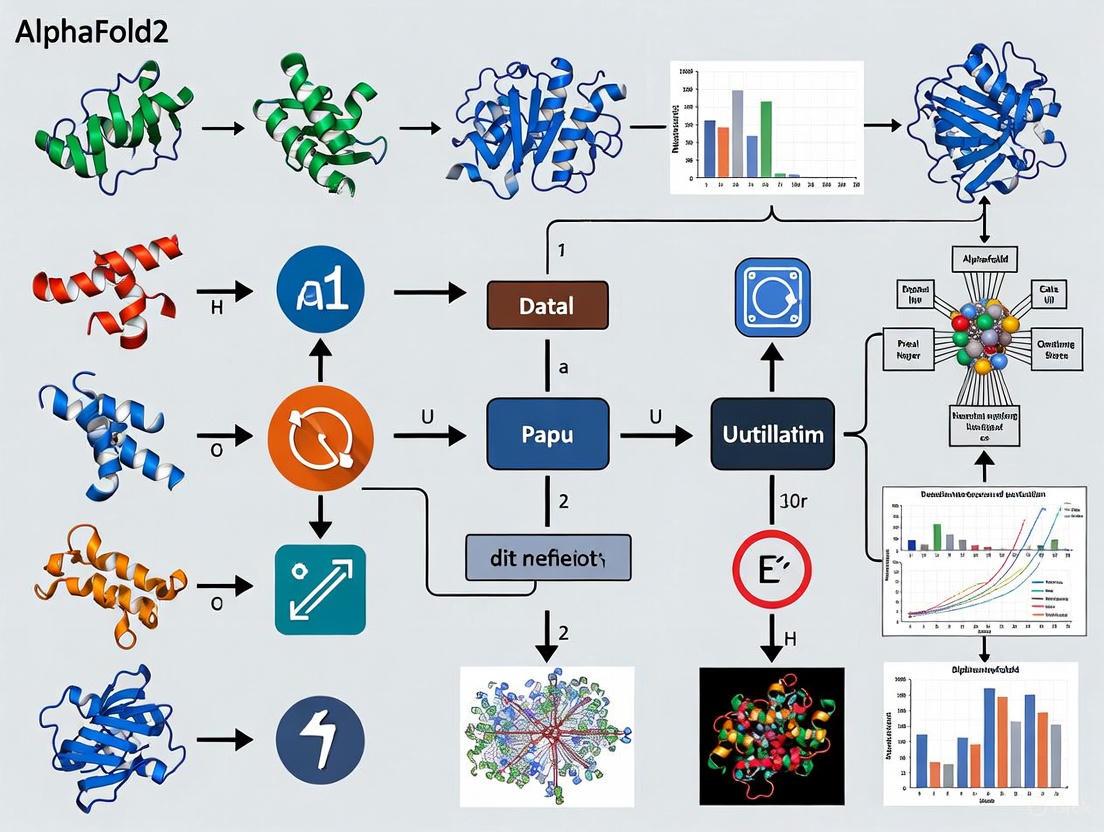
AlphaFold2's success at CASP14, where it achieved a median backbone accuracy of 0.96 Å (a level comparable to experimental error), represented a transformational leap [1]. Its architecture is an end-to-end deep learning model that departs significantly from its predecessor and other traditional methods.
The key innovation lies in its neural network architecture, which jointly embeds two primary inputs:
- Multiple Sequence Alignments (MSAs): A collection of evolutionarily related sequences.
- Pairwise Features: A representation of inferred relationships between residues [1].
These inputs are processed through a novel component called the Evoformer, a neural network block that exchanges information between the MSA and pair representations. This allows the network to reason simultaneously about evolutionary constraints and spatial relationships [1]. The output of the Evoformer is then passed to the Structure Module, which introduces an explicit 3D structure. This module uses an equivariant transformer to iteratively refine the atomic coordinates, starting from a trivial initial state and progressively building a highly accurate model with precise atomic details [1]. A critical feature is "recycling," where the output is recursively fed back into the network for several cycles of refinement, significantly enhancing accuracy [1].
Diagram 1: AlphaFold2's core architecture and workflow for structure prediction.
Performance and Validation
In CASP14, AlphaFold2's predictions were vastly more accurate than any other method, achieving a median backbone accuracy (Cα root-mean-square deviation, RMSD) of 0.96 Å, compared to 2.8 Å for the next best method [1]. This level of accuracy is competitive with many experimentally determined structures. DeepMind subsequently applied AF2 at a massive scale, creating the AlphaFold Protein Structure Database, which expanded the structural coverage of the human proteome from about 17% to over 98%, providing an unprecedented resource for the scientific community [3].
A key feature of AF2 is its internal confidence measure, the predicted Local Distance Difference Test (pLDDT). This per-residue score, ranging from 0 to 100, allows users to assess the reliability of different regions of a predicted model. Generally, pLDDT scores above 90 indicate very high confidence, scores between 70 and 90 are confident, scores between 50 and 70 are low confidence, and scores below 50 should be considered very low confidence, potentially representing unstructured regions [1] [7]. AF2 also provides a Predicted Aligned Error (PAE) matrix, which estimates the confidence in the relative positional alignment of different parts of the model, which is crucial for understanding domain packing and orientations [7].
Application Notes and Protocols
Protocol: Running AlphaFold2 for Single-Chain Protein Prediction
This protocol outlines the steps to predict the structure of a single protein chain using a standard AlphaFold2 implementation, such as the local installation or via ColabFold [7].
Step 1: Input Sequence Preparation
- Obtain the target protein's amino acid sequence in FASTA format. Sequences can be sourced from public databases like UniProt.
- The sequence length should ideally be between 10 and 3,000 amino acids. Very short sequences (<10) may not generate reliable MSAs, while very long sequences may encounter hardware memory limitations [7].
Step 2: Multiple Sequence Alignment (MSA) Generation
- Input the FASTA sequence into the AF2 pipeline.
- The system will automatically query genetic databases (e.g., BFD, MGnify, Uniclust30) to construct an MSA and a pair representation of the target.
- This step identifies evolutionarily related sequences, which provide the co-evolutionary information critical for accurate folding [1] [7].
Step 3: Structure Prediction and Model Generation
- The Evoformer and Structure Module process the MSA and pair representations. No user intervention is required at this stage.
- The system typically generates five models. The iterative "recycling" process (usually 3 cycles by default) is embedded within this step and is key to achieving high accuracy [1].
Step 4: Model Analysis and Selection
- Analyze the generated models using the provided pLDDT and PAE metrics.
- The model with the highest average pLDDT is typically selected as the top-ranked model.
- Visually inspect the model, coloring it by pLDDT to identify low-confidence regions. Use the PAE plot to evaluate inter-domain confidence [7].
Protocol: Benchmarking AlphaFold2 on Peptide Structures
While AF2 excels with globular proteins, its performance on small peptides (10-40 amino acids) requires careful validation. The following protocol is based on the benchmark study by McDonald et al. [8].
Step 1: Dataset Curation
- Select a diverse set of peptides with experimentally determined NMR structures. The benchmark should include different structural classes: α-helical membrane-associated, α-helical soluble, mixed secondary structure membrane-associated, mixed secondary structure soluble, β-hairpin, and disulfide-rich peptides [8].
- A total of 588 peptides were used in the original study.
Step 2: Structure Prediction and RMSD Calculation
- Predict the structure of each peptide in the dataset using AlphaFold2 as described in Protocol 4.1.
- For each peptide, compute the Cα root-mean-square deviation (RMSD) between the AF2-predicted structure and the corresponding experimental NMR structure. The RMSD calculation should be normalized per residue and focused on the secondary structural region to prevent size bias [8].
Step 3: Analysis of Confidence Metrics versus Accuracy
- Compare the pLDDT-ranked order of the five generated models against their actual RMSD to the experimental structure.
- Note any discrepancies where the model with the lowest RMSD (highest accuracy) is not the one ranked highest by pLDDT. This is a known limitation in peptide modeling [8] [7].
- Analyze Φ/Ψ angle recovery and disulfide bond pattern prediction, as these are specific areas where AF2 can show shortcomings for peptides [8].
Table 2: Summary of AlphaFold2 Performance on Different Peptide Classes (based on [8])
| Peptide Class | Number of Peptides | Mean Normalized Cα RMSD (Å per residue) | Key Observations and Shortcomings |
|---|---|---|---|
| α-Helical Membrane-Associated | 187 | 0.098 | Predicted with good accuracy; few outliers. Struggles with helix termini and turn motifs. |
| α-Helical Soluble | 41 | 0.119 | More outliers than membrane-associated; fails to predict helical structure in some cases (e.g., 1AMB). |
| Mixed Sec. Struct. Membrane-Assoc. | 14 | 0.202 | Largest variation and RMSD; correct secondary structure but poor overlap in unstructured regions. |
| β-Hairpin | 176 | Data Not Specified | High accuracy, similar to helical peptides. |
| Disulfide-Rich | 170 | Data Not Specified | High accuracy, but errors in disulfide bond patterns can occur. |
Application in Drug Discovery: Identifying a Kinase Inhibitor
A demonstrated application of AF2 in drug discovery is the rapid discovery of a novel cyclin-dependent kinase 20 (CDK20) inhibitor for hepatocellular carcinoma [3].
Workflow:
- Target Identification: Use a target identification platform (e.g., PandaOmics) to nominate CDK20 as a promising target.
- Structure Retrieval: Download the predicted structure of CDK20 from the AlphaFold Protein Structure Database.
- In Silico Screening & Molecule Generation: Use the AF2 structure with a generative chemistry platform (e.g., Chemistry42) to design and screen thousands of small molecule candidates.
- Compound Filtering: Apply developability filters to select the top 7 candidates for synthesis.
- Experimental Validation: Test the synthesized compounds in biochemical and cellular assays. One compound showed high binding affinity (Kd = 9.2 nM) and selectively inhibited cancer cell proliferation.
This end-to-end process, from target to validated hit, was completed in just 30 days, showcasing the potential of AF2 to dramatically accelerate early-stage drug discovery [3].
The Scientist's Toolkit: Research Reagent Solutions
Table 3: Essential Resources for AlphaFold2-Based Research
| Resource Name | Type | Function and Description | Access Link/Reference |
|---|---|---|---|
| AlphaFold Protein Structure Database | Database | Provides instant, free access to over 200 million pre-computed AF2 protein structure predictions, eliminating the need for local computation. | https://alphafold.ebi.ac.uk/ [3] |
| ColabFold | Software Suite | An open-access, streamlined implementation of AF2 that runs via Google Colab notebooks or locally. It uses the faster MMseqs2 for MSA generation, significantly speeding up predictions. | https://github.com/sokrypton/ColabFold [7] |
| AlphaFold-Multimer | Software Module | A specialized version of AF2 trained to predict the structures of protein complexes (homo- and hetero-multimers), which is crucial for studying protein-protein interactions. | [4] [3] |
| AlphaPullown | Software Tool | A Python package designed for high-throughput screening of protein-protein interactions using AlphaFold-Multimer. | [3] |
| pLDDT & PAE | Analysis Metric | Integrated confidence scores that are essential for interpreting model reliability and guiding experimental design. | [1] [7] |
Critical Limitations and Future Directions
Despite its transformative impact, AlphaFold2 has several important limitations that researchers must consider:
- Static Snapshots: AF2 predicts a single, static structure. It does not model the conformational dynamics, flexibility, or multiple states that are often essential for protein function [7] [9].
- Limitations with Specific Classes: Performance is reduced for non-globular proteins, such as intrinsically disordered regions, and it can struggle with accurate prediction of disulfide bond patterns and membrane protein conformational states [8] [7] [9].
- Peptide Modeling Caveats: As highlighted in Protocol 4.2, the pLDDT score may not reliably rank the most accurate model for peptides, and Φ/Ψ angle recovery can be poor [8].
- Ligand and Cofactor Absence: Standard AF2 predicts apo structures. It does not include ligands, cofactors, post-translational modifications, or ions, which can be critical for function, though AlphaFold3 has begun to address this for some biomolecules [4] [7].
Future directions involve moving beyond static snapshots to predict conformational ensembles and integrating AF2 models with experimental data from techniques like cryo-EM, NMR, and molecular dynamics simulations to model dynamic processes and allosteric mechanisms more accurately [7] [9].
Diagram 2: A hybrid experimental-computational workflow to overcome AF2 limitations.
The prediction of a protein's three-dimensional structure from its amino acid sequence has stood as a monumental challenge in computational biology for over half a century. Often referred to as the "protein folding problem," its solution is crucial for understanding biological function, elucidating disease mechanisms, and accelerating drug discovery. For decades, experimental methods like X-ray crystallography, cryo-electron microscopy (cryo-EM), and nuclear magnetic resonance (NMR) have been the primary means to determine protein structures. However, these techniques are often time-consuming and expensive, resulting in a vast gap between the number of known protein sequences and their experimentally solved structures—a challenge known as the "structural gap" [10] [11].
In November 2020, Google DeepMind's AlphaFold2 (AF2) achieved an unprecedented victory at the 14th Critical Assessment of Structure Prediction (CASP14), demonstrating accuracy competitive with experimental methods for many proteins [4] [10]. This breakthrough represented a transformative moment in structural biology. The subsequent release of the AlphaFold Protein Structure Database, providing over 200 million predicted structures, has since empowered researchers worldwide, offering unprecedented insights into the protein universe and accelerating scientific discovery across biology and medicine [12] [13].
The CASP14 Victory: A Paradigm Shift
The CASP Competition and AlphaFold2's Achievement
The Critical Assessment of Structure Prediction (CASP) is a biennial, double-blind competition that serves as the gold standard for evaluating protein structure prediction methods. In CASP14, AlphaFold2 outperformed all other methods by a significant margin, achieving a median Global Distance Test (GDT) score above 90 for approximately two-thirds of the proteins predicted. The GDT score measures the structural similarity between a prediction and the experimental reference, with a score of 100 representing a perfect match. This level of accuracy was previously attainable only through experimental determination and marked a historic milestone in computational biology [4] [10].
Quantitative Performance at CASP14
Table 1: AlphaFold2 Performance Metrics at CASP14
| Performance Metric | Result | Context and Significance |
|---|---|---|
| Median GDT Score | >90 (for ~2/3 of proteins) | Accuracy considered competitive with experimental methods [4] |
| Overall Performance | Top-ranked by a large margin | Far exceeded all other methods in the competition [12] [4] |
| Key Advancement | Accuracy for "difficult" targets | Dramatically improved predictions for proteins with no known structural templates [10] |
Underlying Architecture and Algorithmic Breakthroughs
AlphaFold2's success stems from a novel, end-to-end deep learning architecture that represents a significant departure from its predecessor, AlphaFold1, and other contemporary methods.
Core Algorithmic Innovations
The system employs an interconnected neural network model that co-evolves representations through two key modules operating on a pair representation (residue-residue relationships) and a single representation (residue-sequence relationships) [4]. A critical innovation is the use of an attention-based mechanism, which allows the network to dynamically focus on the most relevant information when processing sequences and constructing the 3D model. This process iteratively refines the structural prediction, starting from a rough initial topology and progressively improving it while minimizing unphysical bond angles and lengths until a highly accurate structure is produced [4].
Diagram: AlphaFold2's Simplified High-Level Workflow
The AlphaFold Database: Scaling to 200 Million Structures
Following its CASP14 triumph, DeepMind partnered with EMBL's European Bioinformatics Institute (EMBL-EBI) to create the AlphaFold Protein Structure Database. This resource was initially launched with structures for the human proteome and 47 other key organisms, and has since been expanded to contain over 200 million predicted structures, providing broad coverage of the UniProt knowledgebase [12] [13]. This effectively covers nearly the entire catalogued protein universe, a scale that would have been unimaginable using traditional experimental methods. The database is freely and openly available to the global scientific community under a Creative Commons license (CC-BY 4.0) [12].
Database Usage and Impact Metrics
The database has been widely adopted, with over 2 million researchers across 190 countries utilizing it to support their work [13]. It is estimated that the database has potentially saved millions of research years, drastically accelerating the pace of biological inquiry [13].
Experimental Validation and Confidence Metrics
The pLDDT Score: A Measure of Prediction Confidence
AlphaFold2 provides a per-residue confidence score called the predicted Local Distance Difference Test (pLDDT). This metric ranges from 0 to 100 and is a crucial tool for researchers to assess the local reliability of a predicted model [11]. While pLDDT is an internal confidence measure, it has been shown to correlate with model accuracy. It is also increasingly used as an indicator of protein flexibility, with lower scores often corresponding to regions of higher intrinsic disorder or dynamics [14].
Table 2: Interpreting AlphaFold2 pLDDT Confidence Scores
| pLDDT Score Range | Confidence Level | Interpretation and Recommended Use |
|---|---|---|
| > 90 | Very high | High backbone accuracy; suitable for detailed atomic-level analysis [11] |
| 70 - 90 | Confident | Good backbone prediction; reliable for analyzing structural features [11] |
| 50 - 70 | Low | Low confidence; use with caution, potential errors in geometry [11] |
| < 50 | Very low | Very low confidence; likely unstructured or disordered regions [11] |
Practical Protocols for Researchers
This section provides detailed methodologies for accessing and utilizing AlphaFold2 predictions in research workflows.
Protocol 1: Accessing and Analyzing Structures from the AlphaFold Database
The primary method for most researchers is to retrieve pre-computed structures from the AlphaFold Database.
Procedure:
- Access the Database: Navigate to the AlphaFold Protein Structure Database at https://alphafold.ebi.ac.uk/.
- Search for a Protein: Use a UniProt identifier, gene name, or organism name to search for your protein of interest.
- Retrieve the Entry: Select the correct protein entry from the search results.
- Download Data: Download the predicted structure in PDB or mmCIF format. The associated data files, containing per-residue pLDDT scores and predicted aligned error, can also be downloaded.
- Visualization and Analysis:
- Open the PDB file in a molecular visualization tool like PyMOL, UCSF Chimera, or ChimeraX.
- Color the structure by the pLDDT score to visually assess the confidence of different regions. This is critical for interpreting functional domains or active sites.
- Analyze the structure for known functional motifs, binding pockets, or oligomerization interfaces in the context of biological knowledge.
Protocol 2: Running AlphaFold2 for Novel Sequences
For sequences not available in the database (e.g., novel mutants or designed proteins), researchers can run the AlphaFold2 model.
Computational Requirements: Running AlphaFold2 is computationally intensive. The following are general system guidelines, though requirements can vary based on sequence length [15].
Table 3: Recommended System Requirements for AlphaFold2
| Resource Type | Recommended (Best) | Minimum (Poor Experience) |
|---|---|---|
| GPU | NVIDIA A100 | NVIDIA CUDA GPU with >=32GB VRAM |
| CPU Cores | >= 64 | >= 12 |
| RAM | >= 180 GB | >= 64 GB |
| SSD Storage | >= 1.3 TB (NVMe, >3,500 MB/s) | Varies, but fast SSD recommended |
Procedure:
- Software Setup: Install AlphaFold2 from its open-source repository on GitHub, ensuring all dependencies and required genetic databases are installed and configured.
- Input Preparation: Prepare a FASTA file containing the amino acid sequence(s) to be predicted.
- Multiple Sequence Alignment (MSA): Execute the
run_alphafold.pyscript. The system will first generate MSAs using tools like JackHMMER and HHblits against genomic databases. This is the most time-consuming step and performance is highly dependent on CPU cores and disk speed [15]. - Structure Inference: The neural network will process the MSA and template information (if used) to generate the 3D model. This step is heavily dependent on GPU capability. Note that structure prediction time grows exponentially with sequence length [15].
- Output Analysis: The run will produce several PDB files representing top-ranked models, along with JSON files containing pLDDT scores and other confidence metrics. Analyze these outputs as described in Protocol 1.
Protocol 3: Integrating Experimental Data to Guide Predictions (DEERFold)
Advanced protocols are being developed to integrate sparse experimental data to guide AlphaFold2 and model conformational ensembles, addressing a key limitation of predicting single, static structures.
Principle: Methods like DEERFold fine-tune AlphaFold2 to incorporate experimental distance distributions, such as those from Double Electron-Electron Resonance (DEER) spectroscopy, to predict alternative protein conformations [16].
Procedure:
- Experimental Data Collection: Collect DEER data to obtain distance distributions between spin labels site-specifically introduced into the protein.
- Data Representation: Convert the experimental distance distributions into a format compatible with the neural network architecture (e.g., a "distogram").
- Model Guidance: Input the distance constraints into the modified AlphaFold2 model (DEERFold) alongside the protein sequence.
- Ensemble Generation: Run predictions. The incorporation of distance constraints drives the model to fold into conformations consistent with the experimental data, often resulting in a heterogeneous ensemble of structures.
- Validation: Compare the resulting model ensemble with the experimental data and other known structural information to validate the predictions.
Diagram: DEERFold Experimental Workflow
Table 4: Key Resources for AlphaFold2-Based Research
| Resource Name | Type | Function and Application |
|---|---|---|
| AlphaFold Protein Structure Database | Database | Primary repository for accessing over 200 million pre-computed protein structure predictions [12] |
| UniProt | Database | Standard repository of protein sequences and annotations; serves as the foundation for the AlphaFold DB [12] |
| Protein Data Bank (PDB) | Database | Repository for experimentally determined structures; used for validation and comparison with AF2 models [11] |
| AlphaFold2 Open Source Code | Software | Allows researchers to run structure predictions on custom sequences not in the database [12] |
| pLDDT Score | Analytical Metric | Per-residue confidence score essential for interpreting the local reliability of AF2 predictions [11] |
| DEER/EPR Spectroscopy | Experimental Method | Provides distance restraints to guide and validate AF2 models for modeling conformational ensembles [16] |
| Cryo-EM / X-ray Crystallography | Experimental Method | Gold-standard methods for high-resolution structure determination; used to validate AF2 predictions [10] [11] |
Applications in Biology and Medicine
The availability of highly accurate protein structures is revolutionizing numerous fields.
- Drug Discovery: AF2 models are used to understand drug targets, identify binding pockets, and perform structure-based virtual screening, particularly for targets with no or limited experimental structural data [10] [17]. For example, AF2 has been applied to study nuclear receptors, important drug targets, providing models where experimental structures are scarce [11].
- Protein Design: AF2's understanding of structure is inverted to design novel proteins with desired functions, such as therapeutic mini-binders and enzymes [17].
- Understanding Disease Mechanisms: AF2 models help elucidate the structural impact of disease-related mutations, providing insights into conditions like cancer, Parkinson's, and antibiotic resistance [13] [10].
- Membrane Transporters and Enzymes: AF2 has provided structural insights into challenging protein classes like membrane transporters and has been used to engineer plastic-degrading enzymes to address environmental pollution [13] [16].
Limitations and Future Directions
Despite its transformative impact, AlphaFold2 has limitations. It primarily predicts a single, static conformation and may not capture the full spectrum of native conformational dynamics and flexibility that are critical for the function of many proteins [16] [11]. While it can predict some multimeric structures, accurately modeling large protein complexes remains challenging. Furthermore, its performance can be lower for proteins with limited evolutionary information in the multiple sequence alignments [16], and it does not explicitly predict the effects of ligands, ions, or post-translational modifications, though tools like AlphaFold3 are now addressing this [4] [17].
The field continues to evolve rapidly, with new methods like DEERFold demonstrating the integration of experimental data to guide predictions [16], and the recent release of AlphaFold3 expanding capabilities to predict protein interactions with DNA, RNA, and small molecules [4]. The continued development and application of these tools promise to further deepen our understanding of biological systems and accelerate therapeutic development.
AlphaFold2 (AF2) represents a paradigm shift in computational biology, providing a solution to the 50-year-old protein folding problem by predicting three-dimensional (3D) protein structures from amino acid sequences with atomic-level accuracy [1] [10]. Its unprecedented success in the CASP14 assessment demonstrated capabilities competitive with experimental methods, fundamentally transforming structural biology research and therapeutic development [1]. The architectural brilliance of AF2 resides primarily in two interconnected components: the Evoformer, a novel neural network block that processes evolutionary and pairwise relationships, and the Structure Module, which translates these refined representations into accurate atomic coordinates [1] [18]. This application note provides a detailed technical deconstruction of these core components, offering researchers comprehensive insights into their operational mechanisms and implementation protocols.
The Evoformer: A Graph Inference Engine for Evolutionary and Spatial Relationships
The Evoformer serves as the computational trunk of AF2, formulating and continuously refining a structural hypothesis through iterative processing of input data [1]. It operates on two primary representations that are updated in parallel:
- MSA Representation: An Nseq × Nres array (where Nseq is the number of sequences and Nres is the number of residues) that encapsulates evolutionary information from multiple sequence alignments. Each column represents individual residues of the input sequence, while rows represent homologous sequences [1] [19].
- Pair Representation: An Nres × Nres array that models the relationships between every pair of residues in the target protein, encoding information about their spatial and evolutionary constraints [1] [18].
Table 1: Evoformer Input Features and Embedding Process
| Input Type | Representation | Processing Method | Key Innovation |
|---|---|---|---|
| Multiple Sequence Alignment (MSA) | Nseq × Nres array | Clustering by similarity with representative selection | Embedding raw sequences rather than only MSA statistics [18] |
| Template Structures | Nres × Nres distance matrices | Discretization into distogram bins | Integration of known structural homologs when available [1] |
| Primary Sequence | Amino acid residues | Direct embedding with residue features | Preservation of original sequence information [19] |
Core Computational Operations and Information Exchange
The Evoformer's revolutionary design enables sophisticated information exchange through several specialized operations implemented across its 48 blocks [18]:
MSA-Pair Communication Channels:
- Outer Product Mean: The MSA representation updates the pair representation through an element-wise outer product summed over the MSA sequence dimension. Unlike previous approaches, this operation occurs within every Evoformer block, enabling continuous information flow [1].
- Pair Bias Injection: During row-wise attention in the MSA representation, additional logits projected from the pair stack bias the attention mechanism, creating a reciprocal information flow from pairwise to evolutionary data [18].
Triangular Geometric Reasoning: The pair representation undergoes updates inspired by geometric constraints necessary for 3D structural consistency:
- Triangle Multiplicative Update: A symmetric operation that uses two edges of a residue triplet to update the "missing" third edge, enforcing geometric consistency [1].
- Triangular Self-Attention: An attention mechanism augmented with a logit bias to include the third edge of residue triangles, ensuring the pairwise distances satisfy the triangle inequality [1].
Axial Attention Mechanisms:
- Row-wise Attention: Operates across sequences in the MSA to identify which amino acids are evolutionarily related.
- Column-wise Attention: Functions within alignment columns to determine which sequences provide the most relevant structural information [18].
Experimental Protocol: Evoformer Representation Analysis
Purpose: To extract and interpret the intermediate representations generated by the Evoformer for hypothesis generation about protein structure-function relationships.
Materials:
- Protein sequence of interest
- High-performance computing environment with AlphaFold2 implementation
- Multiple sequence alignment tools (Jackhmmer/HHblits)
- Protein structure databases (PDB, UniRef)
Procedure:
- Input Preparation:
- Generate MSAs by querying the target sequence against protein databases using Jackhmmer with default parameters [19].
- Extract template structures if available from the PDB using homology search tools.
Evoformer Activation Extraction:
- Modify the AF2 inference code to save the MSA and pair representations after each Evoformer block.
- Run inference on the target sequence with recycling disabled initially to analyze progressive refinement.
Representation Analysis:
- Apply dimensionality reduction (UMAP/t-SNE) to track the evolution of residue-residue relationships across blocks.
- Compute attention maps from row-wise and column-wise attention heads to identify co-evolutionary patterns.
- Calculate mutual information between MSA and pair representations to quantify their interdependence.
Structural Hypothesis Generation:
- Correlate high-attention residue pairs with known structural motifs or functional sites.
- Identify conserved interaction patterns across Evoformer blocks that may indicate stable structural contacts.
Interpretation: Early Evoformer blocks typically establish coarse-grained residue contacts, while later blocks refine these into precise spatial relationships. Consistent high-attention regions across multiple blocks often correspond to structurally critical elements like active sites or folding nuclei [1].
Diagram 1: Evoformer Information Flow (62 characters)
The Structure Module: From Representations to Atomic Coordinates
Architectural Principles and Invariant Transformations
The Structure Module translates the refined representations from the Evoformer into precise 3D atomic coordinates through a series of equivariant operations [1]. Its design incorporates several key innovations:
- Explicit 3D Structure: Introduces a rotation and translation (rigid body frame) for each residue, initialized trivially with identity rotations and origin positions [1].
- Chain Breakage: Temporarily breaks the chain connectivity to allow simultaneous local refinement of all structure parts, preventing propagation of errors [1].
- Invariant Point Attention (IPA): A novel attention mechanism specifically designed for 3D molecular structures that respects rotational and translational equivariance, meaning predictions are independent of the global orientation [18].
Iterative Refinement through Recycling
A critical aspect of AF2's performance is the iterative refinement process known as "recycling" [1] [19]:
- Initial Prediction: The Structure Module generates an initial 3D structure from the Evoformer outputs.
- Feedback Integration: The predicted structure, along with MSA and pair representations, is fed back into the beginning of the network.
- Progressive Refinement: This recycling process typically repeats three times, with each iteration refining the structural details and improving accuracy [19].
- Confidence Estimation: The model provides per-residue confidence estimates (pLDDT) that reliably predict the local accuracy of the prediction [1].
Table 2: Structure Module Output Metrics and Their Interpretation
| Metric | Calculation | Interpretation | Threshold Values |
|---|---|---|---|
| pLDDT (predicted local distance difference test) | Percentage of atom pairs within distance thresholds of reference [14] | Per-residue confidence estimate | <50: Low confidence, 50-70: Medium, 70-90: High, >90: Very high [1] |
| pTM (predicted TM-score) | Estimated template modeling score for global structure quality [20] | Global fold accuracy assessment | >0.5: Correct fold, >0.8: High accuracy [21] |
| ipTM (interface pTM) | Interface-specific version of pTM for complexes [20] | Protein-protein interface quality | Primary metric for complex assessment [20] |
| PAE (predicted aligned error) | Expected positional error after alignment [20] | Domain orientation confidence | Lower values indicate higher confidence in relative positioning |
Experimental Protocol: Structure Module Ablation Studies
Purpose: To systematically evaluate the contribution of different Structure Module components to prediction accuracy.
Materials:
- Benchmark set of proteins with experimentally determined structures
- Modified AF2 codebase with selective component disabling capability
- Structural comparison tools (TM-score, RMSD calculators)
Procedure:
- Baseline Establishment:
- Select a diverse set of protein targets (different folds, sizes, MSA depths).
- Run standard AF2 prediction with full Structure Module and record accuracy metrics (pLDDT, TM-score, RMSD to experimental structure).
Component Ablation:
- Disable the recycling mechanism by setting recycle count to 1.
- Replace Invariant Point Attention with standard attention mechanisms.
- Modify the residue representation to maintain chain connectivity throughout.
- Remove side chain atoms from the representation, focusing only on backbone.
Controlled Comparison:
- For each ablation condition, run predictions on the same benchmark set.
- Ensure identical initial conditions (MSA, templates, random seeds).
- Quantify accuracy metrics for each condition.
Intermediate Structure Analysis:
- Extract and save intermediate structures during the recycling process.
- Calculate quality metrics for structures after each recycling step.
- Analyze the trajectory of structural refinement.
Interpretation: The recycling process typically contributes significantly to accuracy with minimal extra computational cost. Invariant Point Attention is particularly crucial for proper stereochemistry, while chain breakage enables more effective local refinement [1].
Diagram 2: Structure Module Workflow (46 characters)
Integrated Workflow: From Sequence to Structure
End-to-End Prediction Protocol
Purpose: To provide a comprehensive methodology for utilizing AF2's complete architecture for protein structure prediction.
Materials:
- Protein sequence(s) of interest in FASTA format
- High-performance computing cluster with GPU acceleration
- AlphaFold2 software installation with required databases
- Structural visualization and analysis software (ChimeraX, PyMOL)
Procedure:
- Input Preparation and Feature Extraction:
- Generate MSAs using Jackhmmer against multiple sequence databases (UniRef90, MGnify).
- Search for template structures using HHsearch against the PDB.
- Extract and embed evolutionary and template features.
Evoformer Processing:
- Process inputs through the 48 Evoformer blocks with information exchange between MSA and pair representations.
- Monitor convergence of representations through attention pattern stabilization.
Structure Generation:
- Initialize the structure with trivial frames (identity rotations, origin positions).
- Process through the Structure Module with Invariant Point Attention.
- Perform iterative refinement through three recycling steps.
- Place side chain atoms and refine their positions.
Output and Validation:
- Generate final atomic coordinates in PDB format.
- Compute confidence metrics (pLDDT, pTM, PAE).
- Validate structures using geometric checks (Ramachandran plots, steric clashes).
Troubleshooting:
- Low confidence predictions (pLDDT < 50) often result from shallow MSAs; consider adding homologous sequences.
- Domain misorientation may be addressed by examining the PAE matrix for high inter-domain errors.
- Steric clashes can be relieved using energy minimization tools while preserving the overall fold.
Research Reagent Solutions
Table 3: Essential Research Tools for AlphaFold2 Architecture Studies
| Reagent/Tool | Function | Application Context |
|---|---|---|
| ColabFold | Optimized AF2 implementation with MMseqs2 | Rapid prototyping and predictions without extensive computational resources [20] |
| AlphaFold DB | Repository of precomputed AF2 structures | Benchmarking and comparison of architectural variants [10] |
| ChimeraX with PICKLUSTER | Molecular visualization and analysis | Interpretation of protein complexes and interface scoring [20] |
| ESM-1b | Protein language model | Comparison with evolution-aware representations [22] |
| ATLAS MD Dataset | Molecular dynamics trajectories | Correlation of pLDDT with protein flexibility [14] |
| VoroMQA and VoroIF-GNN | Model quality assessment | Independent validation of interface predictions [20] |
The deconstruction of AF2's core components reveals a sophisticated integration of evolutionary information with physical and geometric constraints. The Evoformer's ability to reason about spatial relationships through triangular updates and the Structure Module's equivariant transformations represent fundamental advances in computational structure prediction. For researchers and drug development professionals, understanding these architectural details enables more informed interpretation of AF2 predictions, appropriate application to biological questions, and targeted modifications for specific use cases. While AF2 has limitations in predicting multiple conformational states and protein-ligand interactions, its core architectural principles provide a robust foundation for future methodological developments in structural bioinformatics.
Multiple Sequence Alignments (MSAs) serve as a fundamental input for accurate protein structure prediction, providing the evolutionary constraints necessary to infer three-dimensional folds. Advanced artificial intelligence systems, most notably AlphaFold2, leverage the co-evolutionary information embedded within MSAs to achieve atomic-level accuracy [1] [23]. By analyzing patterns of correlated mutations across homologous sequences, these systems can identify residue pairs that are spatially close in the native structure, even if they are distant in the primary sequence. This application of evolutionary data addresses the immense complexity of the protein folding problem, acting as a bridge between the amino acid sequence and the final, functional protein architecture. The integration of MSAs has been the cornerstone upon which modern, highly accurate prediction pipelines have been built [24] [2].
The Role of MSAs in AlphaFold2 and Related Systems
Core Architectural Integration
In AlphaFold2, MSAs are processed at the very beginning of the neural network pipeline. The system's Evoformer module, a novel neural network block, is specifically designed to reason about the relationships within the MSA and between residue pairs [1]. The Evoformer treats the prediction as a graph inference problem, where the MSA representation (encoding information across homologous sequences) and the pair representation (encoding relationships between residues in the target sequence) continuously exchange information [1]. This is achieved through several innovative operations:
- MSA-to-Pair Information Transfer: An outer product operation sums over the MSA sequence dimension to update the pair representation in every Evoformer block [1].
- Triangle-shaped Updates within the Pair Representation: These operations enforce geometric consistency, using principles like the triangle inequality to reason about distances between three different residues, thereby making the pair representation more physically plausible [1].
This deep, iterative refinement of co-evolutionary signals allows AlphaFold2 to form a concrete structural hypothesis that is progressively refined throughout the network [1].
MSA-Free Prediction: The Emergence of Protein Language Models
While MSA-based methods set the standard for accuracy, the process of searching and constructing MSAs is computationally expensive, often taking tens of minutes to hours and becoming a bottleneck for high-throughput applications like large-scale virtual screening [24]. This limitation has spurred the development of MSA-free methods that use Protein Language Models (PLMs) [24].
These models, such as the one powering HelixFold-Single, are pre-trained on tens of millions of primary protein sequences using self-supervised learning [24]. During this pre-training, the PLM learns the statistical properties and evolutionary constraints of proteins, effectively embedding co-evolutionary knowledge directly into its parameters [24]. At inference time, the PLM can generate representations for a single sequence that serve as a substitute for the explicit co-evolutionary information found in an MSA. These representations are then fed into a structure module (often adapted from AlphaFold2) to predict 3D coordinates [24]. While their accuracy is particularly strong for proteins with large homologous families, they offer a substantial reduction in prediction time [24].
Table 1: Comparison of MSA-Based and MSA-Free Prediction Approaches
| Feature | MSA-Based (e.g., AlphaFold2) | MSA-Free (e.g., HelixFold-Single) |
|---|---|---|
| Core Input | Multiple Sequence Alignment (MSA) | Single amino acid sequence |
| Source of Co-evolution Data | Explicit retrieval from protein databases | Implicit, learned by a pre-trained Protein Language Model |
| Computational Bottleneck | MSA search and construction | Model inference (forward pass) |
| Typical Prediction Time | Minutes to hours | Seconds to minutes |
| Key Advantage | High accuracy, especially with deep MSAs | Speed, efficiency for high-throughput tasks |
Quantitative Performance and Benchmarking
Accuracy Assessment on Standardized Datasets
The performance of structure prediction methods is rigorously evaluated on blind test datasets like CASP14 and CAMEO. On these benchmarks, MSA-based AlphaFold2 demonstrates exceptional accuracy, producing predictions with a median backbone accuracy of 0.96 Å (Cα root-mean-square deviation at 95% residue coverage), a level competitive with experimental structures [1]. MSA-free methods like HelixFold-Single have shown remarkable progress, achieving competitive accuracy with MSA-based methods on targets that have large homologous families (e.g., those with MSA depths >1,000 sequences) [24]. However, the performance of these MSA-free methods is correlated with the richness of homologous sequences available in nature for the target, underscoring that the underlying source of information remains evolutionary in origin [24].
Table 2: Performance Comparison on CASP14 and CAMEO Benchmarks
| Method | Input Type | Key Metric (CASP14) | Key Finding |
|---|---|---|---|
| AlphaFold2 | MSA | Backbone accuracy: 0.96 Å r.m.s.d.₉₅ [1] | Accuracy competitive with experimental structures [1]. |
| HelixFold-Single | Single Sequence | TM-score (CASP14 & CAMEO) [24] | Competitive with MSA-based methods on targets with large homologous families [24]. |
| AlphaFold2 (Single Sequence Input) | Single Sequence | TM-score [24] | Unsatisfactory accuracy without MSA or PLM assistance [24]. |
| RoseTTAFold | MSA | TM-score (CASP14 & CAMEO) [24] | Outperformed by HelixFold-Single on CAMEO [24]. |
Benchmarking MSA Quality Itself
The quality of an MSA directly impacts the accuracy of the downstream structure prediction. Benchmarks like QuanTest have been developed to objectively evaluate MSA quality by measuring Secondary Structure Prediction Accuracy (SSPA) [25]. The underlying assumption is that a better MSA will lead to more accurate secondary structure predictions. QuanTest can be scaled to test alignments of hundreds or thousands of sequences, providing a flexible framework for evaluating different MSA generation methods [25]. This approach correlates well with traditional benchmarks based on structural alignment, validating its use for assessing this critical input [25].
Experimental Protocols
Protocol 1: Generating an MSA for AlphaFold2 Prediction
This protocol details the steps for constructing a deep MSA to be used as input for AlphaFold2.
Sequence Retrieval: Using the target amino acid sequence, search against large protein sequence databases (e.g., UniRef90, BFD, MGnify) with a sensitive homology search tool like HHblits or JackHMMER.
- Objective: To collect a deep set of homologous sequences.
- Critical Parameters: Use multiple iterations to maximize sensitivity. The depth of the MSA (number of sequences) is a key factor for prediction accuracy.
MSA Construction: Process the search results into a single MSA file.
- Objective: To create a formatted alignment for AlphaFold2.
- Tools: The alignment is typically generated by the search tool itself (e.g., HHblits outputs an A3M format file).
Input to AlphaFold2: The resulting MSA file is fed into the AlphaFold2 neural network.
- Process: Within AlphaFold2, the Evoformer module processes the MSA to extract co-evolutionary signals and generate refined pair and sequence representations, which the Structure module then folds into a 3D atomic model [1].
Protocol 2: MSA-Free Prediction Using a Protein Language Model
This protocol outlines the workflow for high-speed structure prediction using a single sequence and a pre-trained PLM.
Model Pre-training (Typically Pre-computed): A large-scale Protein Language Model (e.g., with billions of parameters) is pre-trained on tens of millions of unlabelled protein sequences using masked language modeling [24].
- Objective: To embed evolutionary and biochemical knowledge into the model's parameters.
- Output: A pre-trained model that can convert a single sequence into informative representations.
Structure Prediction: The target amino acid sequence is input into the prediction pipeline (e.g., HelixFold-Single).
- Step 1: The pre-trained PLM encodes the single sequence into initial single and pair representations [24].
- Step 2: An adapter module may be used to project the PLM's outputs into the format required by the folding module [24].
- Step 3: The EvoformerS (a modified Evoformer) and Structure modules from AlphaFold2 refine these representations and predict the 3D coordinates of all atoms [24].
Protocol 3: Visualizing and Analyzing an MSA with NCBI's MSA Viewer
This protocol describes how to visualize an existing MSA to assess its quality and inspect conservation.
- Upload Alignment: Navigate to the NCBI MSA Viewer. Upload your alignment file (in FASTA or ASN format) via the "Upload Data" dialog [26].
- Navigate and Inspect:
- Use the Panorama view at the top to get an overview of alignment quality and coverage. Red areas indicate regions with a high proportion of mismatches [26].
- Zoom into specific regions by clicking and dragging on the ruler or in the Panorama view [26].
- Hover the mouse over any sequence to see a tooltip with detailed information about the aligned position and sequence ID [26].
- Set a Reference:
- Set a specific sequence (e.g., the target sequence) as the anchor row via the right-click context menu. This re-roots the alignment, and the "% Identity" column will show percent identity of every other sequence to this anchor [26].
- Alternatively, display a consensus sequence as the top row (for nucleotide alignments) to see the most common residue at each position [26].
Table 3: Key Resources for MSA-Based Protein Structure Research
| Resource Name | Type | Function & Application |
|---|---|---|
| UniRef90/BFD/MGnify | Database | Large protein sequence databases used for homologous sequence searches to build deep MSAs. |
| HHblits/JackHMMER | Software Tool | Sensitive homology search tools used for iterative MSA construction from sequence databases. |
| AlphaFold2 Open Source | Software | The open-source code for AlphaFold2, allowing researchers to run predictions with custom MSAs. |
| AlphaFold Protein Structure Database | Database | Repository of over 200 million pre-computed AlphaFold2 structures; allows retrieval of models without local prediction [27]. |
| NCBI MSA Viewer | Web Tool | Visualizes alignments to assess quality, coverage, and conservation; supports custom anchor rows and coloring [26]. |
| Protein Language Models (e.g., ESM-2) | Software/Model | Pre-trained deep learning models that generate evolutionary representations from a single sequence for MSA-free prediction. |
Workflow and Data Visualization
Advanced Applications and Considerations
Special Cases: Predicting Structures of Chimeric Proteins
Predicting the structures of engineered chimeric proteins, such as those created by fusing a structured peptide to a scaffold protein, presents a unique challenge. Standard MSA construction for the entire chimera can lead to significantly reduced prediction accuracy [28]. An effective strategy to restore accuracy is a "windowed MSA" approach, where separate MSAs are generated for the individual components (e.g., the scaffold and the peptide tag) and these alignments are then appended together to form a composite MSA for the full chimeric protein [28]. This technique ensures that the co-evolutionary information specific to each domain is properly represented during the structure prediction process.
Visualization: Creating Optimal Color Schemes for MSAs
Effective visualization of MSAs is crucial for human interpretation. Traditional color schemes are based on manual assignments according to chemical properties. A more quantitative and reproducible approach leverages substitution matrices (e.g., BLOSUM62) to automatically generate color schemes [29]. This method uses an optimization algorithm (e.g., simulated annealing) to assign colors in a perceptually uniform color space (CIE Lab*), so that the perceptual difference between two amino acids' colors corresponds to their evolutionary distance as defined by the substitution matrix [29]. This ensures that visually similar colors are assigned to biochemically similar amino acids, directly aligning the visualization with the principles used to create the alignment itself.
AlphaFold2 represents a transformative advance in protein structure prediction, providing not only atomic coordinates but also essential confidence metrics that estimate the reliability of its predictions. Two scores are paramount for interpreting model quality: the pLDDT (predicted Local Distance Difference Test) and the PAE (Predicted Aligned Error). These metrics provide complementary information, with pLDDT quantifying local per-residue confidence and PAE assessing the relative positional accuracy between different parts of the structure. For researchers in structural biology and drug development, understanding these metrics is crucial for determining which parts of a predicted model can be trusted for further analysis and which require cautious interpretation. Proper utilization of pLDDT and PAE enables informed decision-making regarding model suitability for specific applications such as molecular docking, functional site analysis, and hypothesis generation.
Table 1: Interpretation guide for pLDDT scores
| pLDDT Score Range | Confidence Level | Structural Interpretation |
|---|---|---|
| ≥ 90 | Very high | High backbone and side chain accuracy; reliable for atomic-level analysis [30] |
| 70 - 90 | Confident | Generally correct backbone with potential side chain misplacement [30] |
| 50 - 70 | Low | Low confidence; potentially unstructured or poorly predicted [30] |
| < 50 | Very low | Very low confidence; likely intrinsically disordered or unstructured [30] |
Table 2: Interpretation guide for PAE values
| PAE Value (Å) | Confidence Level | Structural Interpretation |
|---|---|---|
| < 5 | Low error | Confident relative positioning; domains are well-packed [31] [7] |
| 5 - 10 | Moderate error | Some uncertainty in relative positioning [31] |
| > 10 | High error | Low confidence in relative position/orientation; interpret with caution [31] |
Table 3: Key resources for working with AlphaFold2 confidence metrics
| Resource Type | Specific Tool/Resource | Function and Utility |
|---|---|---|
| Database Access | AlphaFold Protein Structure Database | Access pre-computed models with interactive pLDDT and PAE visualizations [31] [32] |
| Software & Libraries | ColabFold | Open-source, accessible platform for running predictions with MMseqs2 for faster homology search [7] [33] |
| Programming Tools | Python Matplotlib library | Custom plotting of confidence metrics from raw AlphaFold2 output files [34] |
| Analysis Tools | AMBER force field | Energy minimization and relaxation of predicted models [34] |
Understanding pLDDT: Local Confidence Metric
Definition and Theoretical Basis
The predicted Local Distance Difference Test (pLDDT) is a per-residue measure of local confidence on a scale from 0 to 100, with higher scores indicating greater confidence and typically more accurate prediction [30]. This metric is AlphaFold2's estimate of how well the prediction would agree with an experimental structure based on the local distance difference test Cα (lDDT-Cα), which assesses the correctness of local distances without requiring structural superposition [30]. The pLDDT score is stored in the B-factor column of output PDB files, replacing the experimental B-factor typically derived from X-ray crystallography [34].
Biological Interpretation of pLDDT Scores
pLDDT scores vary significantly along a protein chain, indicating regions of differential reliability [30]. As summarized in Table 1, scores above 90 indicate very high confidence with both backbone and side chains typically predicted accurately. Scores between 70 and 90 generally correspond to correct backbone predictions with potential side chain placement errors. Regions with scores below 50 indicate very low confidence, which typically arise for two reasons: naturally occurring intrinsic disorder or insufficient information for AlphaFold2 to make a confident prediction [30].
A critical application of pLDDT is identifying intrinsically disordered regions (IDRs), which lack fixed structure under physiological conditions [30]. However, an important caveat exists: some IDRs undergo binding-induced folding upon interaction with molecular partners, and AlphaFold2 may predict these folded states with high pLDDT scores because they were present in the training data [30]. For example, eukaryotic translation initiation factor 4E-binding protein 2 (4E-BP2) is predicted with high confidence in a helical conformation that it only adopts in its bound state [30].
Important Limitations and Caveats
Despite its utility, pLDDT has important limitations. It does not measure confidence in the relative positions or orientations of different protein domains [30]. Additionally, recent research indicates that pLDDT values show no correlation with B-factors from experimental structures, suggesting they do not provide information about local conformational flexibility in globular proteins [33]. Therefore, while low pLDDT may indicate disorder, high pLDDT does not necessarily imply rigidity.
Understanding PAE: Global Confidence Metric
Definition and Theoretical Basis
The Predicted Aligned Error (PAE) is a quantitative measure representing the expected positional error in Ångströms for residue X if the predicted and true structures were aligned on residue Y [31] [35]. This pairwise error metric is visualized as a 2D heatmap where both axes represent residue numbers, and the color at each position (x,y) indicates the predicted error in residue x's position when the structures are aligned on residue y [31]. PAE fundamentally assesses AlphaFold2's confidence in the relative positioning of different structural regions, particularly between domains [31].
Interpretation of PAE Plots
PAE plots provide immediate visual insight into domain architecture and positional confidence. The plot always features a dark diagonal where residues are aligned against themselves, resulting in near-zero error by definition [31]. The biologically relevant information resides in the off-diagonal regions [31]. Well-defined blocks along the diagonal typically represent individual domains with high internal confidence, while the coloring between these blocks indicates confidence in their relative arrangement.
As shown in Table 2, low PAE values (typically <5 Å) between residues from different domains indicate confident relative positioning, while high values (>10 Å) suggest uncertainty in their spatial relationship [31] [7]. For example, the mediator of DNA damage checkpoint protein 1 (AF-Q14676-F1) exhibits two domains that appear close in the 3D model but have high PAE between them, indicating their relative positioning is essentially random and should not be interpreted biologically [31].
Important Limitations and Caveats
PAE has several important limitations. The metric is asymmetric, meaning the PAE value for (x,y) may differ from that for (y,x), particularly between loop regions with uncertain orientations [35]. Additionally, PAE should always be interpreted alongside pLDDT, as the two metrics are sometimes correlated—for instance, disordered regions with low pLDDT typically also exhibit large PAE relative to other protein regions [31].
Integrated Workflow for Metric Interpretation
Diagram 1: Confidence assessment workflow
Protocol for Confidence Assessment
- Retrieve confidence metrics: Access pLDDT scores from the B-factor column of PDB files or directly from resultdict.pkl files, and PAE data from resultdict.pkl files [34].
- Visualize pLDDT: Plot pLDDT scores along the protein sequence and map onto the 3D structure using a color scale (blue: high confidence, red: low confidence) [34].
- Analyze PAE plot: Generate and interpret the PAE heatmap, noting distinct domains along the diagonal and inter-domain confidence levels [31] [34].
- Integrate findings: Combine pLDDT and PAE information to form a comprehensive reliability assessment of different structural regions [31].
- Make informed decisions: Determine model suitability for intended applications based on integrated confidence assessment [7].
Experimental Protocol: Plotting Confidence Metrics
For researchers running local AlphaFold2 predictions, the following Python protocol enables visualization of confidence metrics from output files:
Advanced Applications and Caveats
Special Considerations for Drug Development
For researchers in pharmaceutical applications, several subtle aspects of confidence metrics warrant attention. While high pLDDT (>90) generally indicates reliable atomic positions, certain structural elements may show high confidence but still deviate from experimental structures. These include: enzyme active sites that require co-factors absent in predictions, flexible binding pockets that adopt different conformations upon ligand binding, and post-translationally modified residues that may be predicted in their modified or unmodified state [7].
Additionally, PAE plots are particularly valuable for assessing domain-domain interfaces in multi-domain proteins and protein complexes, which are often important drug targets. Low inter-domain PAE provides confidence in the relative orientation of domains, which is essential for understanding allosteric mechanisms and designing interface inhibitors [31] [7].
Protocol for Experimental Integration
When combining AlphaFold2 predictions with experimental data:
- Use pLDDT to guide model refinement: Focus experimental validation efforts on low pLDDT regions (<70) using techniques such as cryo-EM, SAXS, or NMR [7].
- Utilize PAE for multi-domain proteins: For proteins with multiple domains and high inter-domain PAE, consider solving domain structures individually or using hybrid modeling approaches [31] [7].
- Integrate with experimental data: Use NMR chemical shifts, cryo-EM density maps, or X-ray diffraction data to refine regions with moderate pLDDT (50-70) [7].
- Validate high-confidence predictions: Even regions with pLDDT >90 should be validated when used for critical applications like drug design [7].
pLDDT and PAE provide complementary dimensions of confidence assessment for AlphaFold2 protein structure predictions. pLDDT offers local, per-residue reliability estimates while PAE quantifies the confidence in relative positioning between different structural regions. Through systematic application of the protocols outlined in this document, researchers can make informed decisions about model reliability, identify regions requiring experimental validation, and avoid overinterpretation of low-confidence predictions. As AlphaFold2 continues to transform structural biology, appropriate use of these confidence metrics remains essential for responsible application in research and drug development.
From Sequence to Therapy: Practical Applications of AlphaFold2 in Drug Discovery and Biology
Enhancing Target Identification and Validation for Novel Diseases and Pathogens
The emergence of novel diseases and pathogens presents a significant challenge to global health, with the initial phase of target identification and validation being a critical bottleneck in the drug discovery pipeline. This process involves identifying biomolecules, typically proteins, that play a key role in the disease pathophysiology and confirming that modulating their activity can produce a therapeutic effect. For novel pathogens, the scarcity of experimental structural information has historically hindered rapid therapeutic development. The integration of artificial intelligence (AI)-driven protein structure prediction tools, particularly AlphaFold2 (AF2), is transforming this paradigm by providing immediate, high-accuracy structural models for previously uncharacterized proteins. This Application Note details protocols for leveraging AF2 to accelerate and enhance target identification and validation for novel diseases, providing researchers with a structured framework to prioritize therapeutic targets efficiently [36] [37].
AF2 has demonstrated an accuracy comparable to high-resolution experimental methods for many proteins, providing reliable three-dimensional structural data [10]. This capability is paramount for novel pathogens, where experimental structures are often absent. The AlphaFold database, hosted at EMBL-EBI, provides free access to over 200 million protein structure predictions, dramatically expanding the structural landscape available to researchers [36] [10]. By applying the methodologies outlined herein, scientists can rapidly assess the druggability of potential targets—evaluating their accessibility to small molecules or biologicals—based on predicted structure, thereby de-risking and accelerating the early stages of drug discovery.
AlphaFold2 in the Drug Discovery Workflow
The drug discovery process for a novel pathogen begins with genomic and proteomic data, from which candidate protein targets are selected. The following workflow illustrates the integrated role of AlphaFold2 in the subsequent target identification and validation stages.
Quantitative Assessment of AF2 Models for Target Prioritization
The confidence in an AF2-predicted structure is quantitatively assessed by the predicted Local Distance Difference Test (pLDDT) score, which should be the primary filter for model utility. The following table summarizes the interpretation of pLDDT scores and their implications for different applications in target identification [36].
Table 1: Interpreting AlphaFold2 pLDDT Scores for Target Assessment
| pLDDT Range | Confidence Level | Suitability for SBDD | Recommended Use in Target ID |
|---|---|---|---|
| 90 - 100 | Very High | High | Confident identification of binding pockets; Virtual Screening |
| 70 - 80 | Confident | Moderate | Binding site analysis possible; useful for construct design |
| 50 - 70 | Low | Low | Low confidence for binding sites; identifies domain boundaries |
| 0 - 50 | Very Low | Not Suitable | Poorly modeled regions; indicative of intrinsic disorder |
As a rule-of-thumb, structures with pLDDT > 80 are considered comparable to experimental data and are suitable for in silico modeling and virtual screening purposes. Regions with low pLDDT scores often correspond to flexible loops or linker regions, which can provide vital information for designing protein constructs for subsequent experimental expression and functional studies [36].
Application Example: The Hepatitis E Virus (HEV-3) Replicase
A practical application involved modeling the large replicase polyprotein of the Hepatitis E virus. AF2 generated models for five non-structural proteins with varying confidence levels. These models were then systematically ranked for their potential as drug targets based on: (a) the AF2 confidence (pLDDT) of the predicted structure, (b) the size and accessibility of binding pockets, (c) the existence of ligand-binding data on structurally similar proteins in public databases, and (d) the uniqueness of the predicted protein fold to inform drug selectivity [36]. This structured approach demonstrates how to triage multiple potential targets from a single pathogen.
Experimental Protocols for Target Validation
Once a potential target is identified and a high-confidence AF2 model is obtained, the following experimental protocols can be employed for validation.
Protocol: Binding Pocket Identification and Analysis
This protocol details the steps for identifying and characterizing potential binding pockets on an AF2-predicted structure [36].
I. Objectives
- To identify cavities on the protein surface that could bind small molecules.
- To characterize the physicochemical properties of the pocket (e.g., size, hydrophobicity, charge).
II. Materials and Reagents
- Hardware: A standard desktop computer or high-performance computing (HPC) node.
- Software:
- Molecular Visualization System: UCSF ChimeraX or PyMOL.
- Pocket Detection Tool: FPOCKET, CASTp, or the built-in cavity detection in visualization software.
- Input Data: The AF2-predicted structure file in PDB format and the corresponding pLDDT data file.
III. Procedure
- Model Preparation:
- Load the predicted structure (.pdb file) into your molecular visualization software.
- Analyze the pLDDT scores by coloring the structure according to the per-residue confidence. Disregard regions with very low confidence (pLDDT < 50) during initial analysis.
- Pocket Detection:
- Run a pocket detection algorithm (e.g., FPOCKET) on the prepared structure.
- The software will output a list of predicted pockets, often ranked by a score or volume.
- Pocket Characterization:
- Visually inspect the top-ranked pockets. A promising binding pocket is typically a large, concave cavity with diverse amino acids that can form multiple interactions with a ligand.
- Analyze the residue composition of the pocket (hydrophobic, polar, charged).
- Check public databases (e.g., PDBe, UniProt) for known functional sites or homologous structures with bound ligands that might map to the predicted pocket.
- Output:
- A prioritized list of binding pockets for further investigation.
Protocol: Using AF2 Models for Experimental Structure Determination
High-confidence AF2 models can be used as initial models for molecular replacement in X-ray crystallography or for fitting into cryo-EM density maps, significantly accelerating experimental structure determination [36].
I. Objectives
- To solve the experimental phase problem in X-ray crystallography using an AF2 model.
- To aid in the interpretation and building of a structural model from a cryo-EM density map.
II. Materials and Reagents
- Experimental Data: X-ray diffraction data set or a cryo-EM density map.
- Software:
- Molecular Replacement: Phaser (within Phenix suite), MOLREP.
- Cryo-EM Fitting: COOT, UCSF ChimeraX.
- Input Data: The AF2-predicted structure file (.pdb).
III. Procedure for Molecular Replacement (X-ray Crystallography)
- Prepare the AF2 Model:
- Remove low-confidence regions (e.g., pLDDT < 70) from the AF2 model, as these can hinder successful molecular replacement. These regions can be deleted or converted to poly-alanine.
- Run Molecular Replacement:
- Use the trimmed AF2 model as a search model in a molecular replacement program like Phaser.
- The program will attempt to orient and position your AF2 model within the crystallographic unit cell.
- Refinement and Validation:
- If molecular replacement is successful, proceed with iterative cycles of model building (in COOT) and refinement (in Phenix.refine or REFMAC).
- Validate the final model using standard geometric and statistical criteria.
Protocol: Functional Validation via Assay Development
AF2 models can guide the design of experiments for functional validation, such as the design of expression constructs for stable, active proteins [36].
I. Objectives
- To design protein constructs that are stable and functionally active for biochemical assays.
- To identify potential functional domains and flexible linkers from the predicted structure.
II. Materials and Reagents
- Wet-lab reagents for molecular biology (PCR, cloning), protein expression (e.g., E. coli, insect cell systems), and purification (chromatography systems).
- Assay reagents specific to the hypothesized protein function (e.g., substrates for an enzyme).
III. Procedure
- Domain Identification:
- Visually inspect the AF2 model and its pLDDT plot. Sharp drops in pLDDT between two high-confidence regions often indicate domain boundaries or flexible linkers.
- Construct Design:
- Design DNA constructs that express individual, high-confidence domains (pLDDT > 80) as well as the full-length protein.
- Avoid including long, low-confidence regions (pLDDT < 50) if they are not essential for function, as they may hinder expression and folding.
- Expression and Purification:
- Clone, express, and purify the designed constructs using standard methodologies.
- Functional Assay:
- Develop an activity assay based on the predicted function of the target (e.g., enzymatic assay, binding assay).
- Test the activity of the purified full-length protein and individual domains. Successfully expressing an active domain confirms its functional independence and validates the structural prediction.
The Scientist's Toolkit
The following table lists key reagents and computational tools essential for implementing the protocols described in this document.
Table 2: Research Reagent Solutions for AF2-Driven Target Identification
| Tool/Reagent | Type | Primary Function | Access Link/Reference |
|---|---|---|---|
| AlphaFold DB | Database | Access to pre-computed AF2 structures for a vast number of proteins. | https://alphafold.ebi.ac.uk/ |
| ColabFold | Software Suite | Rapidly run AF2 predictions using MMSeqs2 and Google Colab. | https://github.com/sokrypton/ColabFold |
| ChimeraX | Software | Visualize AF2 structures, analyze pLDDT, and perform basic structural analysis. | https://www.cgl.ucsf.edu/chimerax/ |
| FPOCKET | Software | Open-source tool for detection of protein binding pockets. | https://github.com/DisorderedDev/FPocket |
| Phenix Suite | Software | Software for macromolecular structure determination (e.g., Molecular Replacement). | https://phenix-online.org/ |
| SKEMPI 2.0 | Database | Database of binding free energy changes upon mutation; useful for validation. | [38] |
AlphaFold2 has emerged as a transformative technology for the initial stages of drug discovery against novel diseases and pathogens. By providing high-accuracy structural models, it enables researchers to move swiftly from a pathogen's genome to a prioritized list of validated, druggable targets. The quantitative assessment of model confidence (pLDDT), combined with the structured experimental protocols for binding pocket analysis, functional assay development, and experimental structure determination, provides a robust framework for scientists. Adopting these application notes will significantly enhance the efficiency and success of target identification and validation campaigns, ultimately accelerating the development of new therapeutics for emerging health threats.
Powering Structure-Based Virtual Screening and Hit Identification
The emergence of AlphaFold2 (AF2) has revolutionized structural biology by providing highly accurate protein structure predictions from amino acid sequences alone [1] [2]. This breakthrough has particularly impacted structure-based drug discovery, offering unprecedented access to protein models for targets lacking experimental structures. While initial enthusiasm suggested AF2 structures could directly replace experimental ones in virtual screening (VS), comprehensive evaluations have revealed a more nuanced reality [39] [36] [40]. This application note provides a detailed framework for effectively leveraging AF2 predictions in SBVS and hit identification campaigns, including validated protocols to address limitations and optimize performance.
AF2 achieves remarkable accuracy through a novel neural network architecture that incorporates physical, evolutionary, and geometric constraints of protein structures [1]. The system processes multiple sequence alignments (MSAs) through Evoformer blocks to generate pair representations, followed by a structure module that explicitly constructs 3D coordinates with atomic precision [1]. A key innovation is the iterative refinement process called "recycling," which progressively enhances prediction quality. The model provides per-residue confidence estimates via pLDDT scores, enabling users to assess local reliability [1] [36].
Performance Evaluation of AF2 Structures in Virtual Screening
Comparative Performance Against Experimental Structures
Rigorous benchmarking studies have quantified the performance of AF2 structures in virtual screening across multiple targets and scenarios. The data reveal consistent patterns that should inform screening strategies.
Table 1: Virtual Screening Performance Comparison Across Structure Types
| Structure Type | Average EF1% | Posing Power (RMSD < 2Å) | Screening Power | Key Characteristics |
|---|---|---|---|---|
| Holo Experimental | 24.81 [41] | High | High | Ligand-bound conformation; optimal for screening |
| AF2 Models | 13.16 [41] | Good [42] | Moderate [42] [39] | Often resembles apo state; systematic pocket volume underestimation [43] |
| Apo Experimental | 11.56 [41] | Moderate | Moderate | Ligand-free conformation; similar performance to AF2 |
The table above demonstrates that while AF2 structures perform comparably to apo experimental structures, they show significantly lower early enrichment factors (EF1%) compared to holo structures [41]. This performance gap stems from AF2's tendency to predict single conformational states that may not represent ligand-binding competent configurations [43] [40].
Systematic Limitations for Drug Discovery Applications
Several systematic limitations affect AF2's utility for virtual screening:
- Pocket Geometry: AF2 systematically underestimates ligand-binding pocket volumes by 8.4% on average compared to experimental structures [43]
- Conformational Sampling: The algorithm captures only single conformational states, missing biologically relevant flexibility and dynamics [43]
- Domain-Specific Variability: Ligand-binding domains (LBDs) show higher structural variability (CV = 29.3%) compared to DNA-binding domains (CV = 17.7%) [43]
- Functional Motions: AF2 misses functionally important asymmetry in homodimeric receptors where experimental structures show conformational diversity [43]
Experimental Protocols for AF2 Structure Validation and Preparation
Confidence Metric Assessment Protocol
Before employing AF2 structures in virtual screening, rigorous quality assessment is essential:
pLDDT Score Evaluation
- Download structures from AlphaFold Protein Structure Database or generate custom predictions
- Analyze per-residue pLDDT scores using visualization software (e.g., PyMOL, ChimeraX)
- Threshold Application: Reserve residues with pLDDT > 80 for binding site definition [36] [40]
- Interpretation Guide:
- pLDDT > 90: Very high confidence (backbone accuracy ~1.0 Å)
- pLDDT 70-90: Confident (suitable for binding site analysis)
- pLDDT 50-70: Low confidence (use with caution)
- pLDDT < 50: Very low confidence (avoid for docking)
Predicted Aligned Error (PAE) Analysis
- Generate PAE maps to assess domain orientation reliability
- Identify flexible regions that may affect binding site conformation
- Correlate PAE with pLDDT to identify well-defined binding pockets
Binding Site Comparison
- For targets with experimental structures, calculate RMSD of binding site residues
- Analyze pocket volumes using CASTp or MOE SiteFinder
- Identify critical binding residues and assess their confidence metrics
Structure Refinement Protocol for Virtual Screening
When AF2 structures show suboptimal binding pocket characteristics, apply these refinement techniques:
Induced-Fit Docking Refinement (IFD-MD)
- Application: Use when a known active compound is available
- Methodology:
- Dock known active ligand into AF2 structure using flexible docking
- Perform molecular dynamics simulation to relax protein-ligand complex
- Extract representative snapshots through clustering
- Validate refined structures using enrichment benchmarks
- Performance Gain: EF1% increases from 13.16 to 19.25 on average [41]
MSA Manipulation Strategy
- Rationale: Modified MSAs can generate alternative conformations [44]
- Methodology:
- Identify binding site residues through conservation analysis or known motifs
- Introduce alanine mutations at key positions in the MSA
- Regenerate AF2 predictions with modified MSAs
- Screen alternative conformations using docking studies
- Optimization: Guide mutation strategy using genetic algorithm or random search [44]
Workflow for AF2 Structure Preparation and Refinement
Specialized Applications and Case Studies
GPCR Case Study: Class A Receptors
A focused study on Class A GPCRs demonstrates both capabilities and limitations:
- Sample: 32 representative GPCR targets with experimental (X-ray/Cryo-EM) and AF2 structures [42]
- Validation Metrics: pLDDT, RMSD, MolProbity, Ramachandran favored, QMEAN Z-score
- Docking Performance:
- Key Insight: AF2 models can identify competitive inhibitors despite reduced enrichment [42]
Disordered and Flexible Protein Targets
For disordered proteins and flexible regions, standard AF2 predictions are insufficient:
- Limitation: Individual AF2 structures poorly represent conformational ensembles of disordered proteins [45]
- Solution: AlphaFold-Metainference method combines AF2 distance predictions with molecular dynamics
- Methodology:
- Use AF2-predicted distances as restraints in MD simulations
- Generate structural ensembles representing conformational diversity
- Validate against SAXS data and NMR measurements [45]
- Application: Successfully applied to TDP-43, ataxin-3, and prion proteins [45]
Table 2: The Scientist's Toolkit: Essential Resources for AF2-Based Screening
| Resource Category | Specific Tools | Application in Workflow | Key Function |
|---|---|---|---|
| Structure Databases | AlphaFold Protein Structure Database, PDB | Target identification & validation | Access predicted & experimental structures |
| Quality Assessment | pLDDT, PAE, MolProbity, QMEANDisCo | Structure validation | Evaluate model confidence & stereochemical quality |
| Binding Site Analysis | CASTp, MOE SiteFinder, fpocket | Binding site characterization | Identify & analyze potential ligand binding pockets |
| Molecular Docking | GOLD, Glide, AutoDock | Virtual screening | Pose prediction & scoring of compound libraries |
| Structure Refinement | IFD-MD, Modeller, Rosetta | Model optimization | Improve binding site geometry & flexibility |
| Free Energy Calculations | FEP, AB-FEP | Lead optimization | Predict binding affinities for compound series |
Implementation Framework for Different Drug Discovery Scenarios
Scenario-Based Screening Strategies
Different target scenarios require tailored approaches:
Targets with Holo, Apo, and AF2 Structures Available
- Prioritize holo structures for primary screening [39]
- Use AF2 structures as supplementary models to explore conformational diversity
- Combine results from multiple structures to identify consensus hits
Targets with Only Apo and AF2 Structures
- AF2 and apo structures show comparable VS performance [39]
- Apply refinement protocols to both structure types
- Utilize consensus docking across multiple models
Targets with Only AF2 Structures
Practical Implementation Guidelines
- Template Selection: For targets with only AF2 structures, selecting holo structures from different subtypes within the same protein family can produce comparable results to using AF2 structures [39]
- Multi-Conformer Screening: Generate and screen multiple AF2-derived conformations to account for flexibility
- Consensus Approaches: Combine results from AF2 models with homology models and experimental structures when available
- Validation Cycle: Include experimental validation early to iteratively improve computational models
AlphaFold2 has transformed the landscape of structure-based drug discovery by providing unprecedented access to protein structural information. While direct use of AF2 structures in virtual screening typically yields performance intermediate between apo and holo experimental structures, strategic refinement and validation protocols can significantly enhance their utility. The methodologies outlined in this application note—including confidence-based filtering, induced-fit refinement, MSA manipulation, and ensemble approaches—provide researchers with a robust framework to leverage AF2 predictions effectively across various drug discovery scenarios. As the field advances, the integration of these approaches with experimental validation will continue to bridge the gap between prediction and reality in structure-based hit identification.
Informing Lead Optimization with Free Energy Perturbation (FEP) Calculations
The integration of artificial intelligence (AI)-based protein structure prediction with physics-based computational methods is revolutionizing structure-based drug design. AlphaFold2 (AF2) has emerged as a transformative tool, predicting protein structures from amino acid sequences with atomic-level accuracy [10]. For the critical stage of lead optimization—the process of improving the affinity and properties of a initial "hit" compound—researchers have traditionally relied on high-resolution experimental structures. However, the availability of AF2-predicted structures for virtually any protein target opens new avenues. Free Energy Perturbation (FEP) calculations represent a gold-standard, physics-based approach for predicting the binding affinity of small molecules to their protein targets [36] [46]. This application note details how AF2-predicted structures can be reliably employed in FEP protocols to accelerate lead optimization, with a focus on practical implementation, validation data, and methodological considerations for researchers and drug development professionals.
Validation of AF2 Structures for FEP Calculations
A critical question for the field has been whether AF2-predicted structures are of sufficient quality for the sensitive demands of FEP calculations. Recent studies demonstrate that, under the right circumstances, the answer is affirmative.
Key Performance Studies
Beuming et al. conducted a seminal study applying FEP+ to AF2-predicted structures for a set of well-studied protein-ligand complexes. Their workflow involved generating AF2 models by removing all templates with >30% sequence identity to the target, thus simulating a realistic prospective scenario. The results indicated that for most cases, the calculated ΔΔG values for ligand transformations were comparable in accuracy to those obtained using crystal structures [47]. This finding suggests that AF2-modeled structures are accurate enough for the typical lead optimization stages of a drug discovery program.
A more recent benchmark evaluated HelixFold3 (HF3), a model designed to emulate AlphaFold3's capability to predict protein-ligand complex (holo) structures. The study used eight targets from a standard FEP benchmark set and calculated binding free energies using Flare FEP. The analysis revealed that FEP calculations using both HF3-predicted holo and apo structures achieved accuracy comparable to calculations using crystal structures. The Mean Unsigned Error (MUE) for calculations using HF3 structures was generally below 1.0 kcal/mol for most targets, a level of accuracy sufficient to inform medicinal chemistry decisions [46] [48].
Table 1: Summary of FEP Performance Using AI-Predicted Structures
| Study | Prediction Model | Key Finding | Representative Accuracy (MUE) |
|---|---|---|---|
| Beuming et al. [47] | AlphaFold2 (Apo) | ΔΔG calculations comparable to those from crystal structures. | Comparable to experimental structures |
| Furui & Ohue [46] [48] | HelixFold3 (Holo) | FEP accuracy on par with crystal structures across a full benchmark set. | < 1.0 kcal/mol for most targets |
| Furui & Ohue [46] | HelixFold3 (Apo) | Reliable FEP performance, though binding site accuracy can be lower than holo. | Variable by target |
The Critical Role of pLDDT and Binding Site Assessment
The confidence of an AF2 prediction is quantified by the predicted Local Distance Difference Test (pLDDT) score. As a rule of thumb, regions with a pLDDT > 80 are considered confident to very high confidence and are suitable for in silico modeling and virtual screening purposes, including FEP setup [36] [40]. Low pLDDT scores often indicate unstructured or flexible regions, which can help define domain boundaries and guide the design of protein constructs for experimental validation.
It is important to note that while the global structure may be accurate, the precise conformation of the binding site is paramount for FEP. A study investigating the ability of ColabFold (an AF2 implementation) to predict side-chain conformations found that the error rate increases for higher-level chi (χ) dihedral angles (e.g., χ3), and the model demonstrates a bias toward the most prevalent rotamer states in the Protein Data Bank (PDB) [49]. This underscores the need for careful assessment of the binding site geometry before initiating costly FEP calculations.
Protocols for FEP Calculations Using AF2 Structures
The following diagram illustrates the integrated workflow for using AF2-predicted structures in FEP-driven lead optimization.
Detailed Experimental Protocol
This protocol is adapted from studies that successfully validated FEP on AF2 and HF3 structures [46] [48].
Step 1: Protein Structure Prediction and Selection
- Input: Target protein amino acid sequence.
- Prediction: Use AlphaFold2 or a comparable model (e.g., HelixFold3 for holo-structure prediction) to generate 3D structures.
- Selection Criterion: Select the highest-ranked model and inspect the pLDDT confidence score. For FEP, the binding site region should predominantly have pLDDT > 80 [36] [40]. If the confidence is low in the binding site, consider using template-based modeling or alternative sampling methods.
Step 2: System Preparation for FEP
- Software: Use a standard molecular dynamics package (e.g., AMBER, GROMACS, or commercial platforms like Flare FEP or FEP+).
- Protein Preparation: Add missing hydrogen atoms. Assign protonation states of key residues (e.g., Asp, Glu, His) at the physiological pH of interest, using tools like PDB2PQR or built-in protein preparation wizards.
- Ligand Preparation: Generate 3D structures of the ligand series. Assign partial atomic charges using methods such as AM1-BCC and force field parameters using GAFF2 [46] [48].
- Solvation: Solvate the protein-ligand complex in a pre-equilibrated water box (e.g., TIP3P). Add counterions to neutralize the system's net charge.
Step 3: FEP Map Generation
- Objective: Define the set of alchemical transformations between pairs of ligands in the series.
- Automation: Use tools like LOMAP (or its implementation in Flare FEP) to automatically generate an optimal perturbation map that connects chemically similar ligands, ensuring high phase-space overlap for efficient calculations [46].
- Intermediates: For complex transformations (e.g., large ring changes), the software may intelligently insert intermediate states to improve convergence.
Step 4: FEP Simulation Setup
- Force Fields:
- Simulation Details:
- Ensemble: NPT (constant Number of particles, Pressure, and Temperature).
- Temperature: 298 K.
- Alchemical Pathway: Use multiple λ windows (e.g., 12-16) to gradually transform one ligand into another. Modern protocols may use an adaptive lambda window algorithm to optimize efficiency [46].
- Equilibration: Gradually heat the system and equilibrate with positional restraints on protein heavy atoms, which are subsequently released. A typical equilibration time is 500 ps.
- Production Run: Run simulations for 4–10 ns per λ window to ensure convergence of free energy estimates.
Step 5: Analysis and Validation
- Free Energy Analysis: Use the Multistate Bennet Acceptance Ratio (MBAR) or the Zwanzig equation to compute the relative free energy (ΔΔG) for each transformation.
- Validation: Compare the correlation (R²) and mean unsigned error (MUE) between predicted and experimental ΔΔG values. An MUE < 1.0 kcal/mol is generally considered successful for lead optimization [46] [48].
The Scientist's Toolkit: Essential Research Reagents and Solutions
Table 2: Key Software and Resources for AF2-FEP Workflows
| Category | Tool/Resource | Function | Note |
|---|---|---|---|
| Structure Prediction | AlphaFold2 / ColabFold | Predicts protein 3D structure from sequence. | Standard for apo structures. |
| HelixFold3 / AlphaFold3 | Predicts protein-ligand complex (holo) structures. | Emerging tool for improved binding sites [46] [48]. | |
| FEP Platforms | Flare FEP (Cresset) | Integrated suite for FEP map generation and calculations. | Used in recent validation studies [46]. |
| FEP+ (Schrödinger) | Commercial platform for running FEP calculations. | Validated on AF2 structures [47]. | |
| QresFEP-2 | Open-source, hybrid-topology FEP protocol. | High computational efficiency [50]. | |
| Force Fields | AMBER FF14SB | Parameters for the protein. | Standard for biomolecular simulation. |
| GAFF2 | Parameters for small molecule ligands. | General purpose force field [48]. | |
| Analysis & Validation | pLDDT Score | Metric for local confidence in AF2 predictions. | Focus on >80 for binding sites [36]. |
Limitations and Future Directions
While the combination of AF2 and FEP is powerful, users must be aware of its current constraints.
- Conformational Plasticity: A significant shortfall is that AF2 is designed to predict a single, ground-state conformation. It cannot account for the structural plasticity and multiple metastable states that proteins adopt in solution, which are often critical for ligand binding [36] [51]. For example, AF2 struggles to predict the inactive "DFG-out" conformation of kinases, which is targeted by type II inhibitors [51].
- Advanced Sampling Integration: To overcome the single-conformation limitation, new methods are being developed. The AF2-RAVE framework combines AF2 with enhanced sampling molecular dynamics to systematically explore metastable states and rank them with Boltzmann weights, enabling the docking of ligands into multiple relevant conformations [51].
- Holo-Structure Prediction: The latest generation of models, such as AlphaFold3 and HelixFold3, show promise in directly predicting protein-ligand complex structures, which may lead to more accurate binding site geometries for FEP calculations [46] [48].
The integration of AlphaFold2-predicted structures with Free Energy Perturbation calculations marks a significant advance in computational drug discovery. Validation studies confirm that with careful attention to prediction confidence (pLDDT) and binding site assessment, AF2 models can produce ΔΔG predictions of sufficient accuracy to guide lead optimization campaigns, especially for targets without experimental structures. As methods evolve to better capture protein dynamics and directly predict holo-structures, the synergy between AI-based structure prediction and physics-based free energy methods is poised to become a standard, powerful pillar of modern drug design.
Applications in Multi-Target Drug Design for Complex Diseases
The advent of AlphaFold2 (AF2), a deep learning system developed by DeepMind, has revolutionized structural biology by enabling highly accurate protein structure prediction from amino acid sequences alone [10] [1]. This technology addresses a fundamental challenge in drug discovery: the limited availability of high-resolution protein structures. While experimental methods like X-ray crystallography and cryo-EM have been the gold standard, they are time-consuming, costly, and not always feasible for all proteins or complexes [52]. AF2 has rapidly transitioned from a groundbreaking research tool to a practical instrument in the drug development pipeline, particularly impacting the early stages from target identification to lead optimization [52]. This application note details how AF2's capabilities are being leveraged and enhanced to address the specific challenges of multi-target drug design for complex diseases, providing detailed protocols and analytical frameworks for research scientists and drug development professionals.
AlphaFold2 in the Drug Development Workflow
The drug development process comprises multiple stages, including target identification, target validation, hit identification, hit-to-lead, and lead optimization [52]. AF2 is having a transformative impact across these stages, as outlined in the workflow below.
Key Applications and Methodologies
Enhancing Target Identification and Validation
Application Note: AF2 dramatically expands the universe of druggable targets by providing structural models for proteins previously lacking experimental structures, such as orphan nuclear receptors and novel disease-associated proteins identified through genomic studies [52] [11]. For multi-target design, this allows for the simultaneous structural analysis of multiple proteins within a disease-related pathway.
Protocol 1: Comparative Structural Analysis of a Protein Family
- Input Target Sequences: Compile the amino acid sequences for all proteins of interest (e.g., a kinase family or the nuclear receptor superfamily) from databases like UniProt.
- AF2 Structure Prediction: Run AF2 for each target sequence. Utilize the full database search (UniRef90, MGnify) to generate high-quality Multiple Sequence Alignments (MSAs), which are critical for accuracy [10] [53].
- Confidence Assessment: Examine the per-residue pLDDT scores. Residues with scores >90 are considered highly reliable, while scores <50 indicate low confidence and potential disorder [11].
- Structural Alignment and Comparison: Use structural alignment tools (e.g., in PyMOL or ChimeraX) to superimpose the predicted models.
- Binding Pocket Analysis: Identify and compare the geometry and physicochemical properties of ligand-binding pockets across the protein family to inform the design of selective or multi-target inhibitors [11].
Accounting for Conformational Diversity in Drug Binding
Application Note: A significant limitation of standard AF2 predictions is their focus on a single, ground-state conformation [54]. Proteins are dynamic, and many drugs bind to specific non-native or rare conformational states. This is critical for designing drugs that target specific protein conformations in complex diseases.
Protocol 2: Sampling Non-Native Conformations with AF2-RAVE
- Generate Structural Hypotheses: Starting from the native AF2 structure, apply biophysical constraints or random perturbations to generate thousands of alternative protein conformations.
- AF2 Re-evaluation: Process each of these alternative conformations through AF2. The system is queried repeatedly to assess the likelihood of each hypothetical structure [54].
- Physics-Based Refinement: Evaluate the thermodynamic probability of each AF2-generated structure using all-atom molecular dynamics (MD) simulations based on the laws of statistical mechanics.
- Ranking and Selection: Rank the structures based on their thermodynamic likelihood. This filters thousands of initial hypotheses down to a manageable set of structurally and energetically plausible non-native conformations for docking [54].
Modeling Protein-Protein and Protein-Ligand Interactions
Application Note: Many complex diseases, like cancer, are driven by aberrant protein-protein interactions (PPIs) [52]. Modulating these PPIs is a key strategy in multi-target drug design. While AF2 excels at monomer prediction, its performance on complexes has improved with specialized versions like AlphaFold-Multimer.
Protocol 3: Predicting and Disrupting Protein-Protein Interfaces
- Complex Prediction: Input the sequences of interacting protein chains into the AlphaFold-Multimer pipeline.
- Massive Sampling: Generate a large pool of predictions (e.g., 25 models or more) to increase the probability of capturing the correct bound conformation [55].
- Interface Analysis: Identify key residues at the protein-protein interface ("hot spots") by analyzing the pair representation and inter-residue distances in the predicted complex [52].
- Virtual Screening for PPI Inhibitors: Use the high-confidence model of the interface to perform structure-based virtual screening for small molecules or peptides that can disrupt the PPI.
Quantitative Performance and Limitations
A critical step in employing AF2 for drug discovery is understanding its quantitative performance and systematic limitations, as these factors directly impact experimental design and interpretation.
Table 1: Systematic Analysis of AF2 Performance on Nuclear Receptors [11]
| Metric | DNA-Binding Domains (DBDs) | Ligand-Binding Domains (LBDs) | Implication for Drug Design |
|---|---|---|---|
| Structural Variability (Coefficient of Variation) | 17.7% | 29.3% | LBDs are inherently more flexible; relying on a single AF2 model is insufficient. |
| Ligand-B Pocket Volume | Not Applicable | Systematically underestimated by 8.4% on average | Docking experiments may miss viable hits that require a more open conformation. |
| Capture of Functional Asymmetry | Not Applicable | Fails to capture asymmetry in homodimers | May overlook allosteric mechanisms and important regulatory states. |
| Stereochemical Quality | High | High | Predicted structures have proper bond lengths and angles, suitable for molecular modeling. |
Table 2: Success Rates for Antibody-Antigen Complex Prediction [55]
| Method / Version | Top-1 Success Rate | Top-25 Success Rate | Comment |
|---|---|---|---|
| Initial AlphaFold-Multimer | ~10% | - | Poor performance due to lack of co-evolutionary data for antibody-antigen pairs. |
| AlphaFold-Multimer (v2.2/2.3) | ~60% | ~75% | Massive sampling and improved algorithms significantly enhanced performance. |
| AlphaFold 3 | ~64% | - | Shows improved performance but requires independent benchmarking. |
The Scientist's Toolkit: Essential Research Reagents and Solutions
Table 3: Key Research Reagent Solutions for AF2-Based Drug Discovery
| Item | Function / Application | Example Sources / Tools |
|---|---|---|
| Protein Sequence Databases | Provides the primary amino acid sequence input for AF2 predictions. | UniProt, NCBI Protein |
| Multiple Sequence Alignment (MSA) Databases | Critical for AF2's accuracy. Provides evolutionary constraints used by the Evoformer architecture. | UniRef90, MGnify, Big Fantastic Database (BFD) [53] |
| Template Structure Databases | Provides known structural homologues that AF2 can use as templates, though it can also generate models de novo. | Protein Data Bank (PDB), PDB70/100 [53] |
| Molecular Dynamics (MD) Software | Used for refining AF2 models, assessing stability, and sampling conformational dynamics (e.g., via AF2-RAVE). | GROMACS, AMBER, OpenMM |
| Structure Visualization & Analysis Software | For visualizing predicted structures, calculating RMSD, and analyzing binding pockets and interfaces. | ChimeraX, PyMOL, UCSF Chimera |
| Virtual Screening Platforms | To perform docking of small molecule libraries into AF2-predicted structures and their conformational ensembles. | AutoDock Vina, Glide, FRED |
Integrated Workflow for Multi-Target Drug Design
The following diagram integrates the protocols and tools into a cohesive workflow for a multi-target drug discovery project, emphasizing the iterative use of AF2 and complementary methods.
AlphaFold2 has fundamentally expanded the toolbox for researchers tackling complex diseases through multi-target drug design. By providing rapid access to high-quality protein structures, it accelerates target assessment and enables the structural analysis of entire protein families and pathways. However, practitioners must be cognizant of its limitations, particularly regarding conformational dynamics and ligand-binding pocket geometry. The integration of AF2 with physics-based simulation methods like AF2-RAVE, advanced sampling techniques for complexes, and robust experimental validation creates a powerful, iterative framework for discovering novel therapeutics. This synergistic approach, which leverages the strengths of both AI and classical biophysics, is poised to significantly enhance the efficiency and success of drug discovery for complex, multi-factorial diseases.
Guiding Preclinical Studies Through Cross-Species Protein Comparison
The process of translating preclinical findings into successful clinical applications remains a significant challenge in biomedical research. Cross-species protein comparison has emerged as a powerful strategy to enhance the predictive value of preclinical studies, particularly when integrated with cutting-edge computational tools like AlphaFold2. By analyzing protein conservation and differences across species, researchers can make more informed decisions about appropriate animal models and improve the translatability of their findings to humans.
AlphaFold2 represents a transformative advancement in structural biology, providing highly accurate protein structure predictions from amino acid sequences alone [1] [23]. The system demonstrated unprecedented performance in the CASP14 assessment, achieving atomic-level accuracy competitive with experimental methods [1]. The subsequent release of the AlphaFold database, containing over 200 million protein structure predictions, has provided researchers with an extensive resource for comparative protein analysis [27]. This article outlines practical protocols for leveraging these resources to inform preclinical study design through cross-species protein comparison.
Core Principles of Cross-Species Protein Comparison
Evolutionary and Functional Conservation
Proteins with high sequence and structural conservation across species often perform similar biological functions, making them suitable candidates for cross-species extrapolation. The degree of conservation can indicate whether findings from animal models are likely to translate to humans. Key aspects to evaluate include:
- Primary sequence identity: Higher sequence similarity generally correlates with functional conservation
- Structural conservation: Preserved three-dimensional architecture despite sequence variations
- Active site/binding pocket conservation: Critical for predicting drug-target interactions across species
- Turnover kinetics: Recent evidence shows protein turnover rates correlate with species' lifespan [56]
Integration with Preclinical Study Design
Integrating protein comparison data requires careful consideration of preclinical study design principles. Hypothesis-testing preclinical studies must be designed with rigorous methodologies to reduce experimental bias and enhance reproducibility [57]. Key elements include:
- Clear primary objectives: Defined before experimentation begins
- Appropriate control groups: Vehicle, positive, sham, or naïve controls as relevant
- Proper sample size calculation: Based on the primary outcome measure and minimum effect size of biological relevance
- Randomization: Prevents selection bias in group allocation
- Blinding: Reduces measurement bias during data collection and analysis
Computational Protocols for Cross-Species Protein Analysis
AlphaFold2 Database Mining
Table 1: Key Resources for Cross-Species Protein Comparison
| Resource Name | Type | Primary Function | Access Information |
|---|---|---|---|
| AlphaFold Protein Structure Database | Database | Access predicted structures for >200 million proteins | https://alphafold.ebi.ac.uk/ [27] |
| Protein Data Bank (PDB) | Database | Access experimentally determined protein structures | https://www.rcsb.org/ [23] |
| ESMFold | Prediction Tool | Alternative protein structure prediction method | https://esmatlas.com/ [23] |
| UniProt | Database | Protein sequence and functional information | https://www.uniprot.org/ [27] |
Protocol 1: Comparative Structural Analysis Using AlphaFold2 Predictions
- Identify target protein of interest and obtain human protein sequence from UniProt
- Retrieve homologous sequences from species relevant to your preclinical models (e.g., mouse, rat, non-human primate)
- Access predicted structures through the AlphaFold database using the following workflow:
- Perform structural alignment of human protein with homologs from candidate model species
- Analyze key functional regions (active sites, binding pockets, protein-protein interaction interfaces)
- Evaluate confidence metrics using the predicted Local Distance Difference Test (pLDDT) scores provided with each prediction
- Document regions of high divergence that might impact translatability
Analysis of Protein Turnover Kinetics
Recent research has revealed a significant relationship between protein turnover kinetics and species lifespan. A systematic analysis of proteome turnover kinetics in primary dermal fibroblasts from eight rodent species demonstrated a negative correlation between global protein turnover rates and maximum lifespan [56]. This finding has important implications for preclinical studies of age-related diseases and interventions.
Protocol 2: Assessing Cross-Species Turnover Kinetics
- Select proteins of interest based on your therapeutic area
- Consult existing turnover data from comparative studies such as the rodent fibroblast proteome analysis [56]
- Analyze sequence features that influence turnover rates, including:
- N-terminal residues (N-end rule pathway)
- Presence of degron sequences (rich in proline, glutic acid, serine, threonine)
- Physical properties (isoelectric point, surface area, molecular weight)
- Consider biological context as identical protein sequences can have different half-lives across cell types, tissues, and environmental conditions [56]
Table 2: Cross-Species Comparison of Protein Turnover Kinetics
| Species | Average Protein Half-Life (Days) | Maximum Lifespan (Years) | Correlation with Human Proteome |
|---|---|---|---|
| Mouse | 2.1 | 4 | 0.78 |
| Naked Mole Rat | 3.8 | 32 | 0.82 |
| Human (projected) | 4.5+ | 122 | 1.00 |
| Note: Data adapted from cross-species analysis of proteome turnover in primary dermal fibroblasts [56] |
Experimental Design for Preclinical Translation
Incorporating Protein Comparison Data into Preclinical Protocols
Protocol 3: Preclinical Study Protocol Development
High-quality preclinical study protocols form the foundation for rigorous, reproducible research. The following essential elements should be incorporated [58] [57]:
Lay Summary
- Provide a simple description of the test article and study design
- Explain the disease condition and study rationale in accessible language
- Format for potential use as an executive summary in the final report
Personnel and Credentials
- Document all team members (study director, veterinarians, pathologists, etc.)
- Include credentials to demonstrate qualified personnel as required by 21 CFR Part 58
Objectives
- Define specific primary and secondary objectives
- Include measurement criteria and success parameters for each objective
- Example: "The objective is to evaluate safety determined by: absence of adverse events, ≤5% mortality, absence of thrombi"
Test System Description
- Specify species, strain, age, weight, gender, and number of animals
- Justify species selection based on protein comparison data
- Include source and experimental history of animals
Study Design
- Detail the number of animals, procedures, implantation, explanation, and follow-up
- Use flowcharts or tables for complex designs with multiple timepoints
- Reference the cross-species protein analysis justifying model selection
The Scientist's Toolkit: Essential Research Reagents
Table 3: Research Reagent Solutions for Cross-Species Protein Studies
| Reagent/Resource | Function | Application Notes |
|---|---|---|
| SILAC Media (Stable Isotope Labeling with Amino Acids in Cell Culture) | Metabolic labeling for protein turnover studies | Enables quantitative measurement of protein degradation and synthesis rates [56] |
| Primary Dermal Fibroblasts | Cell culture model for cross-species comparison | Facilitates direct comparison of protein turnover kinetics across multiple species [56] |
| High-pH Reverse-Phase Peptide Fractionation Kit | Peptide separation for mass spectrometry | Improves proteome coverage before LC-MS/MS analysis [56] |
| C18 Chromatography Columns | Peptide separation | Critical component for LC-MS/MS sample preparation [56] |
| Dynamic Isotopic Labeling Reagents | Track protein synthesis and degradation | Enables measurement of protein turnover kinetics across the proteome [56] |
Data Analysis and Interpretation Framework
Assessing Translational Relevance
When analyzing preclinical data, consider the following factors informed by cross-species protein comparison:
- Structural confidence: Prioritize findings related to protein regions with high pLDDT scores (>90) in AlphaFold2 predictions
- Functional conservation: Weight results more heavily when functional domains show high cross-species conservation
- Turnover considerations: For chronic studies, consider the impact of differential protein turnover rates on intervention effects
- Binding site variations: Note any differences in binding pockets that might affect drug-target interactions
Reporting Standards
Comprehensive reporting should include:
- Detailed description of the cross-species comparison methods used to select the model system
- Structural alignment data highlighting regions of conservation and divergence
- Confidence metrics for structural predictions informing experimental interpretations
- Limitations regarding species-specific differences that might affect translatability
Integrating cross-species protein comparison with robust preclinical study design creates a powerful framework for enhancing translational success. AlphaFold2 provides unprecedented access to protein structural information that can guide model selection and interpretation of results. By adopting these protocols, researchers can make more informed decisions throughout the preclinical research pipeline, potentially reducing attrition in later stages of drug development.
The continuing evolution of protein structure prediction databases and tools promises to further refine these approaches, offering increasingly sophisticated methods for bridging the gap between animal studies and human clinical applications.
Integrating AF2 Models with Experimental Data from Cryo-EM and X-Ray Crystallography
The advent of AlphaFold2 (AF2) has marked a transformative period in structural biology, providing an artificial intelligence (AI) system capable of predicting three-dimensional (3D) protein structures from amino acid sequences with atomic-level accuracy [10]. However, a significant limitation is that AF2 is designed to predict a single, static conformation, whereas proteins are dynamic entities that exist as conformational ensembles to perform their functions [59] [7]. This static nature means AF2 can miss alternative biologically relevant states, such as active/inactive conformations or transient intermediate states [43]. Furthermore, for multi-domain proteins, AF2 often accurately predicts individual domain structures but can fail to capture their correct relative orientations [59] [7].
To overcome these limitations, the integration of AF2 predictions with experimental data from cryo-electron microscopy (cryo-EM) and X-ray crystallography has emerged as a powerful synergistic approach. This integration leverages the complementary strengths of computational prediction and experimental observation, enabling researchers to build more accurate, complete, and biologically relevant structural models [60] [61] [62]. This application note details the core protocols and reagents for successfully merging AF2 models with cryo-EM and X-ray crystallographic data, providing a structured guide for researchers and drug development professionals.
Core Integration Protocols
Protocol 1: Generating Conformational Diversity from AF2
A fundamental challenge in using AF2 for modeling protein dynamics is its propensity to generate a single, high-confidence model. The following protocol outlines methods to coax AF2 into producing a diverse set of plausible conformations for subsequent experimental refinement.
Method 1: MSA Manipulation with AFsample2
AFsample2 is a modified inference method that enhances AF2's ability to sample alternative conformations by systematically reducing co-evolutionary signals in the input Multiple Sequence Alignment (MSA) [63].
- Step 1 – MSA Generation: Input the target amino acid sequence and query sequence databases (e.g., Uniref30) to generate a standard MSA.
- Step 2 – MSA Masking: Randomly mask columns in the MSA by replacing residues with an "X" (denoting an unknown residue). A masking probability of 15% is a robust starting point, though optimal performance may require sampling between 5% and 20% for different targets [63]. The first row (target sequence) is never masked.
- Step 3 – Stochastic Inference: Run the AF2 inference process multiple times (e.g., 1250 samples), each time using a newly generated, uniquely masked MSA. Ensure dropout layers are activated during inference to introduce additional stochasticity [63] [61].
- Step 4 – Ensemble Clustering and Analysis: Cluster the resulting models using k-means or k-medoids clustering based on Cartesian coordinates or pairwise internal distances. Select cluster representatives (models closest to cluster centroids) for downstream fitting and analysis [61].
Method 2: Integrating Distance Constraints with Distance-AF
For cases where prior knowledge or low-resolution data suggests specific conformational changes, Distance-AF can be used to bias AF2 models toward desired states [59].
- Step 1 – Constraint Definition: Define distance constraints between pairs of Cα atoms, typically spanning different protein domains or functional sites. These constraints can be derived from biological hypotheses, cross-linking mass spectrometry (XL-MS), NMR, or cryo-EM maps. As few as 6 constraints can be sufficient to guide large-scale domain rearrangements [59].
- Step 2 – Model Generation: Input the protein sequence and the defined distance constraints into the Distance-AF framework. The system incorporates the constraints as an additional loss term during the structure module's iterative optimization.
- Step 3 – Overfitting Regime: Distance-AF operates in an overfitting regime, iteratively updating the network parameters until the predicted structure satisfies the provided distance constraints, all while maintaining proper protein geometry through other AF2 loss terms [59].
The table below summarizes the performance of these advanced sampling methods on established benchmark datasets.
Table 1: Performance of Advanced AF2 Sampling Methods
| Method | Benchmark Set | Key Performance Metric | Result | Reference |
|---|---|---|---|---|
| AFsample2 | OC23 (23 open/closed proteins) | Cases with improved alternate state (ΔTM > 0.05) | 9 out of 23 | [63] |
| AFsample2 | Membrane Transporters (16 proteins) | Cases with improved alternate state | 11 out of 16 | [63] |
| AFsample2 | - | Increase in intermediate conformation diversity vs. standard AF2 | +70% | [63] |
| Distance-AF | Test set (25 challenging targets) | Average RMSD reduction vs. native structure | -11.75 Å | [59] |
Protocol 2: Fitting AF2 Ensembles into Cryo-EM Density Maps
Cryo-EM often produces density maps for states that differ from standard AF2 predictions. This protocol uses density-guided molecular dynamics (MD) to fit a diverse ensemble of AF2 models into a target cryo-EM map.
- Step 1 – Initial Ensemble Generation: Use Protocol 1 (AFsample2) to generate a broad ensemble of initial models (e.g., 1000-1250 models) for the target protein [61].
- Step 2 – Quality Filtering and Clustering: Filter out misfolded models using a structure-quality scoring function like the generalized orientation-dependent all-atom potential (GOAP). Cluster the remaining models using k-means based on Cα coordinates to identify a manageable set of structurally distinct representatives [61].
- Step 3 – Rigid-Body Alignment: Perform a rigid-body fit of each cluster representative into the target cryo-EM density map.
- Step 4 – Density-Guided MD Simulation: Subject each aligned representative to a density-guided MD simulation. A biasing potential is added to the forcefield to steer the model toward the experimental density. Use cross-correlation to monitor the fit to the map and the GOAP score to monitor structural integrity [61].
- Step 5 – Final Model Selection: For each simulation, calculate a compound score that balances the cross-correlation (fit) and the GOAP score (quality). Select the frame with the highest compound score as the final, refined model [61].
The diagram below illustrates this workflow for modeling alternative protein states.
Protocol 3: Integrating AF2 Models with X-Ray Crystallography Data
AF2 models can significantly aid in solving structures via molecular replacement (MR), especially when no suitable homologous template is available.
- Step 1 – Model Selection and Preparation: If the protein is expected to be in a single, stable conformation, the highest-confidence AF2 model from the standard pipeline can be used. For more flexible proteins or domains, use a cluster representative from Protocol 1. Prepare the model by pruning low-confidence regions (pLDDT < 70) to avoid introducing noise into the electron density map [7].
- Step 2 – Molecular Replacement: Use the prepared AF2 model as a search model in standard MR pipelines within software like PHASER.
- Step 3 – Model Building and Refinement: After a solution is found, the AF2 model provides a strong initial framework. Subsequent rounds of manual and automated model building (e.g., in Coot) and refinement (e.g., with Phenix or Refmac) are used to fit the model precisely into the experimental electron density, correcting any local inaccuracies in the AF2 prediction [10] [7].
Application Note: Modeling Short Linear Motifs (SLiMs) for Variant Interpretation
AF2 can model specific protein-protein interactions critical for function, such as those involving Short Linear Motifs (SLiMs), which are often missed in crystallographic studies.
- Objective: Accurately predict the pathogenicity of missense variants located within SLiMs by modeling the SLiM bound to its receptor domain [64].
- Procedure:
- Identify SLiM instances in the human proteome using regular expressions from databases like ELM.
- For each SLiM-receptor pair, use AF2 to predict the 3D structure of the complex. Input the SLiM peptide sequence and the receptor domain sequence simultaneously.
- Use the predicted complex structure as input for the MotSASi algorithm, which uses FoldX to analyze the structural tolerance of every possible single amino acid substitution within the SLiM.
- Integrate the resulting structural tolerance matrix with population allele frequency (gnomAD) and clinical variant (ClinVar) data to classify variants as pathogenic or benign [64].
- Outcome: This integrated approach has been shown to outperform tools like AlphaMissense in predicting variant deleteriousness specific to SLiM regions, providing crucial insights for clinical genomics [64].
The Scientist's Toolkit: Research Reagent Solutions
The following table lists key software and resources essential for implementing the protocols described in this application note.
Table 2: Essential Research Reagents and Software Tools
| Tool/Resource | Type | Primary Function in Integration | Key Feature | Reference/Source |
|---|---|---|---|---|
| AFsample2 | Software | Generates diverse conformational ensembles from AF2. | Random MSA column masking to break co-evolutionary constraints. | [63] |
| Distance-AF | Software | Refines AF2 models using distance constraints. | Incorporates constraints as a custom loss function; no pre-training needed. | [59] |
| GROMACS | Software | Performs molecular dynamics simulations. | Density-guided simulation module for flexible fitting into cryo-EM maps. | [61] |
| MotSASi | Algorithm | Predicts pathogenicity of variants in Short Linear Motifs. | Integrates AF2 models with FoldX energy calculations and clinical data. | [64] |
| AlphaFold DB | Database | Repository of pre-computed structures. | Provides initial models for ~200M proteins, saving computation time. | [10] [7] |
| ModelAngelo | Software | De novo model building from cryo-EM maps. | Useful for comparison but may require completion of models. | [61] |
| PHASER | Software | Molecular replacement for X-ray crystallography. | Uses AF2 models as search models to solve the phase problem. | [10] |
| GOAP Score | Metric | Assesses protein model quality. | Used to filter models and prevent overfitting during MD. | [61] |
The integration of AlphaFold2 with experimental techniques is advancing structural biology from a structure-solving endeavor to a discovery-driven science. By leveraging protocols that enhance AF2's conformational sampling, such as AFsample2 and Distance-AF, and using these ensembles for experimental fitting, researchers can now tackle previously intractable targets, including dynamic membrane proteins and multi-domain complexes with flexible linkers. As these integrative approaches become more automated and robust through deep learning, they promise to significantly accelerate drug discovery and our fundamental understanding of protein function in health and disease.
Beyond the Basics: Advanced Troubleshooting and Optimization of AlphaFold2 Predictions
The advent of AlphaFold2 (AF2) has revolutionized structural biology by providing highly accurate three-dimensional (3D) models of proteins from their amino acid sequences [10] [1]. A critical component of interpreting these models is the predicted Local Distance Difference Test (pLDDT), a per-residue confidence score scaled from 0 to 100, where higher scores indicate greater confidence [30]. While regions with high pLDDT (typically >70) are generally reliable, low-pLDDT regions (pLDDT < 50) present a fundamental interpretation challenge: they may represent genuine intrinsic disorder and protein flexibility, or they may stem from the model's insufficient information to make a confident prediction [30] [14].
Accurately distinguishing between these two scenarios is paramount for researchers and drug development professionals. Misinterpretation can lead to flawed biological hypotheses and wasted experimental resources. This Application Note provides a structured framework and detailed protocols to correctly diagnose the structural implications of low-pLDDT regions in AF2 predictions, enabling more informed downstream research decisions.
Understanding pLDDT and the Spectrum of Low Confidence
The pLDDT score estimates the confidence that a predicted residue's local environment would agree with an experimental structure, based on the local Distance Difference Test Cα (lDDT-Cα) [30] [65]. Its interpretation is stratified into distinct confidence bands, as shown in Table 1.
Table 1: Standard Interpretation Bands for AlphaFold2 pLDDT Scores
| pLDDT Range | Confidence Level | Structural Interpretation |
|---|---|---|
| > 90 | Very high | High backbone and side-chain accuracy; ~80% correct χ1 rotamers [65]. |
| 70 - 90 | Confident | Generally correct backbone, potential side-chain misplacement [30]. |
| 50 - 70 | Low | Caution advised; lower accuracy potential [30]. |
| < 50 | Very low | Indicative of intrinsic disorder or prediction uncertainty [30]. |
Regions with pLDDT below 50 are the primary focus for diagnostic interpretation. AF2 assigns low confidence for two broad classes of reasons [30]:
- Genuine Structural Flexibility: The region is intrinsically disordered and does not adopt a well-defined structure under physiological conditions.
- Prediction Uncertainty: The region has a determinable structure, but AF2 lacks sufficient information (e.g., from co-evolving residues in multiple sequence alignments (MSAs)) to predict it confidently.
A recent large-scale study further categorized low-pLDDT regions into three distinct structural modes, providing a more nuanced framework for interpretation [66] [67]. These are summarized in Table 2.
Table 2: Categorization of Low-pLDDT Regions in AlphaFold2 Predictions
| Prediction Mode | Structural Appearance | Confidence & Accuracy | Biological Correlation |
|---|---|---|---|
| Near-Predictive | Resembles a folded protein chain. | Can be a nearly accurate prediction; "confident" low-pLDDT. | Associated with regions of conditional folding (e.g., upon binding or PTMs) [66]. |
| Pseudostructure | Intermediate; displays isolated, badly-formed secondary-structure-like elements. | Misleading; lacks proper packing and is not a reliable prediction. | Correlates with disorder predictors and is associated with signal peptides [66]. |
| Barbed Wire | Extremely un-protein-like; characterized by wide, looping coils. | No predictive value; conformation is essentially arbitrary. | Strongly correlates with intrinsic disorder annotations (e.g., from MobiDB) [66]. |
Diagnostic Framework and Workflow
To systematically distinguish between intrinsic disorder and prediction uncertainty, we propose the following diagnostic workflow. The schematic below outlines the key decision points and analytical steps.
Diagram 1: Low pLDDT diagnostic workflow. This flowchart guides the user through visual and bioinformatic checks to interpret low-confidence regions.
Protocol 1: Visual Inspection and Categorization
Objective: To classify the low-pLDDT region into one of the three modes (Near-Predictive, Pseudostructure, or Barbed Wire) based on its 3D structural appearance.
Materials:
- Molecular Visualization Software: UCSF ChimeraX, PyMOL, or similar.
- AlphaFold2 Prediction: The protein model of interest, loaded with per-residue pLDDT scores for coloring.
Method:
- Load the predicted structure into your visualization software.
- Color the model by the pLDDT score, typically using a gradient from blue (high confidence) to red (low confidence).
- Visually isolate regions with pLDDT < 50.
- Characterize the structural features of the low-confidence region according to the criteria in Table 2:
- For 'Near-Predictive': Look for a compact, protein-like architecture with elements of secondary structure (helices, sheets) and a seemingly well-packed hydrophobic core.
- For 'Pseudostructure': Identify isolated, short stretches of helices or strands that appear poorly integrated into a globular fold. The overall topology may be "molten" or extended.
- For 'Barbed Wire': Identify the characteristic wide, looping coils that lack any secondary structure and make no inter-residue packing contacts.
Interpretation: This visual classification is the first major branch point in the diagnostic workflow (Diagram 1). A 'Barbed Wire' appearance is a strong indicator of intrinsic disorder.
Protocol 2: Assessing Conditional Folding Potential
Objective: To determine if a 'Near-Predictive' low-pLDDT region may represent a conditionally folded domain.
Materials:
- Literature Databases: PubMed, Google Scholar.
- Protein Interaction Databases: STRING, BioGRID, IntAct.
- Functional Annotation Databases: UniProt, Gene Ontology (GO).
Method:
- Literature Review: Search for known functions, interaction partners, or post-translational modifications (PTMs) of the protein. Focus on whether the low-pLDDT region is a known binding site or is modified (e.g., phosphorylated).
- Database Query:
- Search UniProt for annotations of "Disordered," "Intrinsically Unstructured," or "Conditionally folded."
- Use STRING or BioGRID to identify known protein-protein interactions.
- Check the PDB for structures of homologs or the protein itself in complex with a partner.
- Analyze Binding Partners: If the region is predicted to interact with another molecule, use AlphaFold-Multimer or a similar tool to predict the complex structure. A high-confidence prediction of the complex (i.e., high pLDDT in the bound state) supports the conditional folding hypothesis [30] [68].
Interpretation: If external evidence supports that the region folds upon binding or modification, it can be classified as 'conditionally folded.' Without such evidence, a 'Near-Predictive' region with low pLDDT is more ambiguous and requires further analysis via MSA inspection.
Protocol 3: Interpreting Multiple Sequence Alignment (MSA) Depth
Objective: To determine if low confidence arises from a lack of evolutionary information in the MSA.
Materials:
- AlphaFold Output: The MSA file used for the prediction (e.g.,
.a3m). - Bioinformatics Tools: Scripts to analyze MSA statistics (e.g., in Python/Biopython) or visualization in MSA viewers like Jalview.
Method:
- Calculate MSA Depth: For the low-pLDDT region, compute the number of effective sequences (
Neff) or simply the number of non-gap sequences covering each residue. This information is often part of the standard AlphaFold2 output. - Compare with High-pLDDT Regions: Contrast the MSA depth in the low-confidence region with that of a high-confidence domain in the same protein.
- Visualize MSA Conservation: Open the MSA and inspect the alignment quality and conservation patterns specifically in the low-pLDDT region. Look for a high frequency of gaps, low sequence complexity, or a lack of co-varying residues.
Interpretation: A sparse or shallow MSA over the low-pLDDT region, especially when contrasted with deep MSAs over high-confidence regions, strongly suggests prediction uncertainty. A deep MSA that still results in low pLDDT is a stronger indicator of genuine intrinsic disorder, as the model has sufficient information but infers structural heterogeneity [1].
Table 3: Key Resources for Interpreting Low-pLDDT Regions
| Resource Name | Type | Function in Analysis |
|---|---|---|
| UCSF ChimeraX/PyMOL | Visualization Software | Enables 3D visual inspection and color-coding by pLDDT to categorize prediction modes [66]. |
| Phenix (with AF2 Tool) | Software Suite | Includes tools for annotating and selecting residues based on the near-predictive, pseudostructure, and barbed wire modes [66]. |
| MobiDB | Database | Provides independent annotations of intrinsic disorder from various predictors and experiments, used for validation [66]. |
| AlphaFold Protein Structure DB | Database | Hosts pre-computed AF2 models for the human proteome and other organisms, allowing quick access to pLDDT metrics [65]. |
| UniProt | Database | Provides functional annotations, including information on disordered regions, binding sites, and PTMs, to assess conditional folding [30]. |
| AlphaFold-Multimer | Prediction Tool | Predicts structures of protein complexes to test hypotheses about binding-induced folding in low-pLDDT regions [68]. |
Low pLDDT scores in AlphaFold2 predictions are not a dead end but a starting point for deeper structural biological inquiry. By applying the structured framework and detailed protocols outlined in this Application Note—visual categorization, assessment of conditional folding, and MSA analysis—researchers can transform a binary confidence score into a nuanced biological hypothesis. Correctly distinguishing between intrinsic disorder and prediction uncertainty prevents the dismissal of truly structured regions and prevents over-interpretation of non-physical "barbed wire," ultimately accelerating the pace of discovery in structural biology and drug development.
Managing Multi-Domain Proteins and Understanding Inter-Domain Flexibility via PAE
Proteins are frequently composed of multiple structural domains—compact, independent folding units that cooperate to execute complex biological functions. For researchers and drug development professionals, accurately determining the three-dimensional structure of these multi-domain proteins is crucial, as appropriate inter-domain interactions are essential for function and represent key targets for structure-based drug design [69]. However, multi-domain proteins present a particular challenge for both experimental and computational methods due to their inherent flexibility in inter-domain orientations, which confers a high degree of freedom in the linker or interaction regions connecting the domains [69].
Despite the transformative success of AlphaFold2 (AF2) in predicting single-domain protein structures with high accuracy, its performance on multi-domain proteins remains a significant challenge. This is partly because the Protein Data Bank (PDB), which served as AF2's training set, is structurally biased toward single-domain proteins that are easier to crystallize. Consequently, AF2's predictions for multi-domain proteins are often less accurate at the domain assembly level [69]. This manuscript details application notes and protocols for overcoming these limitations by leveraging AF2's Predicted Aligned Error (PAE) to assess, interpret, and improve models of multi-domain proteins.
Understanding and Interpreting the Predicted Aligned Error (PAE)
What is the PAE?
The Predicted Aligned Error (PAE) is one of AlphaFold2's primary confidence metrics. It is a measure of how confident the model is in the relative position of two residues within the predicted structure. Formally, the PAE between residue x and residue y is defined as the expected positional error (in Ångströms) of residue x when the predicted and true structures are superposed on residue y [31] [7]. In essence, PAE estimates the reliability of the relative placement of different protein segments, making it exceptionally valuable for evaluating inter-domain orientations.
The PAE Plot
The PAE is visualized as a 2D plot where both the x- and y-axes represent the residue indices of the protein. The color of each tile indicates the expected distance error for that residue pair, with dark green signifying low error (high confidence) and light green signifying high error (low confidence) [31]. The plot always features a dark green diagonal where residues are aligned against themselves. For multi-domain proteins, the biologically relevant information is found in the off-diagonal regions that correspond to interactions between different domains. A well-defined, dark green square off the diagonal indicates high confidence in the relative orientation of two domains, while a light green or diffuse area suggests uncertainty.
PAE as a Proxy for Dynamics
Emerging research indicates that the PAE is not merely a static confidence metric but also encodes information about protein dynamics and flexibility. Multiple studies have demonstrated a strong correlation between the PAE matrix and the distance variation (DV) matrix derived from extensive all-atom molecular dynamics (MD) simulations [70] [71]. This correlation suggests that regions of high PAE often correspond to regions of high conformational flexibility, meaning the PAE plot can provide initial insights into the intrinsic dynamics of the protein, particularly the relative mobility of domains [71].
Table 1: Key Confidence Metrics in AlphaFold2 and Their Interpretation
| Metric | What It Measures | Scale | High Confidence | Low Confidence Interpretation |
|---|---|---|---|---|
| pLDDT | Per-residue confidence (local accuracy). | 0-100 | > 90 | Disordered region, flexibility, or low accuracy [7]. |
| PAE | Confidence in relative position of two residues (global arrangement). | 0+ Å | < 5 Å | High flexibility or uncertainty in the relative orientation of domains [7]. |
| pTM | Estimated TM-score for the global structure. | 0-1 | > 0.8 | Likely incorrect global topology [1]. |
Quantitative Assessment of AF2 Performance on Multi-Domain Proteins
Systematic benchmarking reveals specific limitations in AF2's ability to model multi-domain proteins. A comprehensive analysis of AF2 predictions for nuclear receptors, for instance, showed that while AF2 achieves high accuracy for stable conformations, it misses the full spectrum of biologically relevant states. The study found significant domain-specific variations, with ligand-binding domains (LBDs) showing higher structural variability (Coefficient of Variation, CV = 29.3%) compared to DNA-binding domains (DBDs, CV = 17.7%) [43]. Furthermore, AF2 was found to systematically underestimate ligand-binding pocket volumes by 8.4% on average and often missed functionally important asymmetry in homodimeric receptors, capturing only a single conformational state [43].
These limitations have spurred the development of specialized methods. The DeepAssembly protocol, which uses a divide-and-conquer strategy, demonstrates the potential for improvement. As shown in Table 2, DeepAssembly outperforms standard AF2 on multi-domain protein assembly by leveraging domain segmentation and deep learning-predicted inter-domain interactions [69].
Table 2: Performance Comparison: AlphaFold2 vs. DeepAssembly on Multi-Domain Proteins
| Method | Test Set | Average TM-score | Average RMSD (Å) | Key Advantage |
|---|---|---|---|---|
| AlphaFold2 | 219 non-redundant multi-domain proteins [69] | 0.900 | 3.58 | Excellent single-domain accuracy. |
| DeepAssembly | 219 non-redundant multi-domain proteins [69] | 0.922 | 2.91 | 22.7% higher inter-domain distance precision [69]. |
| DeepAssembly | 164 low-confidence AF-DB structures [69] | Improved Accuracy | Improved Accuracy | 13.1% accuracy improvement for low-confidence targets [69]. |
Protocols for Managing Multi-Domain Proteins
Protocol 1: Assessing Multi-Domain Assembly Confidence
This protocol outlines the steps to use PAE for evaluating the quality of an AF2-predicted multi-domain structure.
Workflow Overview
Step-by-Step Methodology
- Generate the PAE Plot: Run AlphaFold2 on your target sequence. The output will include a PAE plot (typically a
.jsonfile). Visualize this plot using the AlphaFold Database interface, ColabFold, or a custom Python script. - Identify Domain Boundaries: Determine the residue ranges for each structural domain in your protein. This can be done using:
- Bioinformatics Tools: Domain prediction servers like Pfam or InterPro.
- AF2 Output: The AF2-predicted structure itself, visualized in software like PyMOL or ChimeraX.
- Literature: Known domain annotations from UniProt.
- Analyze Inter-Domain PAE: On the PAE plot, locate the rectangular regions that correspond to the interaction between two different domains (e.g., residues of Domain A on the x-axis vs. residues of Domain B on the y-axis).
- Interpret Confidence:
- High Confidence (PAE < 5 Å): A dark green square indicates that AF2 is confident in the relative orientation of the two domains. This model can likely be trusted for downstream analysis, such as functional site analysis or drug design [7].
- Low Confidence (PAE > 10 Å): A light green or yellow square indicates low confidence. The relative placement of these domains in the 3D model should be treated as uncertain and may not reflect the biological reality [31] [7]. An example is the mediator of DNA damage checkpoint protein 1, where two domains appear close in the 3D model, but the high PAE indicates their relative position is essentially random [31].
Protocol 2: A Domain Assembly Approach with DeepAssembly
For targets where Protocol 1 reveals low inter-domain confidence, a domain assembly strategy can be employed to generate more accurate models.
Workflow Overview
Step-by-Step Methodology
- Domain Segmentation: Input the full-length protein sequence into a domain boundary predictor (e.g., as integrated in the DeepAssembly protocol) to split the sequence into individual domain sequences [69].
- Single-Domain Structure Prediction: Generate high-accuracy structures for each individual domain sequence. This can be done using AF2 or a template-enhanced version like PAthreader, as used in DeepAssembly. This step capitalizes on AF2's high accuracy for single domains [69].
- Predict Inter-Domain Interactions: Feed features from multiple sequence alignments (MSAs), templates, and domain boundary information into a deep neural network (e.g., DeepAssembly's AffineNet) specifically designed to predict inter-domain interactions and orientations [69].
- Multi-Domain Assembly: Assemble the full-length structure using a population-based evolutionary algorithm. This algorithm performs iterative rigid-body optimization of the rotation angles of each domain, driven by an atomic coordinate deviation potential derived from the predicted inter-domain interactions [69].
- Model Selection: Select the final, optimized full-length model based on a model quality assessment protocol.
The Scientist's Toolkit: Research Reagent Solutions
Table 3: Essential Computational Tools for Multi-Domain Protein Analysis
| Tool / Resource | Type | Primary Function in Multi-Domain Research |
|---|---|---|
| AlphaFold2/ColabFold [7] [1] | Structure Prediction Server | Provides initial 3D models and crucial confidence metrics (pLDDT, PAE) for the full-length sequence and domains. |
| AlphaFold Protein Structure Database [31] | Pre-computed Model Database | Allows instant retrieval of AF2 models for thousands of proteins, including their PAE plots. |
| DeepAssembly [69] | Specialized Assembly Protocol | Used to build more accurate multi-domain structures when standard AF2 shows low inter-domain confidence. |
| PyMOL / ChimeraX | Molecular Visualization | Visualizes 3D structures, defines domain boundaries, and colors models by pLDDT to assess local confidence. |
| Pfam / InterPro | Domain Annotation Database | Predicts domain boundaries from sequence to guide domain segmentation. |
| MD Simulation Software (e.g., NAMD) [70] [71] | Dynamics Simulation | Used to validate PAE-predicted flexible regions and sample alternative domain orientations. |
Within the broader thesis of AlphaFold2's role in structural biology, managing multi-domain proteins requires a nuanced approach that moves beyond accepting the highest-ranked model at face value. The PAE is a critical tool in this endeavor, providing a window into the model's confidence regarding domain packing and, by extension, the protein's potential inter-domain dynamics. As demonstrated, protocols that leverage domain segmentation and specialized assembly algorithms like DeepAssembly can significantly outperform standard AF2 on these challenging targets.
Future developments will likely focus on integrating PAE information more directly with molecular dynamics simulations to explore conformational landscapes [71] and on further refining deep learning methods to better predict the diverse range of biologically relevant, flexible states that multi-domain proteins adopt in solution [43]. For now, a rigorous, PAE-informed workflow is indispensable for researchers and drug developers relying on accurate structural models of multi-domain proteins.
The Multiple Sequence Alignment (MSA) is not merely an input to AlphaFold2 (AF2); it is the foundational source of evolutionary, co-evolutionary, and structural constraints that the deep learning network uses to build accurate atomic models [1]. The standard AF2 pipeline is optimized to produce a single, high-confidence structure, often representing a single conformational state [72]. However, proteins are dynamic entities that sample multiple conformational states to execute their functions. Customizing the MSA—through strategic subsampling, depth control, and template integration—has emerged as a powerful approach to transcend this limitation. These techniques effectively modulate the information content within the MSA, enabling researchers to explore alternative protein conformations, model diverse states, and gain deeper mechanistic insights, thereby unlocking the full potential of AF2 for structural biology and drug discovery.
MSA Subsampling Strategies for Conformational Diversity
Rationale: Reducing Co-evolutionary Constraints
The core hypothesis behind MSA subsampling is that the rich co-evolutionary signals in a deep MSA strongly constrain AF2 to a single, often ground-state, conformation [72]. By strategically reducing these signals, the network is allowed to explore the broader conformational landscape of the protein. Two advanced methods for achieving this are MSA column masking and diversity-based clustering.
MSA Column Masking with AFsample2: This method integrates directly with the AF2 inference code and works by randomly replacing a fraction of columns in the MSA with a masked token ("X"). This partially breaks the covariance information between residues, encouraging the generation of alternative structural hypotheses [72].
Clustering-Based Subsampling: This approach, exemplified by tools like AF-Cluster, uses algorithms such as DBSCAN to cluster sequences in the MSA by sequence similarity. It then selects representative sequences from each cluster to create a subsampled MSA that maximizes evolutionary diversity [73]. This method enhances diversity by covering distant homologs without over-representing similar sequences.
Protocol: Implementing MSA Column Masking with AFsample2
The following protocol details the steps for generating diverse conformational ensembles using the AFsample2 method [72].
- Step 1: MSA Generation. Begin by generating a deep MSA for your target protein sequence using standard databases (e.g., UniRef, BFD) and tools (e.g., HHblits, JackHMMER) as per the default AF2 pipeline.
- Step 2: MSA Masking Parameterization. Determine the optimal masking probability. A probability of 15% is a robust starting point, but performance can be optimized by testing a range between 5% and 20% (see Table 1).
- Step 3: Model Inference with Dropout. Run the AF2 structure prediction multiple times (e.g., 25-100 models). For each model, a uniquely masked version of the MSA is generated on-the-fly. Ensure that dropout in the AF2 neural network is activated during inference to further introduce stochasticity.
- Step 4: Conformational Clustering and Analysis. Cluster all generated models based on structural similarity (e.g., using TM-score or RMSD). Analyze each cluster to identify representative models for different conformational states (e.g., open, closed, intermediate).
The workflow for this protocol is visualized in the diagram below.
Quantitative Performance of MSA Subsampling
The performance of MSA customization strategies is quantified using metrics like TM-score, which measures structural similarity to experimental reference structures. The table below summarizes the performance of AFsample2 on benchmark datasets.
Table 1: Performance of AFsample2 with MSA Column Masking on Benchmark Datasets
| Dataset | Number of Targets | Target State | TM-score Improvement (ΔTM > 0.05) | Notable Example Improvement |
|---|---|---|---|---|
| OC23 (Open-Closed) | 23 | Alternate (Open) | 9 out of 23 targets | TM-score increase from 0.58 to 0.98 |
| OC23 (Open-Closed) | 23 | Preferred (Closed) | No deterioration | Marginal improvement (0.89 to 0.90) |
| Membrane Transporters | 16 | Alternate | 11 out of 16 targets | Significant improvements observed |
Table 2: Effect of MSA Masking Percentage on Prediction Outcomes
| Masking Percentage | Best Alternate State TM-score | Mean Model Confidence (pLDDT) | Recommended Use Case |
|---|---|---|---|
| 0% (No Masking) | 0.80 | ~90 | Standard single-state prediction |
| 15% (Optimal) | 0.88 | ~84 | Robust exploration of alternative states |
| 30% (High) | Performance declines | ~78 | Specialized cases; requires validation |
| >35% (Very High) | Rapid performance drop | Rapid drop | Not recommended |
Advanced MSA Depth and Template Integration
Controlling MSA Depth and Diversity
The depth and diversity of the MSA are critical parameters. While a deep MSA is generally beneficial for accuracy, an overabundance of highly similar sequences can bias the model. The goal of depth control is to curate an MSA that is both rich in evolutionary information and diverse.
- MSA Depth: The number of sequences in the MSA. Very deep MSAs provide strong co-evolutionary signals but can computationally intensive and may over-constrain the model.
- Diversity Maximization: Using clustering algorithms (e.g., via the Subsample MSA API) [73] to select a representative subset of sequences that minimizes redundancy and maximizes phylogenetic diversity. This can lead to more diverse conformational sampling than using the full MSA or a random subset.
- Sequence Weighting: AF2 internally employs sequence weighting to down-weight the contribution of highly similar sequences. Manual subsampling acts as a more aggressive form of this weighting.
Integrating Template Information
AlphaFold2 can incorporate known protein structures from the PDB as templates to guide its predictions [1]. This is a powerful way to introduce strong priors about the protein's fold or specific conformational state.
- Mechanism: Template information is injected into the AF2 network alongside the MSA and pairwise features. The Evoformer block processes these inputs jointly, allowing template-derived structural features to inform the final model [1].
- Strategic Use: When a protein is known to adopt a specific conformation (e.g., from a homolog), providing a template can steer AF2 towards that state. Conversely, to explore novel conformations, one can deliberately omit templates or use templates from different conformational states to introduce conflicting signals that the network must resolve, potentially leading to novel structural insights.
Success in customizing AF2 relies on a suite of computational tools and resources. The table below lists key solutions for implementing the strategies discussed in this note.
Table 3: Research Reagent Solutions for Advanced AF2 Workflows
| Reagent / Resource | Function / Description | Use Case in MSA Customization |
|---|---|---|
| AFsample2 Software [72] | Modified AF2 inference code with integrated MSA column masking. | Primary tool for generating conformational ensembles via MSA masking. |
| Subsample MSA API [73] | A web API that clusters an MSA by sequence similarity using DBSCAN. | For diversity-based subsampling to create non-redundant MSAs. |
| HHblits [74] | A fast, sensitive tool for generating deep MSAs from sequence databases. | The initial step for constructing the input MSA. |
| UniRef & BFD Databases [74] | Large, curated databases of protein sequences. | Source of homologous sequences for building comprehensive MSAs. |
| AlphaFold DB [27] | A repository of pre-computed AF2 predictions for known sequences. | To obtain a baseline model and check for existing structural data. |
Moving beyond the default AlphaFold2 pipeline through strategic MSA customization represents a paradigm shift in computational structural biology. Methodologies such as MSA column masking, as implemented in AFsample2, and diversity-based subsampling have proven highly effective in forcing AF2 to explore beyond its single, high-confidence prediction. As quantified in this note, these approaches can dramatically improve models of alternative conformational states, with TM-score improvements sometimes exceeding 50%, and generate plausible intermediate states. By systematically applying these protocols—carefully tuning masking parameters, subsampling for diversity, and strategically integrating templates—researchers can transform AF2 from a static structure predictor into a dynamic tool for probing the conformational landscapes that underpin protein function and mechanism. This capability is invaluable for foundational research and accelerating drug development by providing structural hypotheses for previously intractable protein states.
Leveraging Recycling and Random Seeds to Improve Model Quality and Sample Conformations
AlphaFold2 (AF2) has revolutionized structural biology by enabling highly accurate protein structure prediction from amino acid sequences. However, a significant limitation of its standard implementation is the production of a single, static structural conformation. This overlooks the intrinsic dynamics of proteins, which often sample multiple conformational states to perform their biological functions. This application note details two powerful and readily implementable methodologies—recycling within the AF2 pipeline and systematic variation of random seeds—to enhance model quality and explore alternative protein conformations. Framed within the broader thesis of advancing AF2 for dynamic structural analysis, these protocols provide researchers and drug development professionals with practical tools to move beyond single-state prediction, thereby enabling studies of state-specific molecular mechanisms and ligand interactions.
Core Concepts and Biological Rationale
The Need for Conformational Ensemble Prediction
Proteins are dynamic entities that populate ensembles of conformations, a property fundamental to their function. The default static snapshot provided by AF2 can obscure biologically relevant states, such as those induced by ligand binding (apo-to-holo transitions) or those accessed during catalytic cycles [44]. Accurately predicting these ensembles is critical for applications in structure-based drug design and mechanistic studies, as a drug may preferentially bind to a specific conformational state that is not the dominant or predicted one [75]. The methods described herein are designed to address this gap by leveraging intrinsic capabilities of the AF2 algorithm.
- Recycling: An iterative refinement process internal to the AF2 network where the initial structure prediction is passed back through the model (Evoformer and Structure Module) multiple times. This allows for progressive refinement of atomic coordinates, improving local geometry and overall model quality, especially for regions of initial low confidence [1].
- Random Seeds: The random seed initializes the stochastic elements of the AF2 algorithm. Using different seeds for parallel predictions from the same input data can lead to the generation of structurally diverse outputs. This variation helps sample the conformational landscape by exploring different trajectories through the model's latent space [76] [77].
Experimental Protocols
Protocol 1: Optimizing Model Quality via Controlled Recycling
This protocol uses the recycling mechanism to refine a protein model, improving its local and global accuracy.
Detailed Methodology:
- Input Preparation: Obtain the target amino acid sequence in FASTA format. Prepare a deep multiple sequence alignment (MSA) using standard tools (e.g., HHblits, JackHMMER).
- Baseline Prediction: Run AF2 (e.g., via ColabFold or local installation) with recycling set to 3 cycles (
--num-recycle=3). This serves as the initial model for comparison. - Iterative Recycling: Perform subsequent AF2 runs, systematically increasing the recycle count (
--num-recycle=6,--num-recycle=9,--num-recycle=12). Note: Excessive recycling (e.g., >12) may lead to over-refinement and structural artifacts; monitoring confidence metrics is crucial. - Model Validation and Selection:
- Evaluate the predicted Local Distance Difference Test (pLDDT) for per-residue confidence.
- Analyze the Predicted Aligned Error (PAE) to assess the relative positioning of domains.
- Select the final model where pLDDT and PAE metrics plateau or show no further improvement, indicating maximal refinement has been achieved.
Protocol 2: Sampling Conformational Landscapes with Random Seeds and MSA Subsampling
This protocol combines random seed variation with MSA manipulation to predict alternative protein conformations, such as those found in fold-switching proteins or proteins with large domain motions.
Detailed Methodology:
- Standard Prediction: Run AF2 with a deep MSA and a default seed to establish the dominant conformational state.
- Ensemble Generation:
- MSA Subsampling: Drastically reduce the depth of the input MSA. Use ColabFold's
--max-seqand--max-extra-seqarguments to specify the number of sequences. The CF-random method demonstrates success with very shallow sampling, sometimes as few as 3 sequences in total (--max-seq=2 --max-extra-seq=1) [77]. - Seed Iteration: For each MSA subsampling depth, execute multiple independent AF2 runs (e.g., 5-25), each with a different random seed (
--seed=0,--seed=1,--seed=2, ...).
- MSA Subsampling: Drastically reduce the depth of the input MSA. Use ColabFold's
- Analysis of Conformational Diversity:
- Cluster all generated models (from deep and shallow MSAs) based on root-mean-square deviation (RMSD).
- Calculate the Template Modeling (TM)-score for different regions of the protein to identify structurally distinct conformations.
- Compare predicted models to any available experimental data for different states (e.g., from DEER spectroscopy [16] or NMR [78]).
Table 1: Key Parameters for Conformational Sampling via CF-random
| Parameter | Recommended Setting | Function |
|---|---|---|
--max-seq |
2 to 16 | Sets the number of cluster centers for MSA sampling. |
--max-extra-seq |
1 to 16 | Sets extra sequences sampled per cluster. |
| Total Sequences | 3 to 192 | Total MSA depth (max-seq + max-extra-seq). |
--num-seeds |
5 to 25 | Number of different random seeds to use per MSA depth. |
--num-recycle |
3 to 6 | Recycling steps; can be kept at default or moderately increased. |
The Scientist's Toolkit: Research Reagent Solutions
Table 2: Essential Computational Tools and Materials
| Item | Function/Description | Example/Reference |
|---|---|---|
| ColabFold | An efficient, cloud-based implementation of AF2, ideal for rapid prototyping and large-scale batch jobs. | [77] [78] |
| OpenFold | A trainable, open-source PyTorch replica of AF2, enabling custom fine-tuning (e.g., DEERFold). | [16] |
| AlphaFold Protein Structure Database | Repository of pre-computed AF2 models for quick reference and baseline comparisons. | [7] |
| Multiple Sequence Alignment (MSA) | A collection of evolutionarily related sequences; the primary source of co-evolutionary information for AF2. | [44] [1] |
| Random Seed | An integer value that initializes the model's random number generators, enabling reproducible yet diverse sampling. | [76] [77] |
Workflow and Data Visualization
The following diagram illustrates the integrated workflow that combines both recycling and random seed strategies to enhance model quality and sample conformational diversity.
Application Case Studies
Case Study 1: Guiding Conformational State Prediction for the hERG Channel
The hERG potassium channel is a critical drug target whose blockade can cause severe cardiotoxicity. Different drugs preferentially bind to specific channel conformations (open, inactivated, closed). Researchers harnessed a template-guided AF2 approach, leveraging random seeds and MSAs, to generate plausible inactivated and closed states of hERG, beyond the default open state. Subsequent molecular dynamics simulations confirmed the non-conductive nature of the predicted inactivated state. Crucially, drug docking simulations into these AF2-predicted states revealed that most drugs bind more effectively to the inactivated state, providing a structural rationale for state-dependent drug trapping and elevated arrhythmia risk [75].
Case Study 2: Large-Scale Discovery of Alternative Folds with CF-random
The CF-random method, which relies on extremely shallow MSA sampling combined with random seed variation, was systematically tested on a benchmark set of 92 experimentally characterized fold-switching proteins. It successfully predicted both the dominant and alternative conformations for 32 proteins (35% success rate), a significant improvement over other AF2-based methods (7-20% success rates). For example, it accurately predicted both conformations of human XCL1, which possess distinct hydrogen bonding networks and hydrophobic cores. This demonstrates the power of this simple protocol to blindly discover large-scale conformational diversity directly from sequence data [77].
Table 3: Performance Metrics of Advanced AF2 Sampling Techniques
| Method / Case Study | Key Metric | Reported Outcome | Biological Impact |
|---|---|---|---|
| CF-random [77] | Success Rate (Fold-switchers) | 35% (32/92 proteins) | Blind prediction of alternative protein folds from sequences. |
| CF-random [77] | Sampling Efficiency | 89% fewer structures generated vs. other methods. | More efficient conformational landscape exploration. |
| MSA Mutation + GA [44] | Virtual Screening | Enhanced performance for targets with poor PDB data. | Generation of drug-friendly protein structures. |
| DEERFold [16] | Distance Restraint Integration | Successful prediction of alternative conformations using DEER data. | Integration of experimental data to guide conformational selection. |
| hERG State Prediction [75] | Drug Docking | Preferential drug binding to AF2-predicted inactivated state. | Explained state-dependent drug block and trapping. |
Addressing Challenges with Membrane Proteins, Peptides, and Ligand-Bound States
AlphaFold2 (AF2) has revolutionized structural biology by providing highly accurate protein structure predictions. However, specific challenges remain when applying this powerful tool to membrane proteins, peptides, and ligand-bound states—areas of critical importance for understanding cellular function and developing therapeutics. This Application Note provides a structured overview of these limitations and offers detailed protocols to address them, enabling researchers to maximize the utility of AF2 predictions in these challenging domains.
Quantitative Assessment of AF2 Performance
Table 1: AlphaFold2 Performance Across Challenging Protein Classes
| Protein Category | Performance Metric | Results | Common Limitations |
|---|---|---|---|
| Membrane Proteins | Topographical accuracy (Human TM proteome) | ~40% Excellent quality (97% accuracy), ~90% with ≥70% accuracy [79] | Membrane plane unawareness, domain orientation errors [80] [79] |
| Peptides (10-40 aa) | Backbone RMSD vs. experimental structures | α-helical MPs: 0.098 Å/residue; α-helical soluble: 0.119 Å/residue; Mixed MPs: 0.202 Å/residue [8] | Poor Φ/Ψ angle recovery, disordered region handling [8] |
| Cyclic Peptides | Backbone heavy atom RMSD | Median RMSD 0.8 Å (58/80 cases <1.5 Å with pLDDT >0.7) [81] | Terminal connection geometry, conformational sampling [81] |
| Ligand-Bound States | Prospective docking hit rates | σ2 receptor: 55% (AF2) vs 51% (experimental); 5-HT2A: 26% (AF2) vs 23% (experimental) [82] | Binding site collapse, apo-conformation bias [82] [44] |
Critical Limitations and Their Structural Basis
AlphaFold2 exhibits several systematic limitations across these challenging protein classes:
Membrane protein orientation: AF2 generates structures without membrane plane awareness, potentially positioning domains to clash with lipid bilayers in reality [80]. This occurs because the algorithm lacks explicit environmental context during structure prediction.
Peptide conformational sampling: Short sequences often exist as dynamic ensembles, but AF2 typically produces single static conformations [7] [8]. This limitation stems from both the training data exclusion of most NMR structures and the inherent averaging nature of the deep learning approach.
Multi-domain protein flexibility: Proteins with multiple domains connected by flexible linkers have accurately predicted individual domains, but their relative orientations are essentially random and biologically uninformative [80]. This uncertainty is reflected in elevated Predicted Aligned Error (PAE) values between domains.
Ligand-binding site inaccuracies: Binding sites often appear "collapsed" in unrefined AF2 models, failing to recapitulate the holo-conformations necessary for ligand recognition [82] [44]. This occurs because AF2 is typically trained on apo-structures or doesn't account for ligand-induced conformational changes.
Experimental Protocols and Methodologies
Protocol 1: Transmembrane Protein Quality Assessment
This protocol ensures reliable interpretation of AF2-predicted transmembrane protein structures using the TmAlphaFold database.
Table 2: TmAlphaFold Quality Assessment Filters
| Filter | Purpose | Interpretation | Follow-up Action |
|---|---|---|---|
| F1: Topography Conflict | Flags conflicts with known topology predictions | Suggests possible membrane embedding errors | Compare with expert-curated topology databases (e.g., HTP) |
| F2: Signal Peptide | Identifies likely signal peptides in TM regions | Potential misassignment of non-TM regions | Mask out low-confidence regions before analysis |
| F3: Globular Domain in Membrane | Detects globular domains incorrectly placed in membrane | Indicates serious structural misplacement | Use domain-aware modeling approaches |
| F4: Protruding Helices | Finds TM helices folded outside bilayer | Suggests local structure inaccuracies | Inspect pLDDT scores in affected regions |
| F5: Low pLDDT | Identifies low-confidence regions (pLDDT < 70) | Highlights potentially unreliable regions | Exercise caution in interpreting these regions |
Procedure:
- Submit query: Input your protein sequence to the TmAlphaFold database (https://tmalphafold.ttk.hu/) to obtain the membrane-embedded structure [79].
- Quality evaluation: Check the provided quality score (Failed to Excellent) based on filter pass rates. Excellent quality indicates 97% topographical accuracy.
- Filter analysis: Examine which specific filters your protein failed (F1-F5) to understand the nature of potential errors.
- Confidence integration: Cross-reference with pLDDT scores; TM regions with pLDDT > 80 typically indicate higher reliability [79].
- Template assessment: Check if experimental templates were available during prediction. Performance improves with ≥5 homologous templates [79].
Protocol 2: Cyclic Peptide Prediction with AfCycDesign
This adapted protocol enables accurate cyclic peptide structure prediction through specialized positional encoding.
Workflow Diagram Title: AfCycDesign Cyclic Peptide Prediction
Procedure:
- Sequence input: Prepare your cyclic peptide sequence in FASTA format.
- Positional encoding modification:
- Implement a custom N×N cyclic offset matrix that modifies AlphaFold2's relative positional encoding [81]
- Set sequence separation between terminal residues to ±1 (rather than length-1 for linear peptides)
- This enforces circularization while maintaining proper bond geometry
- Structure prediction: Run AfCycDesign to generate five structural models.
- Model evaluation:
- Calculate backbone heavy atom RMSD against experimental structures if available
- Analyze pLDDT scores, but note: highest pLDDT doesn't always equal lowest RMSD [81]
- For disulfide-rich peptides, verify correct bond connectivity
- Structure selection: Choose the best model based on both pLDDT and RMSD metrics, examining all five models as the highest pLDDT model may not be most accurate [81].
Protocol 3: Ligand-Binding Site Optimization for Drug Discovery
This protocol modifies AF2 structures to generate conformations more amenable to virtual screening.
Procedure:
- Binding site residue identification: Identify key binding site residues through evolutionary coupling analysis or homology to proteins with known ligands.
- MSA modification strategy:
- Implement a genetic algorithm or random search to introduce mutations in the MSA
- Focus on mutating binding site residues to alanine to induce conformational changes [44]
- Guide mutation strategy with iterative ligand docking simulations
- Structure generation:
- Conformation selection:
- Select structures with expanded, drug-like binding pockets using SiteMap assessment (target score >1.0) [82]
- Prioritize structures that maintain overall fold integrity (global pLDDT > 80)
- Experimental validation:
- Perform prospective docking against optimized structures
- Synthesize and test top-ranked compounds
- Compare hit rates and affinities with experimental structures
The Scientist's Toolkit
Table 3: Essential Research Reagents and Computational Tools
| Tool/Resource | Type | Primary Function | Application Context |
|---|---|---|---|
| TmAlphaFold Database | Database | Membrane embedding & quality assessment | Membrane protein structure validation [79] |
| AfCycDesign | Software | Cyclic peptide structure prediction | Macrocyclic peptide design & engineering [81] |
| ColabDesign | Framework | AF2 customization platform | Implementing custom positional encodings [81] |
| SiteMap | Software | Binding site assessment | Evaluating ligandability of predicted structures [82] |
| DOCK3.8 | Software | Molecular docking | Virtual screening against AF2 structures [82] |
| AlphaFold Database | Database | Pre-computed AF2 models | Starting point for structure optimization [7] |
| PDBTM | Database | Experimental TM structures | Reference for topology validation [79] |
| HHBlits | Software | MSA generation | Template identification & homology detection [79] |
Concluding Recommendations
Successful application of AlphaFold2 to challenging targets requires careful attention to several key principles:
Confidence metric interpretation: Use pLDDT and PAE scores as reliability guides, but recognize that high pLDDT doesn't guarantee biological accuracy, particularly for side-chain conformations and dynamic regions [80] [7].
Multi-model analysis: Always examine all five models generated by AF2, as the highest-ranked model by confidence score may not be the most biologically relevant conformation [81] [8].
Experimental integration: Combine AF2 predictions with experimental data where possible. NMR restraints, cryo-EM densities, and mutagenesis data can refine models and validate predictions [7].
Context awareness: Remember that AF2 predicts static structures, while many biological functions emerge from conformational dynamics. Supplement with molecular dynamics simulations when studying mechanisms involving structural transitions.
These protocols provide a framework for addressing key limitations in membrane protein, peptide, and ligand-binding site prediction. As the field evolves, continued development of specialized methods will further enhance our ability to leverage AlphaFold2 for the most challenging problems in structural biology.
System Requirements and Performance Tuning for Computational Efficiency
In the context of protein structure prediction research, AlphaFold2 has emerged as a transformative tool. Its ability to predict structures with atomic-level accuracy has profound implications for structural biology, drug discovery, and functional protein analysis [10] [83]. However, leveraging this powerful model efficiently requires careful consideration of hardware infrastructure and computational strategies. This application note provides a detailed guide to the system requirements and performance tuning techniques essential for optimizing AlphaFold2 deployments in research environments, enabling researchers and drug development professionals to maximize their computational resources.
System Requirements and Hardware Specifications
Successful deployment of AlphaFold2 requires meeting specific hardware prerequisites that influence both functionality and performance. The following specifications detail the minimum and recommended configurations for effective operation.
Table 1: Minimum and Recommended System Requirements for AlphaFold2
| Component | Minimum Requirements | Recommended Specifications |
|---|---|---|
| GPU | NVIDIA GPU with ≥32GB VRAM, Compute Capability ≥8.0 [84] | NVIDIA A100 80GB [84] |
| CPU | 24 available cores [84] | 36+ available cores [84] |
| System Memory | 64 GB RAM [84] | 128+ GB RAM [84] |
| Storage | 1250 GB free SSD space [84] | 1250 GB free NVMe SSD [84] |
| Software | Docker 23.0.1+, NVIDIA Drivers 535+, NVIDIA Container Toolkit 1.13.5+ [84] | Latest versions with CUDA 12.0+ [85] |
The substantial storage requirement is primarily for biological databases containing evolutionary information and known protein structures [86]. Fast storage media like NVMe SSDs are recommended to handle the significant I/O operations during multiple sequence alignment (MSA) processing. The GPU memory specification is particularly critical, as insufficient VRAM will prevent the model from running altogether, especially for longer protein sequences [84] [85].
Performance Benchmarking and Hardware Selection
Surprisingly, AlphaFold2 demonstrates unique scaling characteristics that differ from many other scientific computing applications. Benchmark tests reveal that increasing the number of GPUs does not significantly improve performance, with 1x, 2x, and 4x GPU configurations showing nearly identical time to completion [87]. Furthermore, performance tests comparing different GPU models (RTX A4500 vs. RTX 6000 Ada) showed minimal differences in runtime despite significant disparities in raw computational power [87].
This suggests that AlphaFold2 performance is likely bottlenecked by specific computational stages rather than overall floating-point operations. Consequently, for laboratories running AlphaFold2 exclusively, investing in high-end multi-GPU systems may not yield proportional benefits. However, if the system will also run other molecular dynamics applications like AMBER, GROMACS, or NAMD—which do scale with additional GPUs—a more robust configuration remains advisable [87].
Performance Tuning and Computational Optimization
Beyond hardware selection, numerous software-based optimizations can significantly enhance AlphaFold2's efficiency and throughput, particularly for large-scale research projects.
Critical Performance Parameters
Table 2: Key Tunable Parameters for AlphaFold2 Performance
| Parameter | Effect on Performance | Recommended Use Cases |
|---|---|---|
MSA Depth (max_msa) |
| Deeper MSA (100s-1000s sequences) generally improves accuracy but increases computation time [88] | Standard predictions with sufficient homologs |
| Recycling (num_recycle) | Increasing recycles (3-20) improves convergence but linearly increases runtime [88] | Challenging targets with low confidence |
| Model Preset (model_preset) | Monomer vs. Multimer models affect resource utilization [86] | Single-chain vs. multi-chain proteins |
| Database Preset (db_preset) | full_dbs vs. reduced_dbs trades accuracy for ~50% speedup [86] | Initial screening vs. final publication |
| Random Seed (random_seed) | Different seeds can generate diverse structures for low-confidence regions [88] | Sampling conformational diversity |
The multiple sequence alignment (MSA) stage represents one of the most computationally intensive phases of AlphaFold2's pipeline. When running multiple predictions concurrently, it is crucial to limit concurrent AlphaFold2 processes per node to a maximum of three to avoid I/O contention [86]. For batch processing of multiple sequences, utilizing parallelization tools like PyLauncher can significantly improve overall throughput by efficiently distributing jobs across available resources [86].
Advanced Optimization Strategies
For specialized research scenarios, several advanced optimization techniques can be employed:
Template Guidance: Providing structural templates (preferably in mmCIF format) can guide predictions, particularly when using a shallower MSA to ensure the template information is not overwhelmed by coevolutionary signals [88].
MSA Subsampling: For proteins with exceptionally deep MSAs (thousands of sequences), stochastic subsampling or clustering by sequence similarity can reduce computational burden while maintaining accuracy, and may even elicit multiple conformations [88].
Custom Constraints: Modified versions of AlphaFold2, such as Distance-AF, incorporate distance constraints from experimental techniques like cryo-EM or NMR to improve accuracy on challenging targets through an iterative overfitting mechanism [89].
Experimental Protocols and Implementation
Single Structure Prediction Protocol
The following workflow details the standard procedure for predicting a single protein structure using AlphaFold2:
Input Preparation: Create a FASTA-formatted file containing the protein sequence(s) of interest [86].
Job Configuration: Prepare a batch submission script specifying resource requirements (see Table 1) and AlphaFold2 parameters (see Table 2).
Execution: Submit the job to the computing environment. Example command for a SLURM-based HPC system:
- Output Analysis: Examine predicted structures and confidence metrics (pLDDT) in the output directory [86].
The following diagram illustrates the computational workflow and parameter optimization strategy:
High-Throughput Batch Processing Protocol
For processing multiple protein sequences efficiently:
Input Organization: Place each FASTA sequence in its own uniquely-named file within a dedicated input directory [86].
Command Generation: Create a
commandlinesfile containing separate AlphaFold2 execution commands for each input sequence, each with a unique output path [86].Parallel Execution: Utilize PyLauncher or similar utilities to distribute jobs across available compute nodes, respecting the three-process-per-node I/O limitation [86].
Resource Monitoring: Track GPU memory utilization and storage I/O to identify potential bottlenecks during large batch operations.
Research Reagent Solutions
Table 3: Essential Computational "Reagents" for AlphaFold2 Research
| Resource | Type | Function | Source |
|---|---|---|---|
| MGnify | Database | Provides metagenomic protein sequences for MSA construction [83] | EMBL-EBI |
| Uniclust30/UniRef90 | Database | Clustered protein sequence databases for efficient homology detection [83] | UniProt Consortium |
| PDB70/100 | Database | Clustered protein structure databases for template-based modeling [83] | RCSB PDB |
| BFD | Database | Big Fantastic Database for comprehensive sequence alignments [83] | Steinegger Lab |
| JackHMMER/HHBlits | Algorithm | Search tools for constructing MSAs from sequence databases [83] | Bioinformatics Toolkits |
Optimizing AlphaFold2 for computational efficiency requires a balanced approach to hardware provisioning, parameter tuning, and workflow design. The unique scaling characteristics of AlphaFold2 make it essential to focus on individual GPU capability rather than multi-GPU parallelization. Strategic adjustment of parameters such as MSA depth, recycling iterations, and database presets enables researchers to balance prediction accuracy with computational cost based on their specific project needs. Implementation of the protocols and optimization strategies outlined in this document will empower research teams to maximize their productivity in protein structure prediction, accelerating discoveries in basic biology and drug development.
Assessing Accuracy and Limitations: A Critical Validation of AlphaFold2 Models
The advent of AlphaFold2 (AF2) represents a paradigm shift in computational structural biology, demonstrating the ability to predict protein structures from amino acid sequences with accuracy often comparable to experimental methods [10] [1]. However, the broad application of these predictions in research and drug development necessitates rigorous and standardized methods to quantify their reliability against experimental benchmarks. This application note provides a detailed framework for researchers to quantitatively assess the accuracy of AF2-predicted structures, focusing specifically on two critical aspects: global backbone accuracy via Root Mean Square Deviation (RMSD) and local side-chain conformational accuracy via dihedral angle analysis. Within the broader thesis of AF2's transformative role in structural biology, this document establishes standardized protocols for validation, enabling scientists to determine where and when AF2 models can be trusted for downstream applications.
Quantitative Accuracy Benchmarks
Systematic benchmarking against experimental structures provides crucial insight into the performance and limitations of AF2 predictions. The data below summarize key accuracy metrics for both overall structures and specific side-chain conformations.
Table 1: Global Backbone Accuracy of AlphaFold2 Structures
| Protein Class/Study Focus | Sample Size | Metric | Average Value | Context & Comparison |
|---|---|---|---|---|
| CASP14 Assessment [1] | Competition Targets | Backbone RMSD (Cα) | 0.96 Å (median) | Vastly outperformed other methods (2.8 Å median) |
| Oncogenic Proteins [90] | 26 Proteins | Backbone RMSD (Cα) | 0.633 Å (range: 0.204 - 1.980 Å) | Direct comparison to experimental structures |
| General Performance [1] | Recent PDB Structures | All-Atom RMSD | 1.5 Å | When backbone prediction is accurate |
Table 2: Side-Chain Conformational Accuracy
| Assessment Parameter | χ1 Angle Error | χ2 Angle Error | χ3 Angle Error | Notes |
|---|---|---|---|---|
| Overall Average Error [49] | ~14% | ~48% | ~47% | Error increases for higher-order χ angles |
| With Structural Templates [49] | ~12% | Information Missing | ~47% | Templates improve χ1 accuracy significantly |
| Without Structural Templates [49] | ~17% | Information Missing | ~50% | Highlights value of template information |
| Bias [49] | Bias towards prevalent PDB rotamers | May miss rare side-chain conformations |
Note: A prediction is typically considered "correct" if within ±40° of the experimental dihedral angle [49].
Experimental Protocols for Accuracy Quantification
Protocol 1: Global Structure Assessment via RMSD
This protocol measures the overall fidelity of a predicted protein backbone to its experimental counterpart.
I. Materials and Software Requirements
- Research Reagent Solutions:
- Experimental Structure File: A high-resolution structure from the PDB (e.g., from X-ray crystallography or cryo-EM).
- Predicted Structure File: The corresponding AF2-predicted model in PDB format.
- Computational Environment: A machine with Python and necessary scientific libraries installed.
II. Step-by-Step Procedure
- Structure Preparation:
- Download both the experimental and predicted PDB files.
- Using a molecular visualization or analysis tool (e.g., PyMOL, Biopython), remove all non-protein atoms (e.g., water, ions, ligands) and hydrogen atoms from both structures to ensure a like-for-like comparison.
- Ensure both structures contain the same amino acid residues and that the sequences are perfectly aligned.
Structural Alignment:
- Perform a sequence-independent structural alignment. This is typically done by superposing the Cα atoms of one structure onto the other using a robust algorithm like the Kabsch algorithm, which minimizes the RMSD between the two coordinate sets. This step corrects for global rotations and translations in 3D space.
RMSD Calculation:
- After optimal superposition, calculate the RMSD using the standard formula:
RMSD = √[ Σ( (x_i - y_i)² ) / N ]
where
x_iandy_iare the coordinates of corresponding Cα atoms from the experimental and predicted structures, respectively, andNis the total number of atoms compared. - The output is a single value in Angstroms (Å), where lower values indicate higher global accuracy.
- After optimal superposition, calculate the RMSD using the standard formula:
RMSD = √[ Σ( (x_i - y_i)² ) / N ]
where
III. Data Interpretation
- An RMSD of < 1.0 Å for the Cα backbone generally indicates a highly accurate prediction, often within the error margin of different experimental determinations of the same protein [49] [90].
- An RMSD of 1.0 - 2.0 Å indicates a good prediction with potential local deviations.
- An RMSD of > 2.0 Å suggests significant structural divergence that warrants careful inspection of specific regions.
Protocol 2: Local Side-Chain Conformation Assessment
This protocol evaluates the precision of individual amino acid side-chain placements, which is critical for understanding functional sites and for applications like drug docking.
I. Materials and Software Requirements
- Research Reagent Solutions:
- Aligned Structures: The experimentally derived and AF2-predicted structure files, pre-aligned as in Protocol 1.
- Analysis Software: Software capable of calculating side-chain dihedral angles (χ angles), such as PyMOL, MDAnalysis, or Bio3D in R.
II. Step-by-Step Procedure
- Identify Residues for Analysis:
- Select the residues of interest. This could be all residues, residues in an active site, or a specific subset (e.g., all arginines and lysines to assess χ4 angles).
Calculate Dihedral Angles:
- For each selected residue in both the experimental and predicted structures, calculate the χ dihedral angles.
- χ1 involves atoms N-Cα-Cβ-Cγ.
- χ2 involves atoms Cα-Cβ-Cγ-Cδ, and so on for higher χ angles.
Compute Angular Deviation:
- For each residue, calculate the absolute difference in each χ angle between the experimental and predicted structures: |χexp - χpred|.
- Account for the circular nature of dihedral angles (e.g., a difference of 359° is actually a 1° deviation).
Categorize and Summarize:
IV. Data Interpretation
- Side-chain accuracy is generally highest for χ1 and decreases for higher-order χ angles due to increased degrees of freedom [49].
- Predictions for non-polar side chains are typically more accurate than for polar ones [49].
- Be aware of AF2's noted bias toward the most prevalent rotamer states in the PDB, which may limit its ability to capture rare but functionally important conformations [49].
The Scientist's Toolkit: Essential Research Reagents
Table 3: Key Resources for AlphaFold2 Accuracy Assessment
| Resource Name | Type | Primary Function in Assessment | Access Link |
|---|---|---|---|
| AlphaFold Protein Structure Database | Database | Source of pre-computed AF2 models for millions of proteins, enabling rapid access for benchmarking. | https://alphafold.ebi.ac.uk |
| Protein Data Bank (PDB) | Database | Primary repository of experimentally determined protein structures, used as the ground truth for comparison. | https://www.rcsb.org |
| ColabFold | Software Suite | A user-friendly, accelerated implementation of AF2 for generating custom predictions, integrated with handy analysis scripts. | https://github.com/sokrypton/ColabFold |
| PyMOL | Software | Molecular visualization system used for structure preparation, visualization, superposition, and measurement. | https://pymol.org |
| MDAnalysis | Software Library | A Python library for structural analysis, capable of performing RMSD calculations and dihedral angle analysis in automated pipelines. | https://www.mdanalysis.org |
Critical Considerations and Limitations
When quantifying AF2's accuracy, researchers must be aware of several important constraints and potential pitfalls:
Confidence Metrics as Guides: AF2's internal confidence metrics, pLDDT (per-residue confidence) and PAE (predicted aligned error between residues), are essential for interpretation [7]. Low pLDDT scores (< 70) often correlate with disordered regions or areas of high flexibility, while high PAE values (> 5 Å) indicate uncertainty in the relative orientation of domains or subunits [7]. However, high confidence scores do not guarantee correctness, and conversely, low-confidence regions are not always inaccurate [7].
Context-Dependent Side-Chain Accuracy: As shown in Table 2, side-chain conformations, especially for χ2 and higher angles, are less reliably predicted than the backbone [49]. This is a critical consideration for drug discovery projects where precise molecular docking requires accurate side-chain positioning in binding pockets. Benchmarking refined and unrefined AF2 structures has shown that while they can be useful for virtual screening, their performance in enriching active compounds can fall behind structures solved with a bound ligand (holo structures) [91].
System-Specific Challenges: AF2 performance can degrade for certain protein classes:
- Peptides and Intrinsically Disordered Regions (IDRs): The lack of a single stable conformation and limited evolutionary information in MSAs challenge accurate prediction [7].
- Proteins with Multiple Conformations: AF2 typically predicts a single, ground-state conformation and may struggle with proteins that adopt multiple distinct biologically relevant states [37] [7].
- Ligand-Bound and Cofactor-Dependent Structures: AF2 models are generated without ligands or cofactors. While they may sometimes resemble the apo state, they can be inaccurate for holo states where ligand binding induces conformational changes [7].
The protocols and benchmarks outlined herein provide a robust framework for researchers to move beyond treating AlphaFold2 models as black-box predictions and toward their informed use as testable structural hypotheses. Quantifying accuracy via RMSD and side-chain dihedral angles is not merely an academic exercise; it is a fundamental step in establishing the reliability of models for downstream applications in mechanistic biology and structure-based drug design. By integrating these quantitative assessments with AF2's internal confidence metrics and a critical understanding of its limitations, scientists can harness the full power of this transformative technology while avoiding its potential pitfalls.
AlphaFold2 has revolutionized structural biology by providing accurate three-dimensional protein structure predictions from amino acid sequences alone [1]. A critical component of its output is the predicted local distance difference test (pLDDT), a per-residue measure of local confidence scaled from 0 to 100 [30] [92]. This metric estimates how well the prediction would agree with an experimental structure and is based on the local distance difference test Cα (lDDT-Cα), which assesses the correctness of local distances without requiring structural superposition [30]. The pLDDT score varies significantly along a protein chain, indicating which regions are predicted with high reliability and which are unlikely to be accurate [30] [92]. For researchers in structural biology and drug development, understanding and applying appropriate pLDDT thresholds is essential for effectively leveraging AlphaFold2 predictions in their experimental workflows.
The pLDDT metric provides more than just a binary reliability indicator; it offers a continuous confidence scale that correlates with specific structural features. Higher pLDDT values indicate greater confidence in both backbone and side chain placements, while lower scores correspond to regions that may be intrinsically disordered or lack sufficient evolutionary information for accurate prediction [30]. This application note establishes a systematic framework for employing the pLDDT > 80 threshold as a benchmark for high-confidence predictions suitable for guiding downstream experimental applications, including structure-based drug design and functional characterization.
Quantitative Interpretation of pLDDT Values
Established pLDDT Classification Scheme
pLDDT values are conventionally categorized into four distinct confidence tiers, each with specific structural implications as detailed in Table 1.
Table 1: Standard pLDDT confidence thresholds and their structural interpretations
| pLDDT Range | Confidence Level | Structural Interpretation |
|---|---|---|
| > 90 | Very high | Both backbone and side chains typically predicted with high accuracy |
| 70 - 90 | Confident | Generally correct backbone prediction with potential side chain misplacement |
| 50 - 70 | Low | Low confidence in prediction, may indicate flexibility or limited data |
| < 50 | Very low | Very low confidence, often corresponds to intrinsically disordered regions |
Rationale for the pLDDT > 80 Threshold
The pLDDT > 80 threshold represents a stringent confidence criterion that selects for regions with high backbone reliability and improved side chain placement compared to the conventional "confident" category (pLDDT > 70). This threshold is particularly valuable for applications requiring high structural precision, such as active site characterization, binding pocket definition, and drug docking studies. At pLDDT > 80, the backbone prediction is typically correct with minimal deviation from experimental structures, and side chain conformations show substantially improved accuracy compared to lower confidence ranges [30].
This threshold effectively balances selectivity and coverage, excluding regions where side chain positioning becomes uncertain while retaining substantial portions of most predicted structures. Proteome-wide analyses demonstrate that applying this threshold maintains coverage of a significant fraction of residues while ensuring high reliability. One large-scale assessment found that AlphaFold2 provides novel, confident (pLDDT > 70) predictions for approximately 25% of residues across 11 model proteomes when compared to homology modeling [93]. The more stringent pLDDT > 80 threshold further refines this set to the most reliable predictions.
Figure 1: Workflow for applying the pLDDT > 80 threshold to guide downstream structural applications. High-confidence regions support various research applications, while low-confidence regions may indicate flexibility or limited data.
Experimental Validation of pLDDT > 80 Threshold
Relationship to Experimental Structural Metrics
Independent validation studies have examined the correlation between pLDDT scores and experimental structural parameters. A systematic investigation comparing pLDDT values to B-factors from X-ray crystallography structures revealed a critical insight: pLDDT values show no correlation with B-factors in globular proteins [33]. This finding indicates that pLDDT primarily reflects prediction confidence rather than inherent protein flexibility. Consequently, the pLDDT > 80 threshold identifies regions where AlphaFold2 can confidently predict structure, regardless of whether those regions are flexible or rigid in experimental conditions.
This distinction is particularly important for proper interpretation of low-confidence regions. While low pLDDT values often correspond to intrinsically disordered regions, they may also indicate regions with insufficient evolutionary information or conditional folding upon binding [94]. The pLDDT > 80 threshold effectively filters for regions where the predicted structure is likely to be accurate based on AlphaFold2's internal confidence metrics.
Performance in Structural Coverage and Accuracy
Large-scale assessments across multiple proteomes demonstrate the value of applying confidence thresholds to AlphaFold2 predictions. These analyses reveal that confident (pLDDT > 70) predictions cover approximately 44% more residues compared to traditional homology modeling approaches, with the pLDDT > 80 threshold capturing a substantial portion of these high-quality predictions [93]. Furthermore, domain-level analyses show that predictions with pLDDT > 80 typically exhibit root-mean-square deviation (RMSD) values below 2 Å when compared to experimental structures [93].
Table 2: Validation metrics for pLDDT thresholds based on large-scale assessments
| Validation Metric | pLDDT > 70 | pLDDT > 80 | pLDDT > 90 |
|---|---|---|---|
| Residue coverage compared to homology modeling | ~44% increase | Moderate | Limited |
| Typical backbone accuracy (RMSD) | < 2.5 Å | < 2.0 Å | < 1.5 Å |
| Side chain reliability | Variable | Good | High |
| Suitability for drug design | Limited | Good | Excellent |
Practical Protocols for Applying pLDDT Thresholds
Protocol 1: Structure Assessment and Quality Control
This protocol provides a standardized workflow for evaluating AlphaFold2 models using the pLDDT > 80 threshold to identify reliable regions for downstream applications.
Step 1: pLDDT Data Extraction
- Obtain per-residue pLDDT scores from AlphaFold2 output files (typically stored in the B-factor column of predicted PDB files)
- Generate a residue-by-residue confidence profile for the entire protein chain
- Compute the mean pLDDT for the entire structure and for individual domains
Step 2: Threshold Application
- Apply the pLDDT > 80 threshold to identify high-confidence regions
- Calculate the percentage of residues exceeding this threshold (coverage metric)
- Segment the structure into continuous high-confidence regions and lower-confidence linkers
Step 3: Structural Annotation
- Map high-confidence regions to functional domains and known motifs
- Identify critical functional sites (active sites, binding pockets) within high-confidence regions
- Flag low-confidence regions that may require experimental validation or alternative approaches
Step 4: Decision Point
- For structures with >70% coverage at pLDDT > 80: Suitable for most applications including drug docking
- For structures with 40-70% coverage: Focus analyses on high-confidence regions only
- For structures with <40% coverage: Consider additional validation or alternative approaches
Protocol 2: Integration with Experimental Structure Determination
AlphaFold2 predictions with pLDDT > 80 can effectively guide experimental structure determination methods, including X-ray crystallography and cryo-EM.
Step 1: Molecular Replacement with AlphaFold2 Models
- Use high-confidence (pLDDT > 80) regions as search models for molecular replacement
- Trim low-confidence regions (pLDDT < 80) to reduce model bias
- Successfully demonstrated for structure determination of novel proteins [33]
Step 2: Cryo-EM Map Interpretation
- Fit high-confidence regions into cryo-EM density maps as structural guides
- Use flexible fitting approaches for lower-confidence regions
- Identify potential conformational differences between prediction and experimental data
Step 3: Model Building and Refinement
- Initialize experimental model building using high-confidence regions
- Exercise caution when interpreting electron density in low-confidence regions
- Validate final refined models against AlphaFold2 predictions
Figure 2: Experimental validation workflow for AlphaFold2 predictions using the pLDDT > 80 threshold to guide structure determination efforts.
The Scientist's Toolkit: Essential Research Reagents
Table 3: Key resources for working with AlphaFold2 predictions and pLDDT metrics
| Resource/Software | Type | Function | Access |
|---|---|---|---|
| ColabFold | Software platform | Implements faster AlphaFold2 with MMseqs2 for homology search, generates pLDDT scores [33] | https://github.com/sokrypton/ColabFold |
| AlphaFold DB | Database | Precomputed predictions for proteomes with pLDDT metrics | https://alphafold.ebi.ac.uk |
| RCSB PDB | Database | Experimental structures for validation, Mol* visualization tool [95] [96] | https://www.rcsb.org |
| pyHCA | Analysis tool | Identifies foldable segments, complements pLDDT analysis [94] | https://github.com/DarkVador-HCA/pyHCA |
| IUPred2A | Analysis tool | Predicts intrinsic disorder, benchmark against pLDDT [93] | https://iupred2a.elte.hu |
| Mol* | Visualization | RCSB's default 3D structure viewer, displays pLDDT via B-factor coloring [95] | Integrated at RCSB PDB |
Limitations and Special Considerations
Interpreting Low pLDDT Regions
While the pLDDT > 80 threshold reliably identifies well-predicted regions, low pLDDT regions require careful interpretation. Regions with pLDDT < 50 may correspond to intrinsically disordered regions, but may also represent conditionally folded domains or regions with limited evolutionary information [94]. Approximately 10% of the human proteome represents a "dark proteome" with features that may not be accurately captured by AlphaFold2, often exhibiting low pLDDT values despite potential structured states [94].
For conditional disorder (where regions fold upon binding to partners), AlphaFold2 may sometimes predict the bound conformation with high pLDDT if the folded state was included in its training set [30]. For example, eukaryotic translation initiation factor 4E-binding protein 2 (4E-BP2) is predicted with high confidence in a helical conformation that corresponds to its bound state, despite being disordered in its unbound form [30].
Domain Orientation and Quaternary Structure
pLDDT exclusively measures local confidence and provides no information about the accuracy of relative domain orientations or quaternary structure. A protein with multiple high-confidence domains (pLDDT > 80 for each domain) may still have incorrect relative positioning of these domains. For multi-domain proteins and complexes, additional metrics such as predicted aligned error (PAE) should be consulted to evaluate inter-domain and inter-chain confidence [30].
The pLDDT > 80 threshold provides a robust, empirically validated benchmark for identifying high-confidence regions in AlphaFold2 predictions. This threshold balances reliability and coverage, selecting regions with accurate backbone geometry and improved side chain placement suitable for most research applications. By implementing the protocols and considerations outlined in this application note, researchers can systematically leverage this threshold to guide experimental design, prioritize functional analyses, and accelerate drug discovery efforts. Proper application of this confidence metric enables more effective integration of computational predictions with experimental structural biology, maximizing the transformative potential of AlphaFold2 in biomedical research.
AlphaFold2 (AF2) has revolutionized structural biology by providing highly accurate protein structure predictions, often achieving accuracy comparable to experimental methods [1] [2]. However, its architecture and training paradigm are fundamentally designed to predict single, static protein conformations, creating a significant limitation for studying dynamic protein processes [97] [7]. This application note examines AF2's inherent constraints in modeling conformational ensembles and plasticity, while providing validated methodological frameworks to extend its capabilities for dynamic structural analysis.
The core limitation stems from AF2's training on individual protein structures from the Protein Data Bank, which biases the system toward predicting single low-energy states rather than the ensemble of conformations that proteins naturally sample in solution [7] [11]. This presents particular challenges for understanding biological mechanisms where conformational dynamics are fundamental to function, including allosteric regulation, signal transduction, and transporter mechanisms [98].
Core Limitations: Conceptual and Technical Foundations
Architectural Constraints in Predicting Plasticity
The AF2 algorithm incorporates evolutionary, physical, and geometric constraints through its Evoformer and structure modules, but these are optimized to converge on a single high-confidence prediction [1]. The system generates a per-residue confidence metric (pLDDT) and predicted aligned error (PAE) that can indicate flexibility, but these measure prediction confidence rather than genuine biological dynamics [7] [99]. Consequently, high-confidence predictions do not guarantee the structure represents the only biologically relevant state [7].
Proteins exhibiting inherent functional plasticity are particularly challenging for standard AF2 implementation. Key examples include:
- Transporters requiring transitions between inward-facing and outward-facing states [98]
- Kinases transitioning between active and inactive conformations [97]
- Nuclear receptors exhibiting ligand-induced conformational changes [11]
- Proteins with fold-switching capabilities or order-disorder transitions [97]
For nuclear receptors, comprehensive analysis reveals that AF2 captures single conformational states even in homodimeric receptors where experimental structures show functionally important asymmetry [11]. Similarly, AF2 systematically underestimates ligand-binding pocket volumes by 8.4% on average, reflecting its limitation in capturing the structural plasticity required for ligand accommodation [11].
The Co-evolutionary Information Bottleneck
AF2's dependence on co-evolutionary information from multiple sequence alignments (MSAs) creates a fundamental constraint. The algorithm interprets residue co-evolution as spatial proximity constraints, effectively predicting a consensus structure that represents the evolutionarily conserved ground state [97] [1]. This approach overlooks functionally important conformational states that may be less evolutionarily conserved but critical for biological function [98].
The MSA depth strongly influences prediction diversity. Deep MSAs with thousands of sequences typically produce conformationally homogeneous predictions, while strategically reduced MSAs can promote alternative conformation sampling [98] [63]. This observation forms the basis for several methodological workarounds discussed in Section 3.
Methodological Frameworks for Ensemble Prediction
Researchers have developed innovative approaches to circumvent AF2's inherent limitations. These methods generally work by modulating the quantity or quality of evolutionary information fed into the network, reducing evolutionary constraints to enable sampling of alternative conformations.
Table 1: Comparison of Methods for Predicting Conformational Ensembles with AlphaFold2
| Method | Core Principle | Key Parameters | Reported Accuracy | Best Applications |
|---|---|---|---|---|
| MSA Subsampling [97] | Randomly selects sequence subsets from master MSA | max_seq:extra_seq values (e.g., 256:512) |
>80% accuracy in predicting state populations | Kinases, proteins with abundant sequence data |
| Stochastic MSA Masking (AFsample2) [63] | Replaces MSA columns with "X" to break covariance | Masking probability (5-20%, optimal ~15%) | TM-score improvements up to 50% (0.58 to 0.98) | Diverse protein families, membrane transporters |
| Shallow MSAs [98] | Uses minimal MSA depths (as few as 16 sequences) | MSA depth (16-128 sequences) | TM-score ≥0.9 for alternative states | Transporters, GPCRs with limited homology |
| DEERFold [16] | Integrates experimental distance distributions | Distribution width (std 2-3 Å) | Successful conformation switching | Systems with existing DEER spectroscopy data |
Protocol: MSA Subsampling for Conformational Distributions
This protocol modifies AF2's input parameters to predict relative populations of protein conformations, based on the method validated for Abl1 kinase and granulocyte-macrophage colony-stimulating factor [97].
Research Reagent Solutions
- Software: Local AlphaFold2 installation or ColabFold server
- Databases: UniRef90, MGnify, Small BFD for MSA construction
- Computing: Modern NVIDIA GPU with ≥16GB memory recommended
Step-by-Step Workflow
- MSA Construction: Generate a comprehensive MSA using JackHMMR against standard databases (UniRef90, MGnify, Small BFD)
- Parameter Configuration: Set
max_seq:extra_seqto 256:512 instead of default values to reduce evolutionary constraints - Stochastic Sampling: Enable dropout during inference (10% for Evoformer, 25% for structure module)
- Extensive Sampling: Generate 160-480 predictions per test case using independent random seeds
- Ensemble Analysis: Cluster resulting structures and calculate relative state populations
Validation Metrics: Compare against NMR-derived state populations [97] or molecular dynamics simulations [97]. The method achieved >80% accuracy in predicting relative state populations for Abl1 kinase and GMCSF [97].
Protocol: AFsample2 with MSA Column Masking
AFsample2 implements a more systematic approach to MSA manipulation by randomly masking columns with "X" residues, directly reducing co-evolutionary signals [63].
Research Reagent Solutions
- Software: Modified AF2 implementation with MSA masking capability
- Implementation: Integrated column masking within AlphaFold codebase
- Analysis: TM-score calculations for quality assessment
Step-by-Step Workflow
- Standard MSA Construction: Generate MSAs using standard databases
- MSA Masking: Apply random column masking with 15% probability (range 5-20% target-dependent)
- Inference: Run predictions with uniquely masked MSA for each model, with dropout activated
- Clustering: Identify state representatives through clustering and confidence metrics
- Ensemble Analysis: Analyze conformational diversity and pathways between states
Performance Characteristics: AFsample2 increases conformational diversity by 70% compared to standard AF2, with TM-score improvements to experimental end states sometimes exceeding 50% (from 0.58 to 0.98) [63]. Optimal masking levels vary by target, with 15% providing the best aggregate performance across diverse test cases [63].
Table 2: Effect of MSA Masking Percentage on Prediction Quality
| Masking Percentage | Alternate State TM-score | Preferred State TM-score | Mean Confidence (pLDDT) | Recommended Use |
|---|---|---|---|---|
| 0% (Standard AF2) | 0.80 | 0.89 | ~90 | Baseline predictions |
| 5% | 0.84 | 0.895 | ~88 | Targets with limited dynamics |
| 15% | 0.88 | 0.90 | ~84 | General purpose ensemble |
| 20% | 0.87 | 0.895 | ~82 | Specific target optimization |
| 30% | 0.85 | 0.89 | ~78 | Limited applications |
| >35% | Rapid decline | Rapid decline | <75 | Not recommended |
Experimental Integration and Validation Frameworks
DEERFold: Integrating Experimental Distance Constraints
DEERFold represents a sophisticated approach that incorporates experimental distance distributions from Double Electron-Electron Resonance (DEER) spectroscopy directly into the AF2 architecture [16].
Research Reagent Solutions
- Platform: OpenFold (trainable PyTorch reproduction of AF2)
- Experimental Data: DEER distance distributions
- Modeling: chiLife for spin label distance modeling
Step-by-Step Workflow
- Distance Distribution Generation: Convert DEER data to distance distributions (128 bins, 2.3125-42 Å range)
- Network Fine-tuning: Train AF2 on structurally diverse proteins with DEER distance constraints
- Inference with Constraints: Generate predictions guided by experimental distributions
- Ensemble Generation: Produce heterogeneous ensembles consistent with experimental data
Key Innovation: DEERFold explicitly handles spin label rotameric freedom, overcoming limitations of direct Cα-Cα distance restraints [16]. The method successfully drives conformational switching in membrane transporters like LmrP and PfMATE using experimental DEER data [16].
Validation Against Experimental Ensembles
Robust validation is essential when employing modified AF2 protocols for ensemble prediction. Recommended approaches include:
- NMR Validation: Compare predicted ensembles with NMR-derived structures and dynamics [97] [8]
- MD Simulation Comparison: Use enhanced-sampling molecular dynamics simulations as reference [97]
- DEER Spectroscopy: Validate against experimental distance distributions [16]
- X-ray/Cryo-EM: Compare alternative states with known experimental conformations [98]
For nuclear receptors, comprehensive analysis reveals that while AF2 achieves high accuracy in predicting stable conformations with proper stereochemistry, it systematically misses functionally important conformational states and asymmetric arrangements observed in experimental structures [11].
Application Guidelines and Best Practices
Target Selection Considerations
The effectiveness of ensemble prediction methods varies significantly by protein class and characteristics:
- Ideal Targets: Proteins with abundant sequence data and documented conformational heterogeneity [97]
- Challenging Targets: Orphan proteins with few homologs, antibodies with hypervariable regions [99]
- Promising Applications: Kinases, transporters, GPCRs, and other allosteric proteins with multiple functional states [97] [98]
Confidence Metric Interpretation
When analyzing conformational ensembles, carefully interpret confidence metrics:
- pLDDT Values: Regions with scores <70 indicate low confidence but may correspond to genuine flexibility [7]
- PAE Graphs: High inter-domain errors may reflect actual biological flexibility rather than prediction failure [99]
- Ensemble Diversity: Genuine conformational heterogeneity should be reproducible across independent runs with different seeds
Implementation Recommendations
- Progressive Sampling: Start with standard AF2, then apply MSA subsampling or masking if conformational diversity is suspected
- Multi-method Approach: Combine MSA manipulation with experimental constraints when possible
- Rigorous Validation: Always validate predicted ensembles against experimental data when available
- Community Standards: Report detailed methodological parameters (MSA depth, masking percentage, sampling number) to ensure reproducibility
While AlphaFold2 represents a transformative advance in protein structure prediction, its inherent limitations in modeling conformational ensembles and plasticity require specialized methodological approaches. The techniques described herein—MSA subsampling, stochastic masking, and experimental integration—provide powerful frameworks to extend AF2 beyond single-state prediction. By implementing these protocols, researchers can leverage AF2's remarkable architectural capabilities while overcoming its constraints for studying dynamic protein processes essential to biological function and therapeutic development. As the field advances, these approaches will continue to evolve, bridging the gap between static structural snapshots and the dynamic conformational landscapes that underlie protein function in living systems.
The advent of artificial intelligence (AI) has catalyzed a paradigm shift in protein structure prediction. This application note provides a comparative analysis of four prominent methodologies: the deep learning-based systems AlphaFold2, RoseTTAFold, and ESMFold, alongside the established computational technique of Traditional Homology Modeling. We frame this analysis within the context of a broader thesis on AlphaFold2, evaluating its performance, architectural innovations, and practical utility against these alternatives for researchers and drug development professionals. The key quantitative findings are summarized in Table 1.
Table 1: Core Characteristics and Performance Comparison of Protein Structure Prediction Methods
| Feature | AlphaFold2 [1] [100] | RoseTTAFold [100] | ESMFold [100] | Traditional Homology Modeling [101] |
|---|---|---|---|---|
| Primary Approach | MSA-based Deep Learning | MSA-based Deep Learning | Protein Language Model | Template-based Modeling |
| MSA Dependency | Required [100] | Required [100] | Not Required [100] | Required |
| Typical Accuracy (CASP14) | ~90% GDT (Near-experimental) [1] | High (Competitive) | High, but lower than MSA-based for proteins with MSAs [100] | High for close homologs; deteriorates with lower sequence identity [101] |
| Inference Speed | Slower (Hours) | Moderate | Very Fast (Up to 60x faster than AlphaFold2 for short sequences) [100] | Fast to Moderate |
| Key Innovation | Evoformer, End-to-end 3D Coordinates | Three-Track Neural Network | Single-Sequence Transformer | Sequence Alignment, Threading |
| Multi-chain & Complex Prediction | Limited (Requires specialized versions like AlphaFold-Multimer, with lower accuracy) [102] | Limited | Limited | Possible with specialized protocols |
| Ability to Model Dynamics/Ensembles | Limited (Static structures) [102] | Limited | Limited | Limited (Static structures) |
| Domain of Applicability | Naturally occurring proteins with MSAs [100] | Naturally occurring proteins with MSAs | Orphan proteins, antibody design, protein engineering [100] | Proteins with identifiable structural homologs |
Understanding a protein's three-dimensional structure is a cornerstone of mechanistic biology and rational drug design. For decades, Traditional Homology Modeling was the primary computational approach, relying on the evolutionary principle that proteins with similar sequences adopt similar structures [101]. This method involves identifying a related protein with a known structure (a template) and threading the target sequence onto it. While reliable for close homologs, its accuracy decreases sharply when sequence identity with the template falls below 20-30%, often resulting in misaligned residues [101].
The field was revolutionized by deep learning. AlphaFold2 demonstrated that an end-to-end deep neural network could achieve atomic accuracy by jointly embedding evolutionary information from Multiple Sequence Alignments (MSAs) and physical constraints into its architecture [1]. Its design incorporates a novel Evoformer block and a structure module that enables iterative refinement. RoseTTAFold adopted a related but distinct approach, employing a three-track neural network that simultaneously processes information on sequence, distance, and coordinates, allowing it to reason about relationships between residues across1D, 2D, and 3D [100]. Both are MSA-dependent, leveraging co-evolutionary signals from genetically related sequences.
In contrast, ESMFold represents a subsequent shift towards MSA-free modeling. It leverages a large protein language model (ESM) pre-trained on millions of sequences, learning structural principles directly from the statistics of single sequences [100]. This eliminates the need for computationally expensive MSA construction, offering a significant speed advantage.
Detailed Architectural and Functional Comparison
AlphaFold2: The Accuracy Benchmark
AlphaFold2's architecture is a masterpiece of bioinformatics-informed deep learning. Its core innovation lies in the Evoformer block, a novel neural network module that operates on both an MSA representation and a pair representation [1]. The Evoformer allows for continuous, bi-directional information exchange between the evolving MSA (capturing evolutionary constraints) and the pair representation (capturing spatial relationships between residues). This is achieved through operations like the triangle multiplicative update, which enforces geometric consistency by reasoning about triangles of edges involving three residues, a key step in ensuring the physical plausibility of the predicted structure [1]. The output from the Evoformer stack is passed to the structure module, which explicitly generates 3D atomic coordinates in a SE(3)-equivariant manner, enabling iterative refinement of the entire predicted structure.
RoseTTAFold: Integrated Three-Track Reasoning
RoseTTAFold also integrates MSA information but does so through a unified three-track neural network. In this architecture, information flows in parallel across:
- 1D Track: Processes the protein sequence.
- 2D Track: Processes residue-pair relationships.
- 3D Track: Processes the atomic coordinates. These tracks are linked by cross-attention, allowing information at one level (e.g., a sequence motif) to directly influence reasoning at another level (e.g., a 3D distance) [103] [100]. This design enables the network to learn the complex mappings between sequence, distance, and structure in an integrated fashion. While highly accurate, its subsequent development, RoseTTAFold sequence space diffusion (ProteinGenerator), demonstrates a move towards more flexible design capabilities, performing diffusion in sequence space to generate novel sequences and structures simultaneously [103].
ESMFold: The Speed Champion
ESMFold bypasses the need for MSAs by leveraging a massive protein language model, ESM (Evolutionary Scale Modeling). The model is trained on millions of protein sequences to predict masked amino acids, learning deep contextual representations of protein sequences in the process [100]. These representations implicitly encode structural information. ESMFold uses the transformer embeddings from ESM to directly predict the 3D structure of a protein from a single sequence, resulting in inference speeds up to 60 times faster than AlphaFold2 for shorter proteins [100]. This makes it ideal for high-throughput applications and for predicting structures of "orphan" proteins with few evolutionary relatives.
Traditional Homology Modeling: The Established Workhorse
Traditional homology modeling is a multi-step process that remains valuable. The critical first step is template identification via sequence similarity search tools like BLAST against the PDB. The next step, target-template alignment, is the major determinant of model quality; errors here propagate directly into the model [101]. The core of the method is model building, which can involve rigid-body assembly, segment matching, or spatial restraint satisfaction. Finally, the model undergoes loop modeling for unaligned regions and energy minimization to relieve steric clashes. Its performance is heavily reliant on the quality and similarity of the available templates.
Table 2: Technical Specifications and Resource Requirements
| Specification | AlphaFold2 | RoseTTAFold | ESMFold | Traditional Homology Modeling |
|---|---|---|---|---|
| Core Architectural Elements | Evoformer, Structure Module, Triangular Updates [1] | Three-Track Network (1D, 2D, 3D) [100] | Transformer-based Protein Language Model [100] | Sequence Alignment Algorithms, Threading, Force Fields |
| Primary Training Data | PDB, MSAs from genomic databases [1] | PDB, MSAs | Millions of protein sequences (UniRef) [100] | PDB |
| Hardware Requirements | High (GPU recommended) [87] | High (GPU recommended) | Moderate to Low | Low to Moderate |
| Inference Scalability | Not scalable with multiple GPUs; single GPU sufficient [87] | Varies | Highly scalable | Highly scalable |
| Key Output Metrics | pLDDT (per-residue confidence), pAE (predicted aligned error) [1] | Confidence scores, RMSD | pLDDT, pTM | RMSD, GDT, MolProbity score |
Experimental Protocols and Validation
Protocol for AlphaFold2 Structure Prediction
This protocol outlines the steps for predicting a protein structure using a local AlphaFold2 installation.
- Input Preparation. Gather the amino acid sequence of the target protein in FASTA format.
- MSA Generation. Use AlphaFold2's provided scripts to search genetic sequence databases (e.g., UniRef90, MGnify) using tools like JackHMMER or HHblits to generate multiple sequence alignments. This is the most computationally intensive step.
- Template Search (Optional). Search the PDB for potential structural templates using the generated MSAs.
- Structure Inference. Run the AlphaFold2 prediction pipeline. The model will process the inputs through its Evoformer and structure modules, typically generating multiple models (e.g., using different random seeds).
- Model Selection and Analysis. The model outputs five predictions ranked by confidence. Select the model with the highest predicted confidence metric, the pLDDT (predicted Local Distance Difference Test). pLDDT scores range from 0-100, where >90 indicates high confidence, 70-90 good confidence, 50-70 low confidence, and <50 very low confidence [1]. Additionally, examine the pAE (predicted Aligned Error) plot to assess the relative positional confidence between residues.
Protocol for Benchmarking and Comparative Analysis
To objectively compare the performance of different prediction tools, a standardized benchmarking protocol is essential.
- Target Selection. Curate a set of protein targets with experimentally solved structures that were not included in the training sets of the deep learning models (e.g., recent PDB deposits). Include targets of varying lengths, fold types, and MSA depths.
- Structure Prediction. Run all tools to be compared (AlphaFold2, RoseTTAFold, ESMFold, a homology modeling server) on the same set of target sequences.
- Ground Truth Comparison. For each prediction, calculate quantitative metrics against the experimental structure:
- RMSD (Root Mean Square Deviation): Measures the average distance between equivalent atoms after superposition. Lower values are better.
- GDT (Global Distance Test): A more robust metric measuring the percentage of residues under a certain distance cutoff [101].
- lDDT (local Distance Difference Test): A reference-free metric that evaluates local consistency, closely correlated with AlphaFold2's pLDDT [1].
- Confidence Metric Correlation. Analyze how well each tool's self-reported confidence score (e.g., pLDDT) predicts the actual observed accuracy (lDDT) on the benchmark set.
The Scientist's Toolkit: Research Reagent Solutions
Table 3: Essential Resources for Protein Structure Prediction Research
| Resource / Reagent | Function / Application | Key Examples & Notes |
|---|---|---|
| AlphaFold2 Software | High-accuracy protein structure prediction. | Available via GitHub; also accessible through ColabFold for easy access [104]. |
| RoseTTAFold Software | Accurate structure prediction; basis for sequence-space diffusion design (ProteinGenerator) [103]. | Available via GitHub. |
| ESMFold Software | Ultra-fast structure prediction from a single sequence. | Available via GitHub; ideal for high-throughput scans and orphan proteins [100]. |
| Homology Modeling Suite | Template-based structure modeling. | SWISS-MODEL, MODELLER, I-TASSER [101]. |
| Protein Data Bank (PDB) | Repository for experimentally determined structures; source of templates and benchmark targets. | Essential for validation and traditional homology modeling [102] [101]. |
| AlphaFold Protein Structure DB | Database of pre-computed AlphaFold2 predictions for numerous proteomes. | Avoids the need for running predictions for many common proteins [100] [102]. |
| Multiple Sequence Alignment Tools | Generate MSAs for MSA-dependent predictors. | JackHMMER, HHblits. Critical input for AlphaFold2 and RoseTTAFold. |
| Structure Visualization Software | Visual inspection and analysis of predicted and experimental structures. | PyMOL, UCSF ChimeraX. |
| Computational Hardware | Running structure prediction algorithms. | A single modern GPU is sufficient for AlphaFold2/RoseTTAFold; CPU-only possible but slower [87]. |
The comparative analysis reveals a nuanced landscape where no single tool is universally superior; rather, they offer complementary strengths. AlphaFold2 remains the gold standard for accuracy when predicting structures of proteins with rich evolutionary information, making it the preferred choice for confident, single-structure prediction where precision is paramount [1] [100].
ESMFold dominates in applications requiring speed and scalability, such as proteome-wide structure prediction or analyzing orphan proteins without deep MSAs [100]. Its MSA-free nature also suggests potential advantages for protein engineering tasks involving novel sequences. RoseTTAFold provides high accuracy comparable to AlphaFold2, and its underlying architecture has proven exceptionally flexible, serving as a foundation for advanced protein design tasks, as demonstrated by the ProteinGenerator model which performs diffusion in sequence space [103].
Despite the AI revolution, Traditional Homology Modeling retains relevance for educational purposes and in scenarios where the relationship to a well-characterized structural template is the primary focus of the research question [104] [101].
Critical Considerations and Limitations
Acknowledging the limitations of these tools is critical for their responsible application.
- Static Structures: Current AI predictors, including AlphaFold2, typically produce a single, static structure. They struggle to capture the inherent dynamics, conformational flexibility, and multiple functional states of proteins [102].
- Multi-Chain Complexes: Predicting the structures of multi-protein complexes remains a significant challenge. While specialized versions exist (e.g., AlphaFold-Multimer), their accuracy is generally lower than for single chains and degrades as complex size increases [102].
- Ligands and Post-Translational Modifications (PTMs): AI-predicted models generally do not include ligands (e.g., drugs, ions, DNA/RNA), co-factors, or PTMs (e.g., phosphorylation, glycosylation), which are often essential for protein function [102].
- Functional Interpretation: A predicted structure is not a direct explanation of function. Deriving mechanistic insights requires integrating the structure with additional biological context, such as domain annotations, mutational data, and experimental functional studies [102].
- Model Confidence: The pLDDT score is an essential reliability metric. Low-confidence regions (pLDDT < 70) should be interpreted with caution as they may be intrinsically disordered or poorly modeled [1] [102].
In conclusion, AlphaFold2's breakthrough performance has firmly established it as a transformative tool in structural biology. However, a researcher's toolkit is most powerful when it contains multiple instruments. The choice between AlphaFold2, RoseTTAFold, ESMFold, and homology modeling should be guided by the specific research question, considering the trade-offs between accuracy, speed, evolutionary context, and the functional insights being sought. Future developments will likely focus on overcoming current limitations, particularly in predicting conformational ensembles, complex structures, and the functional consequences of structural variation.
The advent of AlphaFold2 (AF2) represents a paradigm shift in structural biology, providing an computational tool capable of predicting protein structures with accuracy competitive with experimental methods [1]. This application note details its performance across diverse protein classes—globular proteins, intrinsically disordered proteins (IDPs) and regions (IDRs), and enzymes—framed within a broader thesis on its application in research and drug development. We provide structured quantitative assessments, detailed experimental protocols, and practical toolkits to guide professionals in leveraging AF2 while understanding its current limitations.
Performance Assessment Across Protein Classes
Table 1: Performance Summary of AlphaFold2 Across Different Protein Classes
| Protein Class | Performance Strength | Key Limitations | Key Assessment Metrics |
|---|---|---|---|
| Globular Proteins | High accuracy (backbone RMSD ~0.96 Å); Reliable side-chain placement [1]. | Performance dependent on MSA depth and homology [105]. | pLDDT, RMSD, TM-score [105] [1]. |
| Intrinsically Disordered Proteins/Regions (IDPs/IDRs) | Low pLDDT scores effectively identify disordered regions; Surpasses dedicated disorder predictors like IUPred2 [105] [93]. | Cannot predict dynamical structural ensembles; Generates static, low-confidence models [106] [107]. | pLDDT, SASA (Solvent Accessible Surface Area) [105] [93]. |
| Enzymes (as Globular) | Confident models for catalytic domains; Identifies novel structural features in proteomes [105] [93]. | Ligand binding effects and allosteric regulation may not be captured [107]. | pLDDT, r.m.s.d. vs. reference [105]. |
| Membrane Proteins | Information limited in search results. | Difficulties modelling proteins with unique features or non-standard membrane thicknesses [107]. | pLDDT [107]. |
| Protein Complexes | AF2 can predict structures of complexes it was not explicitly trained on [105] [93]. | Interface accuracy varies; Heterodimers more challenging than homodimers [20]. | ipTM, pDockQ, interface PAE (iPAE) [20]. |
In-Depth Case Studies
Case Study 1: Globular Proteins and Proteome-Wide Coverage
Success: AF2 has demonstrated remarkable success in predicting the structures of well-folded globular proteins. In the CASP14 assessment, AF2 achieved a median backbone accuracy of 0.96 Å RMSD95, far surpassing other methods [1]. A community-wide assessment revealed that for 11 model proteomes, AF2 provided confident (pLDDT > 70) models for an average of 25% more residues compared to traditional homology modeling methods like SWISS-MODEL Repository [105] [93]. This massively expanded structural coverage allows researchers to identify thousands of novel, domain-like structural elements (100-500 residues in length) that were previously absent from the Protein Data Bank [105].
Protocol 1: Predicting and Validating a Globular Protein Structure
- Input Sequence & MSA Construction: Provide the canonical amino acid sequence. Use the AF2 pipeline to search genomic databases (e.g., UniRef, BFD) to build a deep Multiple Sequence Alignment (MSA).
- Structure Prediction: Run the AlphaFold2 network. The process involves the Evoformer block (which reasons about spatial and evolutionary relationships) and the Structure module (which outputs 3D coordinates) [1].
- Model Selection & Confidence Assessment: From the five generated models, select the one with the highest overall pLDDT score. A model with >90% residues having pLDDT > 70 is generally considered high confidence [105] [1].
- Validation:
- Internally: Analyze the predicted Aligned Error (PAE) plot to check for consistent domain packing and low inter-domain error.
- Externally (if possible): Compare the model to a known experimental structure or a trusted domain model (e.g., from Pfam) using root-mean-square deviation (r.m.s.d.) [105].
Case Study 2: Intrinsically Disordered Proteins and Regions
Failure and Opportunity: AF2 is not designed to predict the dynamical ensembles of IDPs/IDRs and consistently generates low-confidence (low pLDDT), seemingly static models for these sequences [106] [107]. However, this "failure" presents an opportunity: the low pLDDT scores are highly effective at identifying disorder. In benchmark tests, AF2-derived metrics (pLDDT and window-averages of SASA) surpassed the performance of IUPred2, a dedicated disorder prediction tool [105] [93]. This makes AF2 a powerful tool for annotating disordered regions in proteomes.
Protocol 2: Identifying and Handling Intrinsically Disordered Regions
- Prediction and Disorder Annotation: Run a standard AF2 prediction. Visually inspect the model in a molecular viewer, coloring by pLDDT score (e.g., blue > 90, yellow ~70, orange ~50, red < 50). Residues with pLDDT < 70 are potentially disordered [105] [93].
- Quantitative Analysis: Extract the per-residue pLDDT data. Regions with a moving average pLDDT consistently below 70 can be confidently annotated as IDRs.
- Alternative Ensemble Generation (for IDPs/IDRs): For a realistic structural ensemble of an IDP/IDR, use specialized generative methods.
- Tool: IDPForge is a diffusion model that creates all-atom IDP ensembles while maintaining folded domains, showing good agreement with experimental NMR, smFRET, and SAXS data [106].
- Workflow: Input the protein sequence into IDPForge to generate a diverse conformational ensemble. The output can be biased with experimental restraints if available [106].
Case Study 3: Enzymes and Protein Complexes
Success in Catalytic Domains, Challenges in Complexes: AF2 reliably produces high-confidence models for the folded catalytic domains of enzymes [105]. However, accurately predicting how enzymes interact with other proteins in complexes remains challenging. Benchmarking studies show that while ColabFold (an AF2 implementation) with templates and AlphaFold3 perform similarly well for heterodimeric complexes, a significant proportion of models ( ~30%) can still be classified as "incorrect" (DockQ < 0.23) [20]. For complexes, interface-specific metrics are more reliable than global scores [20].
Protocol 3: Evaluating Protein Complex Predictions
- Generate Complex Models: Use AlphaFold2-Multimer, ColabFold, or AlphaFold3 to predict the structure of a protein complex.
- Analyze Interface-Specific Metrics: Critically evaluate the model using interface-focused scores rather than global model scores.
- Quality Threshold: A model with an ipTM score above ~0.8 has a high probability of being correct. Use these metrics to rank multiple predictions [20].
The Scientist's Toolkit
Table 2: Essential Research Reagent Solutions for AlphaFold2 Research
| Tool / Reagent | Function / Application | Access / Notes |
|---|---|---|
| AlphaFold Protein Structure Database | Repository of pre-computed AF2 models for major model organisms. | Publicly accessible; quick retrieval of known models [105] [93]. |
| ColabFold | Cloud-based, accelerated version of AF2 with enhanced complex prediction capabilities. | Google Colab notebook; user-friendly for non-experts [20]. |
| pLDDT (predicted lDDT) | Per-residue confidence metric; also used for disorder prediction. | Integral part of AF2 output [105] [1]. |
| PAE (Predicted Aligned Error) | Estimates positional error between residues; critical for assessing domain packing and complex interfaces. | Integral part of AF2 output [20]. |
| ipTM / interface pTM | Confidence metric specifically for the quality of protein-protein interfaces. | Key for evaluating complex predictions from AF-Multimer/ColabFold [20]. |
| IDPForge | Generative model to create all-atom ensembles for IDPs/IDRs. | Open-source resource; complements AF2 for disordered proteins [106]. |
| PICKLUSTER/ C2Qscore | ChimeraX plug-in and command-line tool for scoring protein complex models. | Incorporates a weighted combined score for improved assessment [20]. |
| Geometricus | Algorithm for describing protein structures as comparable "shape-mers". | Useful for large-scale structural comparisons of AF2 models vs. PDB [105] [93]. |
Workflow and Relationship Visualizations
AlphaFold2 Research Workflow
Interpreting AlphaFold2 Confidence Metrics
The release of AlphaFold2 (AF2) in 2021 represented a paradigm shift in structural biology, enabling high-accuracy prediction of protein structures from amino acid sequences alone [108] [37]. This machine learning approach super-charged structural biology by providing insights into protein function and how mutations contribute to disease [108]. In 2022, the AlphaFold Protein Structure Database (AFDB) was launched, providing predictions for nearly all catalogued protein sequences known to science at that time [108]. However, a significant limitation emerged: the database does not automatically update when new protein sequences are discovered or when existing sequences are corrected based on new data [108]. This static nature means the quality of predicted models can decrease over time, leading to out-of-date structures and potentially cascading errors in downstream research applications [108].
The field of protein science exists in a rapidly evolving landscape where new sequence information is constantly generated [108]. This creates a critical challenge for researchers who require access to the most current structural information to ensure their findings are based on accurate models. The synchronization between structure models and rapidly expanding, continuously evolving protein sequence databases remains a major challenge in structural bioinformatics [109]. This application note examines how resources like AlphaSync address this challenge by providing continuously updated structural predictions synchronized with the latest sequence information, ensuring researchers can work with the most current structural data available.
AlphaSync: A Solution for Current Structural Data
AlphaSync is a comprehensive resource developed by scientists at St. Jude Children's Research Hospital that complements the AlphaFold Protein Structure Database by maintaining synchronization with UniProt, the largest database of protein sequences [108] [109]. This free database maintains a collection of 2.6 million UniProt-synchronized structural models across hundreds of species, updating predictions as soon as new or modified sequences become available [108] [109]. The system regularly checks UniProt for new or modified sequences and runs structure predictions for proteins with changed sequence information [108]. When researchers first established AlphaSync, they identified a backlog of 60,000 structures that were outdated, including 3% of human proteins, highlighting the scale of the synchronization problem [108].
Enhanced Features Beyond Structural Updates
Beyond merely updating structures, AlphaSync provides several enhanced features that add significant value for researchers:
- Residue-level annotations: The database includes pre-computed data such as solvent accessibility, dihedral angles, intrinsic disorder status, and over 4.7 billion atom-level noncovalent contacts [109].
- Amino acid interaction networks: Information on which amino acids contact each other within the structure [108].
- Surface accessibility data: Identification of whether amino acids are accessible or buried within the structure [108].
- Conformational state information: Determination of whether amino acids are in structured or unstructured regions [108].
- Simplified data formats: In addition to 3D structural information, the data is presented in a simpler 2D tabular format to enable more insight into individual proteins and facilitate downstream machine learning applications [108].
Table 1: Key Quantitative Features of the AlphaSync Database
| Feature | Specification |
|---|---|
| Total Structural Models | 2.6 million |
| Updated Proteins & Isoforms | 40,016 |
| Species Coverage | 925 species |
| Complete Proteome Coverage | 42 species (including humans, key pathogens, model organisms) |
| Atom-level Noncovalent Contacts | >4.7 billion |
| Initial Outdated Structures Identified | 60,000 |
Comparative Analysis of Protein Structure Databases
Coverage and Synchronization Capabilities
The synchronization capability of AlphaSync differentiates it significantly from the original AlphaFold Protein Structure Database. While AFDB provides structure coverage for over 214 million protein sequences, it does not automatically update when sequence information changes [108] [37]. AlphaSync achieves complete, up-to-date proteome coverage for 42 species, including humans, key pathogens, and model organisms [109]. The database includes predictions for 40,016 updated proteins and isoforms from 925 species, demonstrating its comprehensive approach to maintaining current structural information [109].
Table 2: Comparison of Protein Structure Databases
| Database | Size | Update Frequency | Key Features |
|---|---|---|---|
| AlphaSync | 2.6 million structures | Continuous synchronization with UniProt | Residue-level annotations, interaction networks, simplified 2D format |
| AlphaFold Database (AFDB) | >214 million structures [37] | Static (as of 2022) [108] | Broad coverage but potentially outdated models |
| Big Fantastic Virus Database | >351,000 structures [37] | Information not specified in sources | Virus-specific predictions |
| Computed Human Protein-Protein Interactome | >18,000 structures [37] | Information not specified in sources | Human protein-protein interactions |
Access Methods and Interface
AlphaSync provides both an intuitive web interface and an application programming interface (API), enabling protein research at scale and in detail [109]. The web interface allows researchers to quickly access specific protein structures and their associated annotations, while the API facilitates larger-scale data extraction for bioinformatics pipelines and machine learning applications. This dual approach ensures that both individual researchers and large-scale computational projects can benefit from the updated structural information. The database is freely available at https://alphasync.stjude.org/ [108].
Experimental Protocols for Utilizing AlphaSync
Protocol: Accessing and Utilizing AlphaSync Data for Variant Analysis
Purpose: To provide a methodology for researchers to access current protein structural data from AlphaSync and apply it to the analysis of sequence variants.
Materials and Reagents:
- Computing device with internet access
- Protein sequence or variant of interest
- Standard web browser or API client
Procedure:
- Database Access: Navigate to the AlphaSync web portal at https://alphasync.stjude.org/ using a standard web browser [108].
- Protein Identification: Enter the protein identifier or sequence of interest into the search interface.
- Data Retrieval: Access the most current structural prediction provided, noting the synchronization date with UniProt.
- Annotation Analysis: Examine the residue-level annotations provided, including solvent accessibility, intrinsic disorder status, and interaction networks.
- Variant Mapping: Map the specific sequence variant of interest onto the structural model.
- Structural Impact Assessment: Analyze how the variant affects residues in structured versus unstructured regions, surface accessibility, and potential interaction networks.
- Functional Correlation: Correlate structural findings with known experimental data or literature to form hypotheses about functional impact.
Expected Results: Researchers can expect to obtain a current structural model that reflects the latest sequence information, along with detailed annotations that facilitate understanding of how specific variants might impact protein structure and function.
Protocol: Large-Scale Structural Analysis Using AlphaSync API
Purpose: To enable researchers to perform large-scale structural bioinformatics analyses using the AlphaSync API.
Materials and Reagents:
- Programmatic access environment (Python, R, or similar)
- API client software
- Local storage for structural data
- Data analysis pipelines
Procedure:
- API Authentication: Set up access to the AlphaSync API according to the documentation provided on the website.
- Batch Query Formation: Compile a list of protein identifiers or sequences for analysis.
- Data Request: Submit batch queries to the API following rate limits and usage guidelines.
- Data Retrieval and Storage: Download structural predictions and associated annotations, storing in a local database for analysis.
- Data Parsing: Extract relevant features from the simplified 2D tabular format provided by AlphaSync.
- Machine Learning Application: Utilize the structured data for downstream machine learning tasks such as variant effect prediction, functional site identification, or protein design.
- Validation: Where possible, validate computational findings with experimental data.
Expected Results: Researchers can efficiently process large sets of protein structures with current sequence information, enabling proteome-scale analyses and machine learning applications that benefit from the synchronized nature of the AlphaSync database.
Workflow Visualization
AlphaSync Synchronization Workflow
This diagram illustrates the continuous synchronization process employed by AlphaSync. The system regularly checks UniProt for new or modified protein sequences [108]. When changes are detected, AlphaSync runs structure predictions to generate updated models [108]. These synchronized structures are then made available to researchers, who can utilize them for various biomedical research applications with confidence that they reflect the latest sequence information [108] [109].
Research Reagent Solutions
Table 3: Essential Research Resources for Protein Structure Prediction
| Resource | Function | Application in Structural Research |
|---|---|---|
| AlphaSync Database | Provides synchronized protein structure predictions | Access to current structural models based on latest sequence data |
| UniProt Knowledgebase | Primary protein sequence database | Source of canonical and variant sequences for synchronization |
| AlphaFold2 Software | Protein structure prediction algorithm | Generation of structural models from sequence data |
| ColabFold | Accessible protein folding pipeline | Rapid generation of custom structural predictions |
| Foldseck | Rapid structural similarity search | Identification of structurally similar proteins |
| PyLauncher Utility | Batch job management on HPC systems | Large-scale parallel structure prediction |
| MMSeq2 | Rapid sequence search tool | Multiple sequence alignment generation for co-evolutionary analysis |
The importance of continuous updates in protein structure databases cannot be overstated in a rapidly evolving scientific landscape. Resources like AlphaSync address a critical need by ensuring that predicted protein structures stay continuously updated and enriched with key information such as amino acid interaction networks, surface accessibility, and disorder status [108]. This enables researchers to move from sequence to insight faster than ever before, minimizing structural and sequence inaccuracies from propagating through the research literature and accelerating the development of better treatments and cures [108]. As the field of structural biology continues to advance, synchronized resources like AlphaSync will play an increasingly vital role in ensuring that researchers have access to the most current and accurate structural information for their scientific investigations.
Conclusion
AlphaFold2 represents a paradigm shift in structural biology, providing researchers with immediate access to highly accurate protein models that are revolutionizing target selection, drug design, and functional annotation. While its predictions for high-confidence regions are on par with experimental structures, users must critically employ its built-in confidence metrics and understand its limitations regarding dynamics, ligand binding, and specific protein classes. The future of AF2 lies in its integration with experimental data, the development of methods to predict multiple states and complexes, and the emergence of continuously updated databases like AlphaSync. For the biomedical community, the thoughtful application of AF2 promises to dramatically accelerate the pace of discovery, from fundamental biological insights to the development of new therapeutics for human disease.
References
- 1. Highly accurate protein structure prediction with AlphaFold [https://www.nature.com/articles/s41586-021-03819-2]
- 2. Before and after AlphaFold2: An overview of protein ... [https://www.frontiersin.org/journals/bioinformatics/articles/10.3389/fbinf.2023.1120370/full]
- 3. & Protein Discovery with... | Medium Structure Prediction Drug [https://medium.com/@khang.pham.exxact/protein-structure-prediction-drug-discovery-with-alphafold-and-rosettafold-c433cc8814c8]
- 4. AlphaFold [https://en.wikipedia.org/wiki/AlphaFold]
- 5. Overview of AlphaFold2 and breakthroughs in overcoming ... [https://www.sciencedirect.com/science/article/abs/pii/S0010482524007054]
- 6. Challenges and limitations in computational prediction of ... [https://pmc.ncbi.nlm.nih.gov/articles/PMC10796696/]
- 7. The power and pitfalls of AlphaFold2 for structure ... [https://pmc.ncbi.nlm.nih.gov/articles/PMC11956457/]
- 8. on Peptide Benchmarking - PMC AlphaFold 2 Structure Prediction [https://pmc.ncbi.nlm.nih.gov/articles/PMC9883802/]
- 9. Computational drug for membrane development targets protein [https://www.nature.com/articles/s41587-023-01987-2?error=cookies_not_supported&code=d1691c14-b810-4c87-9ad5-4b8748672d23]
- 10. AlphaFold2 and its applications in the fields of biology and ... [https://www.nature.com/articles/s41392-023-01381-z]
- 11. Comprehensive analysis of experimental and AlphaFold 2 ... [https://pmc.ncbi.nlm.nih.gov/articles/PMC12149446/]
- 12. AlphaFold Protein Structure Database [https://alphafold.ebi.ac.uk/?ref=warpnews.se]
- 13. AlphaFold - Google DeepMind [https://deepmind.google/science/alphafold/]
- 14. Flexibility or uncertainty? A critical assessment of ... [https://www.sciencedirect.com/science/article/abs/pii/S0969212625003442]
- 15. AlphaFold2 NIM Performance [https://docs.nvidia.com/nim/bionemo/alphafold2/latest/performance.html]
- 16. Modeling protein conformational ensembles by guiding ... [https://www.nature.com/articles/s41467-025-62582-4]
- 17. AI Models Enhance the Drug Discovery Portfolio [https://www.genengnews.com/topics/artificial-intelligence/ai-models-enhance-the-drug-discovery-portfolio/]
- 18. AlphaFold 2: Attention Mechanism for Predicting 3D ... [https://piip.co.kr/en/blog/AlphaFold2_Architecture_Improvements]
- 19. AlphaFold2: A high-level overview | AlphaFold [https://www.ebi.ac.uk/training/online/courses/alphafold/inputs-and-outputs/a-high-level-overview/]
- 20. Assessing scoring metrics for AlphaFold2 and AlphaFold3 ... [https://pmc.ncbi.nlm.nih.gov/articles/PMC12516916/]
- 21. AlphaFold 2: Why It Works and Its Implications for ... [https://pmc.ncbi.nlm.nih.gov/articles/PMC8592092/]
- 22. Using AlphaFold2 Evoformer for protein function prediction [https://medium.com/@lifengyi_6964/using-alphafold2-evoformer-for-protein-function-prediction-1ae4afa5ddbf]
- 23. AlphaFold2 and its applications in the fields of biology and ... [https://pmc.ncbi.nlm.nih.gov/articles/PMC10011802/]
- 24. A method for multiple-sequence-alignment-free protein ... [https://www.nature.com/articles/s42256-023-00721-6]
- 25. Protein multiple sequence alignment benchmarking ... [https://pmc.ncbi.nlm.nih.gov/articles/PMC5408826/]
- 26. Guide to Using the Multiple Sequence Alignment Viewer - NCBI [https://www.ncbi.nlm.nih.gov/tools/msaviewer/tutorial1]
- 27. AlphaFold Protein Structure Database [https://alphafold.ebi.ac.uk/]
- 28. Improving prediction accuracy in chimeric proteins with ... [https://www.sciencedirect.com/science/article/pii/S2001037025003034]
- 29. Substitution matrix based color schemes for sequence ... [https://bmcbioinformatics.biomedcentral.com/articles/10.1186/s12859-020-3526-6]
- 30. pLDDT: Understanding local confidence | AlphaFold [https://www.ebi.ac.uk/training/online/courses/alphafold/inputs-and-outputs/evaluating-alphafolds-predicted-structures-using-confidence-scores/plddt-understanding-local-confidence/]
- 31. PAE: A measure of global confidence in AlphaFold2 ... [https://www.ebi.ac.uk/training/online/courses/alphafold/inputs-and-outputs/evaluating-alphafolds-predicted-structures-using-confidence-scores/pae-a-measure-of-global-confidence-in-alphafold-predictions/]
- 32. AlphaFold | EMBL-EBI Training [https://www.ebi.ac.uk/training/online/courses/alphafold]
- 33. pLDDT Values in AlphaFold2 Protein Models Are ... [https://www.mdpi.com/2073-4352/13/11/1560]
- 34. how to plot the prediction quality metrics with AlphaFold2 [https://www.mindwalkai.com/blog/explained-how-to-plot-the-prediction-quality-metrics-with-alphafold2]
- 35. Predicted Aligned Error [https://en.wikipedia.org/wiki/Predicted_Aligned_Error]
- 36. Alphafold2 protein structure prediction : Implications for ... [https://pmc.ncbi.nlm.nih.gov/articles/PMC7614146/]
- 37. Emerging frontiers in protein structure prediction following ... [https://pmc.ncbi.nlm.nih.gov/articles/PMC11999738/]
- 38. Benchmarking AlphaFold3's protein-protein complex ... [https://pmc.ncbi.nlm.nih.gov/articles/PMC11177964/]
- 39. Evaluation of AlphaFold2 Structures for Hit Identification ... [https://pubmed.ncbi.nlm.nih.gov/38630855/]
- 40. AlphaFold2 protein structure prediction: Implications for ... [https://www.sciencedirect.com/science/article/abs/pii/S0959440X22002056]
- 41. Benchmarking Refined and Unrefined AlphaFold2 ... [https://chemrxiv.org/engage/chemrxiv/article-details/62ac0e7b04a3a9682d49ce98]
- 42. Reliability of AlphaFold2 Models in Virtual Drug Screening [https://pubmed.ncbi.nlm.nih.gov/39337622/]
- 43. Bridging prediction and reality: Comprehensive analysis of ... [https://www.sciencedirect.com/science/article/pii/S2001037025001734]
- 44. Leveraging AlphaFold2 structural space exploration for ... [https://www.sciencedirect.com/science/article/pii/S2405580825001979]
- 45. AlphaFold prediction of structural ensembles of disordered ... [https://www.nature.com/articles/s41467-025-56572-9]
- 46. Validating AI protein structure prediction models using Free ... [https://cresset-group.com/science/science-resources/validating-ai-protein-structure-prediction-models-using-free-energy-perturbation/]
- 47. Are Deep Learning Structural Models Sufficiently Accurate ... [https://pubmed.ncbi.nlm.nih.gov/36099477/]
- 48. Benchmarking HelixFold3-Predicted Holo Structures for ... [https://pmc.ncbi.nlm.nih.gov/articles/PMC11947795/]
- 49. Using AlphaFold2 to Predict the Conformations of Side ... [https://pmc.ncbi.nlm.nih.gov/articles/PMC11844428/]
- 50. Accurate predictions of protein mutational effects ... [https://www.nature.com/articles/s42004-025-01771-0]
- 51. Empowering AlphaFold2 for protein conformation selective ... [https://elifesciences.org/articles/99702]
- 52. The rise of AlphaFold in drug design [https://www.sciencedirect.com/science/article/abs/pii/S0079646825000025]
- 53. AlphaFold2 in biomedical research: facilitating the ... [https://www.frontiersin.org/journals/molecular-biosciences/articles/10.3389/fmolb.2024.1414916/full]
- 54. University of Maryland Scientists 'Cautiously Optimistic' About ... [https://cmns.umd.edu/news-events/news/university-maryland-scientists-cautiously-optimistic-about-ais-role-drug-discovery]
- 55. Beyond AlphaFold 2: The next frontier in macromolecular ... [https://www.mindwalkai.com/blog/beyond-alphafold-2]
- 56. Cross-species Comparison of Proteome Turnover Kinetics [https://pmc.ncbi.nlm.nih.gov/articles/PMC5880112/]
- 57. General Principles of Preclinical Study Design - PMC [https://pmc.ncbi.nlm.nih.gov/articles/PMC7610693/]
- 58. Critical Elements of High-Quality Preclinical Study Protocols [https://veranex.com/blog/critical-elements-of-high-quality-preclinical-study-protocols]
- 59. Distance-AF improves predicted protein structure models by ... [https://pmc.ncbi.nlm.nih.gov/articles/PMC12484874/]
- 60. High-resolution protein modeling through Cryo-EM and AI [https://pmc.ncbi.nlm.nih.gov/articles/PMC12590954/]
- 61. Modeling cryo-EM structures in alternative states with ... [https://www.nature.com/articles/s42004-025-01751-4]
- 62. AlphaFold Integration with Cryo-EM: Synergizing AI ... [https://www.linkedin.com/pulse/alphafold-integration-cryo-em-synergizing-ai-data-hilton-shomron-n2ype]
- 63. AFsample2 predicts multiple conformations and ensembles ... [https://www.nature.com/articles/s42003-025-07791-9]
- 64. Integrating AlphaFold2 models and clinical data to improve ... [https://pmc.ncbi.nlm.nih.gov/articles/PMC12338786/]
- 65. Highly accurate protein structure prediction for the human proteome | Nature [https://www.nature.com/articles/s41586-021-03828-1]
- 66. Categorizing prediction modes within low- pLDDT regions of... [https://pubmed.ncbi.nlm.nih.gov/40937679/]
- 67. Categorizing prediction modes within low-pLDDT regions of AlphaFold2 structures - PubMed [https://pubmed.ncbi.nlm.nih.gov/40501571/]
- 68. How is AlphaFold 2 used by scientists? [https://www.ebi.ac.uk/training/online/courses/alphafold/validation-and-impact/how-is-alphafold-used-by-scientists/]
- 69. Multi-domain and complex protein structure prediction using ... [https://pmc.ncbi.nlm.nih.gov/articles/PMC10692239/]
- 70. AlphaFold2 models indicate that protein sequence ... [https://www.nature.com/articles/s41598-022-14382-9]
- 71. Effective Molecular Dynamics from Neural Network-Based ... [https://pmc.ncbi.nlm.nih.gov/articles/PMC11181330/]
- 72. AFsample2 predicts multiple conformations and ensembles ... [https://pmc.ncbi.nlm.nih.gov/articles/PMC11882827/]
- 73. Subsample MSA API [https://support.levitate.bio/api/api-subsample-msa/]
- 74. protein structural property prediction using MSA transformer [https://www.nature.com/articles/s41598-022-18205-9]
- 75. Harnessing AlphaFold to reveal hERG channel ... [https://elifesciences.org/reviewed-preprints/104901v1]
- 76. Severe deviation in protein fold prediction by advanced AI [https://www.nature.com/articles/s41598-025-89516-w]
- 77. Large-scale predictions of alternative protein ... [https://pmc.ncbi.nlm.nih.gov/articles/PMC12219334/]
- 78. High-throughput prediction of peptide structural conformations ... [https://pubmed.ncbi.nlm.nih.gov/39677766/]
- 79. How AlphaFold2 shaped the structural coverage of ... [https://www.nature.com/articles/s41598-023-47204-7]
- 80. How accurate are AlphaFold 2 structure predictions? [https://www.ebi.ac.uk/training/online/courses/alphafold/validation-and-impact/how-accurate-are-alphafold-structure-predictions/]
- 81. Cyclic peptide structure prediction and design using ... [https://www.nature.com/articles/s41467-025-59940-7]
- 82. AlphaFold2 structures guide prospective ligand discovery - PMC [https://pmc.ncbi.nlm.nih.gov/articles/PMC11253030/]
- 83. AlphaFold2 in biomedical research [https://pmc.ncbi.nlm.nih.gov/articles/PMC11319189/]
- 84. Prerequisites — NVIDIA NIM for AlphaFold2 [https://docs.nvidia.com/nim/bionemo/alphafold2/latest/prerequisites.html]
- 85. Alphafold - RCC User Guide [https://docs.rcc.uchicago.edu/software/apps-and-envs/alphafold/]
- 86. AlphaFold2 at TACC [https://docs.tacc.utexas.edu/software/alphafold/]
- 87. AlphaFold Benchmarks & Hardware Recommendations [https://www.exxactcorp.com/blog/benchmarks/alphafold2-gpu-benchmarks-and-hardware-recommendations]
- 88. Customising AlphaFold2 structure predictions | AlphaFold [https://www.ebi.ac.uk/training/online/courses/alphafold/advanced-modeling-and-applications-of-predicted-protein-structures/customising-alphafold-structure-predictions/]
- 89. Distance-AF: Modifying Predicted Protein Structure Models ... [https://pmc.ncbi.nlm.nih.gov/articles/PMC10723377/]
- 90. Comparison Of 3D Structures Generated by AlphaFold2 to ... [https://www.jsr.org/hs/index.php/path/article/view/5532]
- 91. Benchmarking Refined and Unrefined AlphaFold2 ... [https://pubmed.ncbi.nlm.nih.gov/36897766/]
- 92. Predicted local distance difference test (pLDDT) - Fastfold | Docs [https://docs.fastfold.ai/learn-about/glossary-of-terms/predicted-local-distance-difference-test-plddt]
- 93. A structural biology community assessment of AlphaFold2 ... [https://www.nature.com/articles/s41594-022-00849-w]
- 94. Digging into the 3D Structure Predictions of AlphaFold2 with ... [https://pmc.ncbi.nlm.nih.gov/articles/PMC9599455/]
- 95. Structure [https://www.rcsb.org/docs/exploring-a-3d-structure/structure]
- 96. RCSB PDB: Homepage [https://www.rcsb.org/]
- 97. High-throughput prediction of protein conformational ... [https://www.nature.com/articles/s41467-024-46715-9]
- 98. Sampling alternative conformational states of transporters ... [https://elifesciences.org/articles/75751]
- 99. Strengths and limitations of AlphaFold 2 [https://www.ebi.ac.uk/training/online/courses/alphafold/an-introductory-guide-to-its-strengths-and-limitations/strengths-and-limitations-of-alphafold/]
- 100. Decoding Protein Structures: From AlphaFold to Beyond [https://www.recursion.com/news/demystifying-protein-structure-prediction-models-alphafold-rosettafold-esmfold-and-beyond]
- 101. Protein structure prediction and model quality assessment [https://pmc.ncbi.nlm.nih.gov/articles/PMC2808711/]
- 102. Challenges in bridging the gap between protein structure ... [https://pmc.ncbi.nlm.nih.gov/articles/PMC11623436/]
- 103. Multistate and functional protein design using ... [https://www.nature.com/articles/s41587-024-02395-w]
- 104. Cracking AlphaFold2: Leveraging the power of artificial ... [https://pmc.ncbi.nlm.nih.gov/articles/PMC11210786/]
- 105. A structural biology community assessment of AlphaFold2 ... [https://pmc.ncbi.nlm.nih.gov/articles/PMC9663297/]
- 106. Deep Learning of Proteins with Local and Global Regions ... [https://arxiv.org/html/2502.11326v1]
- 107. AlphaFold3 — What's next in computational drug discovery? [https://medium.com/@leowossnig/alphafold3-whats-next-in-computational-drug-discovery-2da534c0845e]
- 108. AlphaSync database keeps protein structure prediction ... [https://www.stjude.org/media-resources/news-releases/2025-medicine-science-news/alphasync-database-keeps-protein-structure-prediction-resource-up-to-date.html]
- 109. AlphaSync is an enhanced AlphaFold structure database ... [https://pubmed.ncbi.nlm.nih.gov/41219581/]