CADD in 2025: Integrating AI, Physics, and Data to Revolutionize Drug Discovery
This article provides a comprehensive overview of modern Computer-Aided Drug Design (CADD) for researchers and drug development professionals.
CADD in 2025: Integrating AI, Physics, and Data to Revolutionize Drug Discovery
Abstract
This article provides a comprehensive overview of modern Computer-Aided Drug Design (CADD) for researchers and drug development professionals. It explores the foundational principles of CADD, details cutting-edge methodological advancements including the powerful synergy of machine learning and physics-based simulations, and addresses critical troubleshooting and optimization challenges. The content further examines rigorous validation frameworks and comparative analyses of computational methods, synthesizing key insights to guide the effective application of these tools in accelerating therapeutic development for areas like oncology and infectious diseases.
The Foundations of CADD: Core Principles and the Current Landscape
Computer-Aided Drug Design (CADD) represents a transformative force in modern pharmaceuticals, marking the field's evolution from traditional, empirical methods to a rational, targeted discovery process [1] [2]. Historically reliant on serendipitous discoveries and resource-intensive trial-and-error methodologies, drug discovery has been fundamentally reshaped by CADD's integration [1]. This interdisciplinary computational approach leverages principles from computational chemistry, molecular biology, bioinformatics, and cheminformatics to model, predict, and optimize interactions between small molecules and biological targets [2]. By understanding these atomic and molecular interactions, researchers can predict binding affinities, selectivity, and pharmacological effects before synthesizing and testing compounds in the laboratory [2]. The primary objective of CADD is to accelerate the drug discovery process by helping medicinal chemists guide the strategic choices of drug candidates, significantly reducing research costs and development cycles while improving the precision of hit identification and lead optimization [3] [4]. CADD now provides support for experiments throughout the research process of a drug candidate, from the identification of biological targets to the first pre-clinical studies, establishing itself as a core pillar of contemporary drug discovery pipelines [4] [2].
Core Methodologies and Experimental Protocols
The versatility and effectiveness of CADD arise from a suite of sophisticated computational techniques, which are broadly categorized into two complementary approaches: Structure-Based Drug Design (SBDD) and Ligand-Based Drug Design (LBDD) [1] [4]. The choice between these methodologies depends primarily on the availability of either the three-dimensional structure of the biological target or known active ligand information.
Structure-Based Drug Design (SBDD)
SBDD relies on the three-dimensional structural information of biological targets, such as proteins or nucleic acids, to design or optimize drug candidates [2]. This approach begins with obtaining a reliable 3D structure of the target, either through experimental means or computational modeling when experimental data is unavailable [2].
Protocol 2.1.1: Molecular Docking and Virtual Screening
- Objective: To predict the binding orientation of small molecules within a target's binding pocket and identify potential hit compounds from large chemical libraries.
- Materials:
- Target protein structure (from PDB, AlphaFold, etc.)
- Library of small molecule compounds (in SDF or MOL2 format)
- Docking software (AutoDock Vina, Glide, DOCK, MOE)
- Procedure:
- Target Preparation:
- Obtain the 3D structure of the protein target from the Protein Data Bank (PDB) or via computational prediction tools like AlphaFold [1] [4].
- Remove water molecules and co-crystallized ligands, unless critical for binding.
- Add hydrogen atoms and assign appropriate protonation states to amino acid residues at physiological pH.
- Define the binding site coordinates, typically based on known ligand binding locations or through binding site detection algorithms.
- Ligand Preparation:
- Obtain 3D structures of compounds from chemical databases (e.g., ZINC, PubChem).
- Generate plausible tautomers and stereoisomers.
- Minimize ligand energy using molecular mechanics force fields to ensure proper geometry.
- Molecular Docking:
- Configure docking parameters (search space, exhaustiveness).
- Execute the docking simulation to generate multiple binding poses for each ligand.
- Score each pose using the software's scoring function to estimate binding affinity.
- Pose Analysis and Selection:
- Visually inspect top-scoring poses for key interactions (hydrogen bonds, hydrophobic contacts, pi-stacking).
- Select compounds with favorable binding modes and scores for experimental validation.
- Target Preparation:
Protocol 2.1.2: Binding Free Energy Calculation using Molecular Dynamics
- Objective: To achieve a more accurate, quantitative prediction of protein-ligand binding affinity by accounting for dynamic effects and solvation.
- Materials:
- Docked protein-ligand complex structure
- Molecular dynamics software (GROMACS, NAMD, AMBER)
- High-performance computing (HPC) resources
- Procedure:
- System Setup:
- Solvate the protein-ligand complex in a water box (e.g., TIP3P water model).
- Add ions to neutralize the system and achieve physiological salt concentration.
- Energy Minimization:
- Perform steepest descent or conjugate gradient minimization to remove steric clashes.
- Equilibration:
- Conduct gradual equilibration under NVT (constant Number, Volume, Temperature) and NPT (constant Number, Pressure, Temperature) ensembles to stabilize temperature and pressure.
- Production MD Simulation:
- Run an extended MD simulation (nanoseconds to microseconds) to sample conformational space.
- Save trajectory frames at regular intervals for analysis.
- Free Energy Analysis:
- Use methods like MM-PBSA/GBSA or free energy perturbation (FEP) on the trajectory to calculate binding free energies [2].
- Analyze results to rank compounds by predicted binding affinity.
- System Setup:
Ligand-Based Drug Design (LBDD)
LBDD is applied when the 3D structure of the target is unknown, leveraging the information from a set of ligands with known biological activity under the hypothesis that structurally similar molecules exhibit similar pharmacological properties [5] [2].
Protocol 2.2.1: Quantitative Structure-Activity Relationship (QSAR) Modeling
- Objective: To build a predictive model that correlates chemical structure features with biological activity.
- Materials:
- A curated dataset of compounds with associated bioactivity data (e.g., IC50, Ki)
- Chemoinformatics software (KNIME, MOE, Python/R with appropriate libraries)
- Procedure:
- Data Curation:
- Collect structures and biological activity data for a congeneric series of compounds.
- Ensure data quality by removing duplicates and correcting errors.
- Molecular Descriptor Calculation:
- Compute a wide range of molecular descriptors (e.g., physicochemical, topological, electronic) for all compounds.
- Dataset Division:
- Split the dataset into training set (~70-80%) for model building and test set (~20-30%) for model validation.
- Model Building:
- Use machine learning algorithms (e.g., Partial Least Squares, Random Forest, Support Vector Machines) on the training set to relate descriptors to activity.
- Model Validation:
- Apply the model to the test set to predict the activity of unseen compounds.
- Assess model performance using statistical metrics (e.g., R², Q², RMSE).
- Application:
- Use the validated model to predict the activity of new, untested compounds and prioritize them for synthesis.
- Data Curation:
Protocol 2.2.2: Pharmacophore Modeling and Virtual Screening
- Objective: To identify the essential structural features responsible for biological activity and use this model to screen compound libraries.
- Materials:
- A set of known active compounds and (optionally) inactive compounds
- Pharmacophore modeling software (MOE, LigandScout)
- Procedure:
- Conformational Analysis:
- Generate a set of low-energy conformers for each active ligand.
- Model Generation:
- Superimpose the active conformers and identify common chemical features (e.g., hydrogen bond donors/acceptors, hydrophobic regions, aromatic rings, charged groups).
- Create a pharmacophore hypothesis that defines the spatial arrangement of these features.
- Model Validation:
- Test the model's ability to discriminate between known active and inactive compounds.
- Virtual Screening:
- Use the validated pharmacophore model as a 3D query to search large compound databases.
- Retrieve and visually inspect hits that match the pharmacophore features.
- Select promising hits for experimental testing.
- Conformational Analysis:
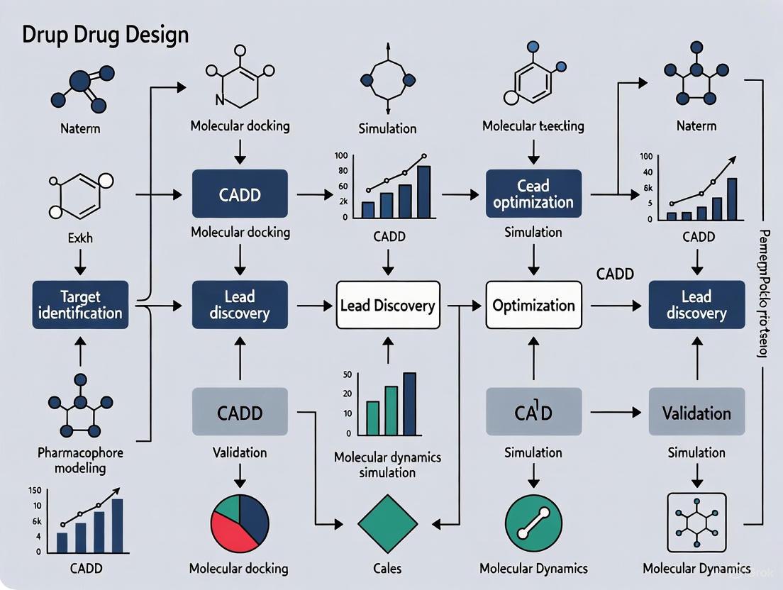
The following workflow diagram illustrates the integrated application of these SBDD and LBDD methodologies within a modern drug discovery pipeline.
Diagram 1: Integrated CADD Drug Discovery Workflow. This diagram outlines the strategic decision-making process between SBDD and LBDD approaches based on data availability, converging on virtual screening for hit identification.
Essential Research Reagents and Computational Tools
The effective application of CADD methodologies relies on a sophisticated toolkit of software platforms, databases, and computational resources. The table below catalogs key research reagent solutions essential for executing the protocols described in this document.
Table 1: Essential Research Reagent Solutions for CADD
| Tool/Resource Name | Type | Primary Function | Application in Protocol |
|---|---|---|---|
| AlphaFold [1] [4] | Structure Prediction | Predicts 3D protein structures from amino acid sequences with high accuracy. | SBDD: Provides reliable target structures when experimental ones are unavailable. |
| AutoDock Vina [1] | Docking Software | Fast, accurate molecular docking and virtual screening. | Protocol 2.1.1: Predicting ligand binding poses and affinities. |
| GROMACS [1] | Molecular Dynamics | High-performance MD simulation package for simulating biomolecular systems. | Protocol 2.1.2: Running production MD simulations for free energy calculations. |
| MOE (Molecular Operating Environment) [4] | Integrated Software Suite | Comprehensive platform for structure- and ligand-based design, QSAR, and simulations. | Multiple: Used across SBDD and LBDD protocols for docking, pharmacophore modeling, and QSAR. |
| KNIME [4] | Workflow Platform | Visual platform for creating data science workflows and automating computational tasks. | Protocol 2.2.1: Building and validating QSAR models; automating virtual screening pipelines. |
| ZINC/ChEMBL | Compound Database | Publicly accessible databases of commercially available and bioactive compounds. | Protocol 2.1.1 & 2.2.2: Source of compound libraries for virtual screening. |
| Protein Data Bank (PDB) | Structure Repository | Central repository for experimentally determined 3D structures of biological macromolecules. | SBDD: Primary source of target structures for docking and analysis. |
Quantitative Data and Performance Metrics
The impact of CADD is demonstrated through both its predictive accuracy in specific tasks and its overall contribution to streamlining the drug discovery pipeline. The following tables summarize performance metrics for key computational tools and techniques.
Table 2: Performance Comparison of Molecular Docking Tools [1]
| Tool | Primary Application | Key Advantages | Common Limitations |
|---|---|---|---|
| AutoDock Vina | Predicting binding affinities and orientations. | Fast, accurate, and easy to use. | May be less accurate for highly flexible systems. |
| AutoDock GOLD | Predicting binding for flexible ligands. | High accuracy for handling ligand flexibility. | Requires a license and can be expensive. |
| Glide | High-accuracy docking and virtual screening. | Highly accurate and integrated with Schrödinger suite. | Requires commercial Schrödinger suite. |
| DOCK | Versatile docking and virtual screening. | Versatile; can be used for both docking and virtual screening. | Can be slower than other tools. |
| SwissDock | Web-based docking predictions. | Easy to use and accessible online. | May not be as accurate for complex systems. |
Table 3: Summary of Key CADD Techniques and Applications
| Computational Method | Theoretical Basis | Output/Deliverable | Impact on Discovery Process |
|---|---|---|---|
| Molecular Docking [1] [2] | Molecular mechanics, scoring functions. | Predicted binding pose and affinity score. | Rapid identification of potential hits from large libraries; suggests initial binding hypotheses. |
| Molecular Dynamics (MD) [1] [6] | Statistical mechanics, Newtonian physics. | Time-evolution trajectory of the system; free energy estimates. | Provides dynamic insight into binding stability, mechanisms, and more accurate affinity predictions (MM-PBSA/GBSA, FEP). |
| QSAR [1] [5] | Statistical modeling, Machine Learning. | Predictive model linking chemical descriptors to activity. | Guides lead optimization by predicting the activity of unsynthesized analogs. |
| Pharmacophore Modeling [5] [2] | Chemical feature perception and alignment. | 3D query representing essential interactions for bioactivity. | Enables scaffold hopping and identification of novel chemotypes via virtual screening. |
| Virtual Screening [1] [3] | Docking, pharmacophore, or similarity searching. | A prioritized list of candidate molecules for experimental testing. | Dramatically reduces the number of compounds requiring costly experimental HTS. |
Computer-Aided Drug Design has unequivocally transitioned from a supplementary tool to a central pillar of drug discovery [1] [7]. By integrating computational methodologies across the entire discovery pipeline—from target analysis and hit identification to lead optimization and ADMET prediction—CADD provides a rational framework that significantly reduces the time and cost associated with bringing new therapeutics to market [5] [4]. The synergistic application of structure-based and ligand-based approaches, powered by advancements in artificial intelligence and ever-increasing computational resources, ensures that CADD will continue to be a critical driving force in the development of safer and more effective medicines [3] [2].
In the field of Computer-Aided Drug Design (CADD), two principal paradigms have emerged as cornerstones for modern therapeutic discovery: structure-based drug design (SBDD) and ligand-based drug design (LBDD) [8] [9]. These methodologies represent complementary approaches to the same fundamental challenge: efficiently identifying and optimizing chemical compounds that effectively modulate biological targets. SBDD relies on the three-dimensional structural information of the target protein, typically obtained through experimental methods like X-ray crystallography or computational predictions, to guide the design of molecules that complement the binding site [8] [10]. In contrast, LBDD leverages information from known active compounds to infer properties of new potential drugs when the target structure is unknown or difficult to obtain [8] [11].
The strategic selection and integration of these approaches have become increasingly critical in pharmaceutical research, as they offer pathways to reduce discovery timelines and costs while improving the quality of candidate compounds [12] [13]. This article provides a comprehensive comparison of these dominant methodologies, detailing their respective use cases, experimental protocols, and emerging integration strategies that leverage the strengths of both paradigms.
Core Principles and Comparative Analysis
Structure-Based Drug Design (SBDD)
SBDD is fundamentally rooted in the principle of molecular recognition, designing compounds that sterically and chemically complement the target binding site [8] [10]. This approach requires detailed knowledge of the three-dimensional architecture of the biological target, typically a protein or nucleic acid involved in a disease process [14]. The process begins with obtaining a high-resolution structure of the target protein, which can be achieved through experimental techniques including X-ray crystallography, nuclear magnetic resonance (NMR) spectroscopy, and cryo-electron microscopy (cryo-EM), or through computational predictions using tools like AlphaFold [10] [11] [14].
Once the structure is obtained, researchers analyze the binding site characteristics including shape, electrostatic properties, and hydrogen-bonding capabilities [14]. This structural information enables rational drug design through computational techniques such as molecular docking, where potential drug candidates are virtually screened for their ability to bind the target, and molecular dynamics simulations, which assess the stability of proposed protein-ligand complexes [8] [11]. The primary advantage of SBDD lies in its ability to provide atomic-level insights into drug-target interactions, facilitating the design of highly specific compounds with optimized binding affinity [8] [10].
Ligand-Based Drug Design (LBDD)
LBDD approaches are employed when three-dimensional structural information of the target is unavailable, but data about molecules that interact with the target exist [8] [11]. This methodology operates on the similarity-property principle, which posits that structurally similar molecules are likely to exhibit similar biological activities [11] [12]. LBDD techniques analyze the physicochemical properties and structural features of known active compounds to build models that predict the activity of new molecules [8].
Key LBDD methods include Quantitative Structure-Activity Relationship (QSAR) modeling, which establishes mathematical relationships between molecular descriptors and biological activity, and pharmacophore modeling, which identifies the essential steric and electronic features necessary for molecular recognition [8] [11]. These approaches enable virtual screening of compound libraries to identify novel candidates that share critical characteristics with known actives, even when their molecular scaffolds differ significantly (a process known as "scaffold hopping") [11]. The major strength of LBDD is its independence from target structure, making it applicable to targets that are difficult to characterize structurally, such as membrane proteins [8].
Comparative Analysis of SBDD and LBDD
Table 1: Comparative Analysis of Structure-Based and Ligand-Based Drug Design Approaches
| Aspect | Structure-Based Drug Design (SBDD) | Ligand-Based Drug Design (LBDD) |
|---|---|---|
| Primary Requirement | 3D structure of target protein [8] | Known active ligands [8] |
| Key Techniques | Molecular docking, molecular dynamics simulations, free energy perturbation [8] [11] | QSAR, pharmacophore modeling, similarity searching [8] [11] |
| Typical Applications | Rational design of novel scaffolds, affinity optimization, selectivity engineering [8] [14] | Scaffold hopping, lead expansion, analog series optimization [11] |
| Key Advantages | Provides atomic-level interaction details; enables de novo design [8] [10] | Fast and computationally efficient; no need for protein structure [8] |
| Major Limitations | Dependent on quality and relevance of protein structure; computationally intensive [8] [14] | Limited by chemical space of known actives; may miss novel mechanisms [8] [12] |
Table 2: Structural Biology Techniques for SBDD
| Technique | Resolution Range | Sample Requirements | Key Applications in SBDD |
|---|---|---|---|
| X-ray Crystallography | 1.5-3.5 Å [10] | High-quality protein crystals [8] [10] | Atomic-level binding site analysis; ligand co-crystallization [8] |
| Cryo-EM | 3.0-3.5 Å (typically) [10] | Vitrified protein solutions [10] | Membrane proteins; large complexes [8] [10] |
| NMR Spectroscopy | 2.5-4.0 Å [10] | Isotopically labeled proteins in solution [8] [10] | Studying protein dynamics; flexible systems [8] |
| Computational Prediction | Variable (e.g., AlphaFold) [11] [14] | Protein sequence [14] | Targets resistant to experimental structure determination [11] |
Experimental Protocols
Protocol 1: Structure-Based Virtual Screening
Objective: To identify novel hit compounds through molecular docking against a known protein structure.
Materials and Reagents:
- Target Protein Structure: Experimentally determined (PDB format) or computationally predicted [11] [14]
- Compound Library: Commercially available (e.g., ZINC, Enamine) or proprietary collections in SDF or PDBQT format [15]
- Docking Software: AutoDock Vina, Glide, GOLD, or similar [15]
- Computational Resources: High-performance computing cluster for large-scale screening [13]
Procedure:
- Target Preparation:
- Obtain the 3D structure of the target protein from the Protein Data Bank or through prediction tools like AlphaFold [11] [14].
- Remove water molecules and extraneous ligands, except those critical for binding [15].
- Add hydrogen atoms and optimize hydrogen bonding networks using tools like MolProbity [14].
- Define the binding site coordinates based on known ligand positions or predicted active sites [15].
Ligand Library Preparation:
Molecular Docking:
Post-Docking Analysis:
Validation:
Protocol 2: Ligand-Based Virtual Screening Using QSAR
Objective: To predict compound activity using quantitative structure-activity relationship models.
Materials and Reagents:
- Training Set Compounds: Known active and inactive compounds with measured biological activities [8] [11]
- Test Compounds: Database of compounds to be screened and prioritized [15]
- Chemical Descriptor Software: PaDEL-Descriptor, RDKit, or similar [15]
- Modeling Environment: Python/R with machine learning libraries (scikit-learn, TensorFlow) [16] [15]
Procedure:
- Data Set Curation:
- Compile a diverse set of compounds with reliable activity data (IC50, Ki, or EC50 values) [12].
- Apply chemical standardization to normalize structures (tautomer standardization, salt removal) [12].
- Divide data into training (80%) and test (20%) sets using rational splitting methods to ensure chemical space coverage [15].
Molecular Descriptor Calculation:
Model Development:
Virtual Screening:
Model Interpretation and Validation:
Research Reagent Solutions
Table 3: Essential Research Reagents and Computational Tools for CADD
| Reagent/Tool | Function | Example Applications |
|---|---|---|
| Protein Data Bank (PDB) | Repository of experimentally determined protein structures [10] | Source of target structures for SBDD [10] |
| ZINC Database | Curated collection of commercially available compounds [15] | Virtual screening compound libraries [15] |
| AutoDock Vina | Molecular docking software [15] | Predicting ligand binding modes and affinities [15] |
| PaDEL-Descriptor | Molecular descriptor calculation software [15] | Generating chemical features for QSAR modeling [15] |
| AlphaFold | Protein structure prediction tool [11] | Generating models when experimental structures are unavailable [11] |
Integrated Workflows and Combined Approaches
Sequential Integration Strategies
The sequential integration of LBDD and SBDD methods represents a powerful funnel-based approach that maximizes efficiency in virtual screening [11] [17]. In this workflow, large compound libraries are first processed using fast ligand-based methods to reduce the chemical space, after which the pre-filtered subset undergoes more computationally intensive structure-based analysis [11] [12]. This strategy is particularly valuable when dealing with ultra-large chemical libraries containing billions of compounds, where exhaustive structure-based screening would be prohibitively resource-intensive [12].
A typical sequential workflow proceeds through these stages:
- Initial Filtering: Large compound collections are screened using 2D or 3D similarity searches against known active compounds or through pre-trained QSAR models [11] [17].
- Property-Based Filtering: Compounds passing the initial screen are evaluated for drug-like properties, including physicochemical characteristics, potential toxicity, and synthetic accessibility [12].
- Structure-Based Prioritization: The refined compound set undergoes molecular docking against the target structure [11] [17].
- Consensus Scoring: Final candidate selection incorporates results from both ligand-based and structure-based approaches [11].
This sequential approach was effectively demonstrated in the CACHE Challenge #1, where participants sought ligands for the LRRK2-WDR domain [12]. Successful teams typically employed initial ligand-based filtering to narrow the enormous chemical space before applying structure-based methods to the reduced compound sets, highlighting the practical utility of this integrated strategy in real-world drug discovery scenarios [12].
Parallel and Hybrid Screening Approaches
Advanced screening pipelines increasingly employ parallel implementation of LBDD and SBDD methods, where compounds are simultaneously evaluated using both approaches [11] [17]. The independent results are subsequently combined using consensus scoring frameworks that leverage the complementary strengths of each method [11] [12]. This strategy helps mitigate the limitations inherent in individual approaches and increases the probability of identifying authentic active compounds [17].
Key parallel implementation strategies include:
- Parallel Scoring: Selecting the top-ranked compounds from both ligand-based similarity rankings and structure-based docking scores without requiring consensus between them [17]. This approach increases sensitivity and the likelihood of recovering potential actives, particularly when one method underperforms due to technical limitations [11].
- Hybrid Scoring: Multiplying or averaging normalized scores from different methods to create a unified ranking system [11] [17]. This approach favors compounds ranked highly by both methodologies, thereby prioritizing specificity and increasing confidence in selected candidates [17].
- Ensemble Methods: Using multiple protein conformations or diverse ligand sets to capture the dynamic nature of binding sites and the heterogeneity of active chemical series [11]. These ensembles provide a more comprehensive representation of the drug-target interaction landscape compared to single-structure or single-template approaches [11].
Diagram 1: Combined LBVS and SBVS workflow. This diagram illustrates a parallel virtual screening approach where ligand-based and structure-based methods are applied simultaneously, with results combined through consensus scoring to identify high-confidence hit compounds [11] [17].
Case Study: Identification of Natural Tubulin Inhibitors
A recent study exemplifies the powerful integration of structure-based and ligand-based approaches in the discovery of natural inhibitors targeting the human αβIII tubulin isotype, a protein implicated in cancer drug resistance [15]. This research employed a comprehensive methodology that leveraged the complementary strengths of both paradigms to identify promising therapeutic candidates.
The integrated workflow proceeded through these key stages:
- Target Preparation: Researchers developed a homology model of the human αβIII tubulin isotype using Modeller software, based on a bovine tubulin template with 100% sequence identity [15].
- Structure-Based Virtual Screening: The team screened 89,399 natural compounds from the ZINC database against the Taxol-binding site of tubulin using AutoDock Vina, selecting the top 1,000 hits based on binding energy [15].
- Machine Learning Classification: A supervised machine learning approach was implemented to distinguish between active and inactive compounds based on chemical descriptor properties [15]. The model was trained on known Taxol-site targeting drugs (actives) and non-Taxol targeting drugs (inactives) with decoys generated by the DUD-E server [15].
- ADMET and Biological Property Prediction: The top candidates were evaluated for absorption, distribution, metabolism, excretion, and toxicity (ADMET) properties, along with Prediction of Activity Spectra for Substances (PASS) analysis to anticipate potential biological activities [15].
- Validation through Molecular Dynamics: The final four hit compounds underwent molecular dynamics simulations to assess complex stability and binding modes, with binding affinity calculations confirming strong interactions with the target [15].
This case study demonstrates how the sequential application of structure-based and ligand-based methods, augmented by machine learning, can efficiently identify promising drug candidates with specific target affinity [15]. The integrated approach allowed researchers to leverage the precision of structure-based docking while incorporating the pattern recognition capabilities of ligand-based modeling, ultimately identifying natural compounds with potential to overcome drug resistance in cancer therapy [15].
Structure-based and ligand-based drug design represent complementary paradigms in modern computational drug discovery, each with distinctive strengths, limitations, and optimal application domains [8] [11]. SBDD provides atomic-level insights into drug-target interactions but requires high-quality structural information, while LBDD offers efficient screening capabilities based on known active compounds without requiring target structure [8]. The strategic integration of these approaches through sequential, parallel, or hybrid implementation creates synergistic workflows that enhance hit identification efficiency and quality [11] [17].
Future directions in CADD point toward increasingly sophisticated integration of these methodologies, powered by advances in artificial intelligence and machine learning [12] [16]. Deep learning models that simultaneously leverage both structural and ligand information, such as the DRAGONFLY framework for de novo drug design, represent the next frontier in computational drug discovery [16]. Furthermore, the growing availability of predicted protein structures through tools like AlphaFold is expanding the applicability of SBDD to previously inaccessible targets [11] [14]. As these computational approaches continue to evolve, the strategic combination of structure-based and ligand-based methodologies will remain essential for addressing the complex challenges of modern drug discovery and development.
The Drug Development Burden: A Quantitative Analysis
The traditional drug discovery and development process is characterized by immense financial investment and extended timelines, presenting significant market challenges that Computer-Aided Drug Design (CADD) aims to address.
Table 1: Drug Development Cost and Timeline Analysis
| Development Phase | Average Duration | Average Cost (USD) | Probability of Success |
|---|---|---|---|
| Discovery & Preclinical | 1 - 6 years [18] | $15 - $100 million [18] | Preclinical to Phase I Transition: ~10% [18] |
| Clinical Trials (Phases I-III) | 6 - 7 years [18] | $435 million (Phase I: $25M; Phase II: $60M; Phase III: $350M) [18] | Phase I to Approval: ~12% [19] |
| FDA Review & Approval | 0.5 - 2 years [18] | $2 - $3 million (application fee) [18] | N/A |
| Total | 10 - 15 years [19] [18] | $2.6 billion (incl. cost of failures) [20] [19] | 1 in 5,000 compounds from preclinical stage [18] |
The primary drivers of the $2.6 billion cost include the high failure rate of drug candidates (approximately 90% fail in clinical trials) and the prolonged development cycle requiring consistent funding over a decade or more [18]. CADD emerges as a strategic solution to rationalize and expedite this process, offering a more efficient and cost-effective approach by leveraging computational power to predict compound behavior before costly synthetic and experimental work begins [13].
CADD Methodologies and Protocols
CADD encompasses two primary computational approaches: Structure-Based Drug Design (SBDD) and Ligand-Based Drug Design (LBDD). The application of these methods integrates into a streamlined workflow for lead identification and optimization.
Core CADD Workflow
The following diagram illustrates the logical relationship and workflow between the major CADD methodologies.
Structure-Based Drug Design (SBDD) Protocol
SBDD relies on the knowledge of the three-dimensional structure of the biological target, typically a protein [1].
Protocol 1: Target Modeling and Binding Site Characterization
- Objective: Generate an accurate 3D model of the target protein and identify the putative binding site.
- Software Tools:
- Homology/Comparative Modeling: SWISS-MODEL [21], MODELLER [21] (Used when an experimental structure is unavailable but a homologous template exists).
- Deep Learning-Based Structure Prediction: AlphaFold [21], ESMFold [1], I-TASSER [21] (Used for ab initio modeling without a clear template).
- Binding Site Prediction: CASTp [21], Active Site Prediction Tool [21] (Identifies cavities and pockets on the protein surface suitable for ligand binding).
- Methodology:
- Sequence Alignment: For homology modeling, align the target protein sequence with the template sequence(s).
- Model Building: Generate 3D coordinates for the target based on the template structure, modeling missing loops and side chains.
- Model Refinement: Use energy minimization and molecular dynamics (MD) simulations (e.g., with GROMACS [1] [21]) to relax steric clashes and improve stereochemistry.
- Model Validation: Assess the quality of the generated model using tools like PROCHECK or MolProbity to verify Ramachandran plot outliers and rotamer geometry.
- Binding Site Identification: Run binding site prediction algorithms to define the coordinates and volume of the active site.
Protocol 2: Molecular Docking and Virtual Screening
- Objective: Predict the binding orientation (pose) and affinity of small molecules within the target's binding site and screen large compound libraries in silico.
- Software Tools: AutoDock Vina [1] [21], Glide [1] [21], GOLD [1], DOCK [1].
- Methodology:
- Preparation of Structures:
- Protein: Add hydrogen atoms, assign partial charges (e.g., using Kollman or AMBER force fields), and define protonation states of key residues (e.g., His, Asp, Glu).
- Ligand Library: Obtain structures from databases like ZINC [21] or PubChem [21]. Prepare ligands by generating 3D conformations, optimizing geometry, and assigning charges (e.g., using Gasteiger-Marsili).
- Grid Generation: Define a 3D grid box encompassing the binding site. The box size should be large enough to accommodate ligand flexibility.
- Docking Execution: Run the docking algorithm. For virtual screening, this is performed on thousands to millions of compounds.
- Pose Scoring and Ranking: The docking software scores each pose using a scoring function (knowledge-based, force-field based, or empirical). Rank compounds based on their predicted binding affinity (e.g., kcal/mol).
- Post-Docking Analysis: Visually inspect top-ranked complexes (using PyMOL, Maestro [21]) to analyze key interactions (H-bonds, hydrophobic contacts, pi-stacking). Cluster similar poses to identify consensus binding modes.
- Preparation of Structures:
Ligand-Based Drug Design (LBDD) Protocol
LBDD is employed when the 3D structure of the target is unknown, and the design is based on known active molecules (ligands) [1].
Protocol 3: Pharmacophore Modeling and 3D Database Screening
- Objective: Create an abstract model of the essential molecular features responsible for biological activity and use it to identify new scaffolds.
- Software Tools: LigandScout [21], Phase [21], Pharmer [21].
- Methodology:
- Data Set Curation: Compile a set of known active compounds and, if possible, inactive decoys with diverse structures but similar properties.
- Conformational Analysis: Generate a representative set of low-energy conformers for each molecule in the training set.
- Pharmacophore Hypothesis Generation:
- Constructive Phase: Superimpose active molecules and identify common chemical features (e.g., hydrogen bond donors/acceptors, hydrophobic regions, aromatic rings, charged groups).
- Model Building: Create a spatial model (hypothesis) defining the geometric and chemical constraints common to active compounds.
- Hypothesis Validation:
- Use statistical methods (e.g., Fischer's randomization test [21]) to assess the significance of the model.
- Test the model against a validation set of compounds with known activity to evaluate its predictive power.
- Virtual Screening: Use the validated pharmacophore model as a 3D query to search large chemical databases and retrieve compounds that match the feature arrangement.
Protocol 4: Quantitative Structure-Activity Relationship (QSAR) Modeling
- Objective: Develop a quantitative model that correlates numerical descriptors of chemical structures with their biological activity.
- Software Tools: Various commercial and open-source QSAR packages (e.g., within Schrödinger suite, KNIME, Python/R libraries).
- Methodology:
- Data Set Preparation: Assay a congeneric series of compounds for a specific biological endpoint (e.g., IC50, Ki).
- Molecular Descriptor Calculation: Compute numerical descriptors for each compound, which can be 0D (molecular weight), 1D (substructure counts), 2D (topological indices), or 3D (molecular surface area, volume).
- Model Development:
- Split data into training and test sets.
- Use machine learning algorithms (e.g., Random Forest, Support Vector Machines, Partial Least Squares regression) on the training set to build a model linking descriptors to activity.
- Model Validation: Assess the model's internal consistency (cross-validation on training set) and, more importantly, its predictive ability using the external test set. Key metrics include R² and Q².
- Model Application: Use the validated QSAR model to predict the activity of new, untested compounds and prioritize them for synthesis.
The Scientist's Toolkit: Essential Research Reagent Solutions
Table 2: Key Research Reagents, Databases, and Software for CADD
| Item Name / Category | Function / Application | Specific Examples |
|---|---|---|
| Commercial Software Suites | Integrated platforms for molecular modeling, simulation, and data analysis. | Schrödinger Suite (Maestro, Glide) [21], BIOVIA Discovery Studio [21] |
| Open-Source Molecular Dynamics | Simulate the time-dependent behavior of biomolecules in physiological conditions. | GROMACS [1] [21], AMBER [21], OpenMM [1] |
| Docking & Virtual Screening Tools | Predict ligand binding pose and affinity; screen compound libraries in silico. | AutoDock Vina [1] [21], DOCK [1], SwissDock [1] |
| Public Compound Databases | Sources of chemical structures for virtual screening and lead discovery. | ZINC [21] (for purchasable compounds), PubChem [21] (bioactivity data) |
| Protein Structure Databases | Sources of experimental and predicted protein structures for SBDD. | Protein Data Bank (PDB), AlphaFold Protein Structure Database [1] |
| Specialized Hardware | Accelerate computationally intensive calculations like MD and AI model training. | High-Performance Computing (HPC) Clusters, Graphics Processing Units (GPUs) [22] |
| Machine Learning Frameworks | Develop and train custom predictive models for QSAR, de novo design, etc. | TensorFlow, PyTorch (often integrated into broader CADD platforms) [13] |
Application Note: Quantitative Market Landscape of Computer-Aided Drug Design (CADD)
The global Computer-Aided Drug Design (CADD) market is experiencing transformative growth, propelled by the integration of advanced computational technologies such as Artificial Intelligence (AI) and Machine Learning (ML). CADD utilizes computational methods to discover, design, and optimize drug candidates, significantly accelerating the drug discovery pipeline and reducing associated costs [23]. This application note provides a detailed quantitative analysis of the key players driving innovation and the distinct regional landscapes shaping the global CADD market, with a focus on North America's dominance and the rapid emergence of the Asia-Pacific region.
Key Global Players and Competitive Landscape
The CADD market features a dynamic ecosystem of established technology firms, specialized software providers, and agile startups. Their contributions are fundamental to the methodologies described in subsequent experimental protocols.
Table 1: Key Players and Technological Contributions in the CADD Market
| Company/Organization | Primary Role/Contribution | Key Technologies/Services | Recent Strategic Developments (2024-2025) |
|---|---|---|---|
| Schrödinger, Inc. [23] | Software Provider & Service Provider | Physics-based computational platforms, Molecular modeling | Key player in the dominant North American market. |
| BIOVIA (Dassault Systèmes) [23] [24] | Software Provider | Scientific software for molecular modeling, simulation, and data management | Part of a key player segment in a dominant market. |
| Absci Corporation [25] | AI-Driven Drug Discovery | Generative AI for de novo protein and drug design | Collaborated with AMD to deploy AI accelerators for drug discovery workloads. |
| NVIDIA [26] | Technology Enabler | Advanced GPUs, AI platforms (Clara) for biomedical research | Partnered with IQVIA to boost clinical research with AI agents. |
| Google/Google Cloud [26] | Technology Enabler | Cloud AI tools for biomedical image analysis and data processing | Expanded collaboration with Recursion to leverage cloud technologies for drug discovery. |
| Insilico Medicine [25] | AI-Driven Drug Discovery | Generative AI platform for target identification and molecule design | Its AI platform identified a drug target and created a drug for fibrosis. |
| Chai Discovery [25] | Biotech Startup | AI-powered platform for novel antibody design | Secured $70M to evolve its Chai-2 platform for designing new antibodies. |
| Latent Labs [25] | AI-Driven Discovery | AI foundation models for programmable biology and protein design | Secured $50M in funding to establish generative AI models for developing new proteins. |
| Rowan [22] | CADD Platform Provider | Integrated platform for benchmarking, validation, and workflow management | Aims to reduce the "invisible work" in CADD, such as software integration and model validation. |
Regional Market Analysis: Quantitative Dynamics
Regional dominance in the CADD market is influenced by factors including technological infrastructure, R&D investment, government initiatives, and the local presence of pharmaceutical and biotech industries.
Table 2: Regional Analysis of the CADD Market (2024-2034 Projections)
| Region | Market Share (2024) | Projected CAGR (2025-2034) | Key Growth Drivers | Noteworthy Regional Initiatives |
|---|---|---|---|---|
| North America | ~45% [25] [23] | Not explicitly stated | Presence of key players, state-of-the-art R&D infrastructure, high healthcare technology investments, focus on personalized medicine [25] [23] [26]. | US FDA issued guidelines on AI for regulatory decision-making [25]. |
| Asia-Pacific (APAC) | Not the largest share | Fastest Growing [25] [23] | Rapid industrialization, government-driven innovation programs, expanding pharmaceutical sector, rising disease burden, growing investments in R&D [25] [23] [26]. | China's "AI + Medicine" plan (2025-2027); Japan's MHLW funding for AI-enabled drug discovery [25]. |
| Europe | Substantial share [27] | Not explicitly stated | Stringent quality standards, sustainability goals, increasing R&D initiatives [27]. | Not specified in search results. |
| Latin America, Middle East & Africa | Gradual progression [27] | Gradual progression [27] | Improving economic conditions, rising urbanization, growing awareness of advanced solutions [27]. | Not specified in search results. |
The CADD landscape is characterized by robust growth in North America, led by technological innovation and a strong biopharmaceutical ecosystem, while the Asia-Pacific region presents the highest growth potential due to strategic governmental support and rapid market expansion. The synergy between key players advancing AI/ML technologies and supportive regional policies is defining the future of efficient and effective drug discovery.
Protocol: Computational Methods for Structure-Based Drug Design
Scope
This protocol outlines a standard workflow for Structure-Based Drug Design (SBDD), the dominant segment in the CADD market which accounted for approximately 55% share in 2024 [25] [23]. SBDD relies on the 3D structural information of a biological target to identify and optimize potential drug molecules [25].
Principle
SBDD utilizes the atomic-level structure of a target protein, often obtained from X-ray crystallography, Cryo-EM, or NMR, to guide the discovery of ligands that bind with high affinity and specificity. This approach allows for the rational design of novel therapeutics and was notably applied in the development of protease inhibitors for treatments like Paxlovid [25].
Experimental Procedures
Target Preparation and Selection
- Objective: To obtain a clean, biophysically relevant 3D structure of the target protein for computational studies.
- Methods:
- Source Structure: Obtain the target structure from the Protein Data Bank (PDB) or through homology modeling.
- Structure Refinement: Remove water molecules, co-crystallized ligands, and ions not involved in the binding site. Add missing hydrogen atoms and assign correct protonation states for residues (e.g., His, Asp, Glu) using software like BIOVIA Discovery Studio [23] or Schrödinger's Protein Preparation Wizard.
- Binding Site Definition: Define the active site or allosteric pocket of interest based on known catalytic residues or the location of a co-crystallized native ligand.
Molecular Docking for Virtual Screening
- Objective: To computationally screen large libraries of compounds and predict their binding pose and affinity within the target site.
- Methods:
- Library Preparation: Prepare a database of small molecule structures (e.g., from ZINC database) by energy minimization and generating possible tautomers and stereoisomers.
- Docking Execution: Perform docking simulations using software such as AutoDock Vina [25]. This involves sampling possible conformations (poses) of the ligand within the defined binding site and scoring them based on a scoring function.
- Pose Analysis: Visually inspect the top-ranked poses for key interactions like hydrogen bonds, hydrophobic contacts, and pi-stacking. Prioritize compounds with strong complementary interactions for further analysis.
Binding Affinity Refinement using Molecular Dynamics (MD)
- Objective: To assess the stability of the protein-ligand complex and obtain more accurate binding free energy estimates.
- Methods:
- System Setup: Solvate the protein-ligand complex in a water box (e.g., TIP3P model) and add ions to neutralize the system.
- Simulation Run: Perform MD simulations using packages like GROMACS or AMBER on high-performance computing (HPC) systems or cloud platforms (e.g., Google Cloud AI, NVIDIA Clara) [26]. A typical production run may be for 100 nanoseconds.
- Trajectory Analysis: Analyze the root-mean-square deviation (RMSD) of the protein and ligand to check for complex stability. Calculate binding free energies using methods like Molecular Mechanics/Generalized Born Surface Area (MM/GBSA).
Experimental Validation and Iterative Design
- Objective: To synthesize and test top-ranked computational hits, using experimental data to refine the models.
- Methods:
- Compound Acquisition/Synthesis: Acquire or synthesize the computationally identified hit compounds.
- In vitro Assays: Test compounds for binding affinity (e.g., Surface Plasmon Resonance) and functional activity in biochemical or cell-based assays.
- Cycle of Learning: Use the experimental results to validate the computational predictions. If a co-crystal structure is obtained with a hit compound, use it to refine the docking protocol and initiate further rounds of design and optimization in an iterative cycle.
Workflow Visualization
The Scientist's Toolkit: Research Reagent Solutions
Table 3: Essential Research Reagents and Tools for SBDD
| Item/Tool | Function/Description | Example Providers/Platforms |
|---|---|---|
| Protein Structure Database | Repository of experimentally determined 3D protein structures for target selection and preparation. | Protein Data Bank (PDB) |
| Compound Library | Large collections of small molecules for virtual screening to identify initial hits. | ZINC Database |
| Molecular Docking Software | Predicts the preferred orientation and binding affinity of a small molecule to a protein target. | AutoDock Vina [25], Schrödinger Suite [23] |
| Molecular Dynamics Software | Simulates the physical movements of atoms and molecules over time to study complex stability and dynamics. | GROMACS, AMBER |
| AI/ML Drug Design Platform | Uses generative models and predictive algorithms to design novel molecules and optimize properties. | Insilico Medicine Platform [25], Absci Corp. AI [25] |
| Integrated CADD Platform | Streamlines workflows by combining benchmarking, validation, and computation in a unified environment. | Rowan [22] |
| High-Performance Computing (HPC) | Provides the computational power required for demanding tasks like MD simulations and AI model training. | NVIDIA GPUs [26], Google Cloud AI [26] |
The field of computer-aided drug design (CADD) is undergoing a profound transformation, moving beyond traditional structure-based modeling to embrace a new era defined by artificial intelligence (AI), cloud-native infrastructure, and novel therapeutic modalities. This paradigm shift is accelerating the entire drug discovery value chain, from initial target identification to clinical trials, enabling researchers to address biological targets once considered "undruggable" [28]. The integration of these three powerful trends is compressing discovery timelines that traditionally spanned years into months, while simultaneously improving the precision and success rates of new therapeutic candidates [29] [30]. This document provides detailed application notes and experimental protocols for leveraging these converging technologies within modern CADD research frameworks.
Quantitative Landscape: Market Data and Performance Metrics
The adoption of advanced technologies in drug discovery is reflected in robust market growth and distinct performance advantages. The tables below summarize key quantitative data for strategic planning.
Table 1: Computer-Aided Drug Design (CADD) Market Segmentation (2024) [23]
| Segmentation Category | Dominant Segment (Market Share) | Highest Growth Segment (CAGR) |
|---|---|---|
| Type | Structure-Based Drug Design (SBDD) (~55%) | Ligand-Based Drug Design (LBDD) |
| Technology | Molecular Docking (~40%) | AI/ML-Based Drug Design |
| Application | Cancer Research (~35%) | Infectious Diseases |
| End-User | Pharmaceutical & Biotech Companies (~60%) | Academic & Research Institutes |
| Deployment Mode | On-Premise (~65%) | Cloud-Based |
Table 2: Performance Metrics of Leading AI-Driven Drug Discovery Platforms [29]
| Company / Platform | Key AI Approach | Reported Efficiency Gain | Example Clinical Candidate |
|---|---|---|---|
| Exscientia | Generative AI, Centaur Chemist | ~70% faster design cycles; 10x fewer compounds synthesized | CDK7 inhibitor (GTAEXS-617), LSD1 inhibitor (EXS-74539) |
| Insilico Medicine | Generative AI | Target to Phase I in 18 months for IPF drug | Idiopathic Pulmonary Fibrosis drug (Phase I) |
| Recursion | Phenotypic Screening, AI | Integrated platform with Exscientia post-merger | Multiple oncology programs |
| BenevolentAI | Knowledge Graphs, Target ID | AI-derived targets advancing to clinic | Multiple undisclosed programs |
| Schrödinger | Physics-Based Simulations, FEP+ | Platform for rapid in-silico candidate optimization | Multiple partnered and internal programs |
Application Note: Implementing AI-Driven Discovery Workflows
Core Principles and Workflow
AI is revolutionizing CADD by automating complex design tasks and extracting insights from large-scale multimodal data. Leading platforms demonstrate that AI can compress the early-stage discovery and preclinical timeline from a typical 5 years to under 2 years in some cases [29]. The core applications include generative chemistry for de novo molecular design, predictive models for ADMET (Absorption, Distribution, Metabolism, Excretion, Toxicity) properties, and target identification through biological network analysis.
Figure 1: AI-Driven Drug Discovery Cycle. This workflow illustrates the iterative "design-make-test-analyze" loop accelerated by AI, where experimental feedback continuously refines the computational models.
Experimental Protocol: AI-Guided Lead Optimization
Protocol Title: Iterative Lead Optimization Using a Closed-Loop AI Design Platform
Objective: To optimize a hit compound into a preclinical candidate with desired potency, selectivity, and ADMET properties using an integrated AI-driven workflow.
Materials:
- AI Software Platform: Access to a generative AI chemistry platform (e.g., Exscientia's Centaur Chemist, Insilico Medicine's Chemistry42) [29].
- Initial Compound: Confirmed hit molecule from HTS or virtual screening.
- Target Product Profile (TPP): A defined set of criteria for the desired candidate (e.g., IC50 < 100 nM, >30x selectivity, CLhep < 10 mL/min/kg).
- Automation Studio: Robotic synthesis and high-throughput screening infrastructure [29].
Methodology:
- Data Curation and Model Priming (Week 1):
- Curate all existing SAR data for the hit series and related chemotypes.
- Input the TPP into the AI platform as optimization constraints.
- Train or fine-tune platform-specific models on the proprietary dataset.
Generative Design Cycle (Week 2):
- The AI platform proposes a focused library of novel compounds (typically 50-200) predicted to meet the TPP.
- A medicinal chemist reviews and approves the proposed structures for synthesis.
Automated Synthesis and Testing (Weeks 3-4):
- Approved compound designs are sent to an automated synthesis platform (e.g., Exscientia's AutomationStudio) [29].
- Synthesized compounds are purified and subjected to a predefined assay cascade (e.g., primary potency, cytotoxicity, microsomal stability).
Data Integration and Model Retraining (Week 5):
- Experimental results are fed back into the AI platform.
- The predictive models are retrained on the new data, improving their accuracy for the next cycle.
Iteration:
- Repeat steps 2-4 until a compound meeting all TPP criteria is identified.
Key Performance Indicator: Success is measured by the number of design cycles and total compounds synthesized to reach the candidate. AI platforms have demonstrated the ability to achieve this with 10x fewer compounds than traditional medicinal chemistry [29].
Application Note: Leveraging Cloud-Native CADD Infrastructures
Core Principles and Architecture
Cloud computing delivers scalable, collaborative, and cost-effective computational resources, overcoming the limitations of traditional on-premise HPC clusters. It democratizes access to state-of-the-art CADD tools for smaller biotechs and academic labs [31]. The cloud service models relevant to CADD are:
- IaaS (Infrastructure as a Service): Provides virtualized computing resources (e.g., AWS, Google Cloud) for running complex molecular dynamics simulations or virtual screening campaigns [32].
- PaaS (Platform as a Service): Offers a development environment for building custom drug discovery applications and workflows, such as specialized data analytics platforms [32].
- SaaS (Software as a Service): Delivers ready-to-use CADD applications via a web browser (e.g., Schrödinger's LiveScope, BIOVIA Discovery Studio) [32].
Figure 2: Cloud Collaboration Architecture for CADD. This diagram shows how a centralized cloud platform enables seamless collaboration and data integration across different roles and locations.
Experimental Protocol: Large-Scale Virtual Screening on the Cloud
Protocol Title: Cloud-Based High-Throughput Virtual Screening of Billion-Compound Libraries
Objective: To rapidly screen an ultra-large chemical library against a protein target to identify novel hit compounds.
Materials:
- Cloud Provider Account: An account with a major cloud provider (e.g., AWS, Google Cloud, Microsoft Azure).
- Target Structure: A prepared 3D structure of the target protein (e.g., from PDB or homology model).
- Chemical Library: A commercially available or proprietary compound library in a suitable format (e.g., ZINC20, Enamine REAL).
- Docking Software: A licensed or open-source molecular docking software (e.g., AutoDock Vina, Glide, FRED) configured as a cloud-native solution.
Methodology:
- Infrastructure Setup (Day 1):
- Use a pre-configured cloud formation template (e.g., AWS CloudFormation) to launch a virtual HPC cluster with hundreds to thousands of CPU cores.
- Configure parallel file storage (e.g., AWS FSx for Lustre) for high-speed I/O during the screening.
Data and Software Deployment (Day 1):
- Upload the target structure and chemical library to the cloud storage.
- Deploy the docking software across the compute cluster using a container orchestration service (e.g., Kubernetes).
Job Execution and Orchestration (Days 2-5):
- Use a job scheduler (e.g., AWS Batch) to split the chemical library into chunks and distribute docking tasks across all compute nodes.
- Monitor job progress through a cloud-based dashboard.
Post-Processing and Analysis (Day 6):
- Collate all results into a centralized cloud database.
- Use cloud-based data analytics tools (e.g., Jupyter Notebooks on Google Colab) to rank compounds by docking score and interaction patterns.
- Apply AI/ML models to further filter and prioritize top hits for purchase and testing.
Key Considerations:
- Cost Management: Utilize spot/transient instances for significant cost savings and auto-scaling to shut down resources when not in use [31].
- Security: Ensure the cloud configuration complies with relevant data protection regulations (e.g., HIPAA, GDPR) through encryption and access controls [31] [32].
Application Note: Designing for Emerging Therapeutic Modalities
Emerging modalities represent a shift from traditional small molecules and antibodies to therapies that act on DNA, RNA, or through engineered cellular machinery. They now account for $197 billion, or 60%, of the total pharma projected pipeline value [33]. Key modalities include:
- RNA Therapeutics: Including mRNA, siRNA, and antisense oligonucleotides (ASOs) that modulate gene expression [28] [34].
- Targeted Protein Degraders: Such as PROTACs (PROteolysis TArgeting Chimeras) that use the cell's ubiquitin-proteasome system to eliminate specific proteins [28] [34].
- Cell & Gene Therapies: Including CAR-T and CRISPR-based gene editing that offer potential cures for genetic diseases [33] [28].
Experimental Protocol: In-Silico Design of a PROTAC Molecule
Protocol Title: Computational Design and Optimization of a Bifunctional PROTAC Degrader
Objective: To design a novel PROTAC molecule that mediates the degradation of a protein of interest (POI) by recruiting an E3 ubiquitin ligase.
Materials:
- Structures: High-resolution crystal structures or high-quality AlphaFold2 models of the POI and the E3 ligase (e.g., Cereblon, VHL).
- Known Ligands: Structures of known small-molecule binders for the POI and the E3 ligase.
- Software: Molecular docking software (e.g., Glide, GOLD), molecular dynamics (MD) simulation package (e.g., GROMACS, Desmond), and a linker database.
Methodology:
- POI and E3 Ligase Ligand Analysis (Week 1):
- Identify solvent-exposed attachment vectors on the known POI and E3 ligase ligands where a linker can be connected without disrupting binding.
- Perform molecular docking to confirm the binding pose and identify optimal attachment points.
Linker Screening and PROTAC Assembly (Week 2):
- Screen a database of flexible and rigid linkers of varying lengths.
- In silico connect the POI ligand and E3 ligase ligand with selected linkers to generate a library of putative PROTAC molecules.
Ternary Complex Modeling and Assessment (Week 3):
- Model the full ternary complex (POI:PROTAC:E3 Ligase) using protein-protein docking guided by the PROTAC structure.
- Run short MD simulations to assess the stability of the ternary complex and the proximity between the POI's lysine residues and the E3 ligase's catalytic cysteine.
PROTAC Property Prediction (Week 4):
- Use AI/ML models or physicochemical calculations to predict the cellular permeability, solubility, and metabolic stability of the top-designed PROTACs, as their large size and complexity often pose challenges.
Key Consideration: The choice of E3 ligase is critical. While most PROTACs use a limited set of E3 ligases (Cereblon, VHL), research is actively expanding this toolbox to include others like DCAF16 and KEAP1 to access new targets and tissues [34].
The Scientist's Toolkit: Essential Research Reagent Solutions
Table 3: Key Research Reagents and Platforms for Advanced CADD
| Reagent / Solution | Function / Application | Examples / Vendor |
|---|---|---|
| Generative AI Chemistry Platforms | De novo design of novel small molecules optimized for multiple parameters. | Exscientia's Centaur Chemist, Insilico Medicine's Chemistry42 [29] |
| Digital Twin Software | Creates AI-generated virtual control patients in clinical trials to reduce placebo group size and accelerate timelines. | Unlearn.ai [30] [34] |
| Cloud-Based CADD Platforms | Provides scalable computing, SaaS tools, and collaborative workspaces for distributed teams. | Schrödinger LiveSuite, BIOVIA Discovery Studio on Cloud [31] [23] |
| PROTAC-Specific Design Suites | In-silico tools for modeling bifunctional degraders and ternary complexes. | Specific modules in Schrödinger Suite, Cresset Flare [34] |
| CRISPR Design Tools | AI-powered design of guide RNAs for gene editing therapies with minimized off-target effects. | Tools from Broad Institute, MIT [28] [34] |
| LNPs & Delivery Design Software | Computational modeling of lipid nanoparticles and other delivery vehicles for RNA/protein-based therapeutics. | Various academic and commercial molecular dynamics packages |
Methodologies in Action: A Deep Dive into Modern CADD Tools and Applications
The Synergy of Machine Learning and Physics-Based Simulations
In the field of Computer-Aided Drug Design (CADD), a new paradigm is emerging from the integration of machine learning (ML) and physics-based simulations. This synergy aims to overcome the individual limitations of each approach: ML models can struggle with generalization and physical realism, while purely physics-based methods are often computationally prohibitive for large-scale exploration [35] [36]. The combination creates a powerful framework that leverages the predictive speed of ML with the rigorous physical foundation of simulation methods, ultimately accelerating drug discovery [37] [38].
This integration is particularly valuable for addressing complex challenges in modern drug discovery, including the design of novel molecular scaffolds, targeted protein degradation, and the development of biologics [37]. By harnessing both data-driven insights and fundamental physical principles, researchers can generate drug candidates with higher predicted affinity, improved synthetic accessibility, and greater novelty [39]. This document provides detailed application notes and experimental protocols for implementing these synergistic approaches, complete with quantitative data comparisons and visual workflows.
Quantitative Performance Comparison
The integration of ML and physics-based methods has demonstrated quantitatively superior performance across multiple drug discovery benchmarks, from molecular generation efficiency to binding affinity prediction accuracy.
Table 1: Performance Metrics of Physics-Informed AI in Drug Discovery
| Method/System | Key Innovation | Test System | Performance Results | Comparison to State-of-the-Art |
|---|---|---|---|---|
| NucleusDiff [36] | Manifold-constrained diffusion model accounting for atomic distances | CrossDocked2020 (100 complexes) | Significant improvement in binding affinity prediction; Reduced atomic collisions to nearly zero | Outperformed state-of-the-art models in binding affinity |
| NucleusDiff [36] | Same as above | COVID-19 3CL protease | Increased prediction accuracy | Reduced atomic collisions by up to two-thirds compared to other leading models |
| VAE-AL Workflow [39] | Variational autoencoder with nested active learning cycles | CDK2 | 9 molecules synthesized, 8 with in vitro activity, 1 with nanomolar potency | Successfully generated novel scaffolds distinct from known templates |
| VAE-AL Workflow [39] | Same as above | KRAS | 4 molecules with potential activity identified via in silico methods | Explored sparsely populated chemical space effectively |
Table 2: Classification Performance of Machine Learning Methods Under varying Data Conditions [40]
| Method | Best For | Worst For | Key Performance Characteristics |
|---|---|---|---|
| Linear Discriminant Analysis (LDA) | Smaller number of correlated features (not exceeding ~half sample size) | Large feature sets | Most stable (precise) error estimates under optimal conditions |
| Support Vector Machines (SVM) with RBF kernel | Larger feature sets (sample size ≥20) | Small sample sizes | Clear outperformance over LDA, RF, and kNN as feature set grows |
| k-Nearest Neighbour (kNN) | Growing number of features | High variability data with small effect sizes | Performance improves with feature growth, outperforms LDA and RF unless data variability is high |
| Random Forests (RF) | Highly variable data with small effect sizes | Many common scenarios | Outperforms only kNN in specific high-variability, small-effect-size cases |
Experimental Protocols
Protocol 1: VAE with Active Learning for Molecular Generation
This protocol implements a generative AI workflow combining a variational autoencoder (VAE) with nested active learning cycles to generate novel, synthetically accessible molecules with high predicted binding affinity [39].
Materials and Reagents:
- Chemical Databases: Target-specific training sets (e.g., known CDK2 or KRAS inhibitors)
- Software: VAE architecture with encoder/decoder networks, molecular docking software (AutoDock Vina, Glide, DOCK, or SwissDock [1]), chemoinformatics toolkits for similarity assessment
- Computational Resources: High-performance computing cluster with GPU acceleration
Procedure:
- Data Preparation and Initial Training
- Represent training molecules as SMILES strings, then tokenize and convert to one-hot encoding vectors
- Pre-train VAE on a general molecular dataset to learn viable chemical space
- Fine-tune VAE on target-specific training set to increase target engagement
Nested Active Learning Cycles
Inner Cycle (Chemical Optimization):
- Sample the VAE to generate new molecules
- Evaluate generated molecules for drug-likeness, synthetic accessibility, and similarity to training set using chemoinformatic oracles
- Add molecules meeting threshold criteria to a temporal-specific set
- Use this set to fine-tune the VAE in subsequent training iterations
Outer Cycle (Affinity Optimization):
- After set number of inner cycles, subject accumulated molecules in temporal-specific set to docking simulations
- Transfer molecules meeting docking score thresholds to permanent-specific set
- Use permanent-specific set to fine-tune VAE for subsequent cycles
Repeat inner and outer cycles for predetermined iterations (typically 3-5 outer cycles with multiple inner cycles each)
Candidate Selection and Validation
- Apply stringent filtration to molecules in permanent-specific set
- Perform intensive molecular modeling simulations (e.g., PELE, absolute binding free energy calculations)
- Select top candidates for synthesis and experimental validation
Troubleshooting:
- If generated molecules lack diversity, adjust similarity thresholds in inner AL cycle
- If synthetic accessibility is poor, increase weighting of SA oracle in evaluation step
- If binding affinity plateaus, incorporate more sophisticated physics-based scoring in outer cycle
Protocol 2: Physics-Informed Diffusion Model for Binding Affinity Prediction
This protocol implements NucleusDiff, a diffusion model that incorporates physical constraints to reduce unphysical atomic collisions while maintaining high binding affinity prediction accuracy [36].
Materials and Reagents:
- Training Data: CrossDocked2020 dataset (~100,000 protein-ligand binding complexes)
- Software: NucleusDiff implementation, standard molecular visualization tools
- Computational Resources: GPU-enabled workstations or compute cluster
Procedure:
- Model Configuration
- Implement diffusion model architecture with manifold constraints
- Establish anchoring points on molecular manifold to monitor atomic distances
- Configure repellant force parameters to prevent atomic collisions
Training Protocol
- Train model on CrossDocked2020 dataset using standard training epochs
- Validate on held-out test set of 100 complexes
- Monitor both binding affinity accuracy and atomic collision metrics
Inference and Prediction
- Input novel protein-ligand complexes for binding affinity prediction
- Generate predictions with confidence intervals based on model uncertainty
- Visualize results to verify physical plausibility of predicted binding modes
Validation:
- Test model on external datasets not included in training (e.g., COVID-19 3CL protease)
- Compare performance against state-of-the-art models without physics constraints
- Quantify reduction in atomic collisions while maintaining binding affinity accuracy
Workflow Visualization
VAE-AL Generative Molecular Design Workflow
Physics-Informed AI Model Architecture
Research Reagent Solutions
Table 3: Essential Research Reagents and Computational Tools
| Category | Specific Tool/Resource | Function/Application | Key Features |
|---|---|---|---|
| Molecular Docking | AutoDock Vina [1] | Predicting binding affinities and orientations of ligands | Fast, accurate, easy to use |
| AutoDock GOLD [1] | Predicting binding affinities, especially for flexible ligands | Accurate for flexible ligands, requires license | |
| Glide [1] | Predicting binding affinities and orientations | Accurate, integrated with Schrödinger suite | |
| Structure Prediction | AlphaFold2 [1] [35] | Protein structure prediction from sequence | AI-driven high-accuracy prediction |
| ESMFold [1] | Protein structure prediction | Alternative to AlphaFold2 | |
| SWISS-MODEL [1] [35] | Homology modeling | Automated server, comparative modeling | |
| Molecular Dynamics | GROMACS [1] | Simulating behavior of proteins over time | Classical mechanics simulations |
| OpenMM [1] | Molecular dynamics simulations | Customizable, GPU acceleration | |
| Generative Models | VAE-AL Framework [39] | Generating novel molecules with desired properties | Combines variational autoencoder with active learning |
| NucleusDiff [36] | Structure-based drug design with physical constraints | Manifold-constrained diffusion model | |
| Specialized Databases | CrossDocked2020 [36] | Training dataset for structure-based drug design | ~100,000 protein-ligand binding complexes |
Computer-Aided Drug Design (CADD) has become an indispensable pillar in modern pharmaceutical research, dramatically accelerating the discovery and optimization of therapeutic agents [41]. Among its various methodologies, structure-based drug design (SBDD) leverages three-dimensional structural information of biological targets to guide the identification and development of small molecule drugs [42]. This article details three core SBDD techniques—molecular docking, molecular dynamics (MD) simulations, and free-energy perturbation (FEP)—that form a synergistic pipeline for predicting and optimizing protein-ligand interactions. Molecular docking provides initial binding mode and affinity predictions, MD simulations introduce critical dynamics and flexibility, and FEP calculations deliver highly accurate, quantitative binding affinity predictions [43] [42] [44]. The convergence of increased computational power, sophisticated algorithms, and integration of machine learning (ML) is continually enhancing the accuracy, efficiency, and scope of these methods, solidifying their role in reducing the time and cost associated with bringing new drugs to market [45] [42] [41].
Molecular Docking: Predicting Binding Poses and Affinities
Molecular docking is a foundational SBDD technique used to predict the optimal binding conformation (pose) of a small molecule (ligand) within a target's binding site and to estimate its binding affinity [43].
Key Methodologies and Algorithms
Docking algorithms comprise two main components: a conformational search algorithm and a scoring function [43].
Conformational Search Methods: These algorithms explore the vast conformational space of the ligand within the protein's binding site.
- Systematic Methods: These exhaustively explore conformations by systematically rotating rotatable bonds. Examples include Systematic Search (used in Glide and FRED) and Incremental Construction (used in FlexX and DOCK) [43].
- Stochastic Methods: These use random sampling and probabilistic rules to explore conformational space. Prominent examples include Monte Carlo (MC) methods and Genetic Algorithms (GA), the latter being used in AutoDock and GOLD [43].
Scoring Functions: These are mathematical functions used to rank docking poses by predicting the binding affinity, typically aiming to reproduce binding thermodynamics (ΔG = ΔH - TΔS) [43]. The development of more general and accurate scoring functions remains an active area of research, with machine learning-based functions showing significant promise [46].
Application Notes and Protocol
A robust molecular docking protocol involves several critical steps to ensure biologically relevant and reproducible results [43].
- Target Preparation: Obtain a high-quality 3D structure of the target protein from experimental sources (PDB) or predictive models (AlphaFold2). Prepare the structure by adding missing atoms, assigning protonation states, and optimizing hydrogen-bonding networks [43] [47].
- Ligand Preparation: Generate 3D structures of small molecules from their chemical representations. Assign correct bond orders, ionization states, and generate possible tautomers and stereoisomers [43].
- Docking Grid Generation: Define the spatial coordinates and dimensions of the binding site on the target protein to focus the conformational search [43].
- Pose Prediction and Scoring: Execute the docking run using a selected search algorithm and scoring function to generate a set of predicted ligand poses, each with an associated score [43].
- Post-Docking Analysis: Critically evaluate the top-ranked poses. Prioritize those with favorable interaction patterns (e.g., hydrogen bonds, hydrophobic contacts) and consider using MD simulations for further pose refinement and validation [43] [44].
Table 1: Common Conformational Search Algorithms in Molecular Docking
| Method | Description | Representative Software |
|---|---|---|
| Systematic Search | Systematically rotates all rotatable bonds by fixed intervals. | Glide, FRED [43] |
| Incremental Construction | Fragments the ligand, docks rigid fragments, and rebuilds linkers. | FlexX, DOCK [43] |
| Monte Carlo (MC) | Makes random changes to conformations, accepting/rejecting based on energy/probability. | Glide [43] |
| Genetic Algorithm (GA) | Evolves populations of ligand conformations based on a fitness score (e.g., docking score). | AutoDock, GOLD [43] |
Molecular Dynamics: Incorporating Flexibility and Dynamics
Molecular Dynamics simulations address a key limitation of static docking by modeling the time-dependent behavior of proteins and ligands, treating atoms as particles that move according to Newton's laws of motion [43] [42].
Key Applications in Drug Discovery
MD simulations provide deep insights into biomolecular systems that are inaccessible through static structures alone [42] [44].
- Sampling Protein Flexibility and Cryptic Pockets: MD can capture conformational changes, loop movements, and the opening of transient "cryptic" pockets that are not visible in crystal structures but can be targeted for drug design [42] [44].
- The Relaxed Complex Scheme (RCS): This method uses representative protein conformations (snapshots) extracted from MD simulations for ensemble docking. RCS accounts for target flexibility and increases the probability of identifying true binders that might not dock well into a single, rigid crystal structure [42].
- Pose Refinement and Validation: Short MD simulations can be used to assess the stability of a docked protein-ligand complex. A correctly posed ligand typically remains stable, while an incorrect pose may drift away from its initial position [44].
Application Notes and Protocol
A typical MD-based analysis protocol involves the following stages:
- System Setup: Embed the protein-ligand complex in a solvation box (e.g., water), add ions to neutralize the system's charge, and assign a molecular mechanics force field parameters to all atoms [44].
- Energy Minimization: Remove any steric clashes and relax the system to a local energy minimum [44].
- Equilibration: Gradually heat the system to the target temperature (e.g., 310 K) and equilibrate the density and pressure under the desired ensemble (e.g., NPT). This prepares the system for production MD [44].
- Production Simulation: Run a long, unrestrained simulation to collect trajectory data. The required length depends on the biological process of interest, ranging from nanoseconds to microseconds or beyond [44].
- Trajectory Analysis: Analyze the saved trajectories to extract meaningful insights. This can include:
- Root Mean Square Deviation (RMSD): Measures structural stability.
- Root Mean Square Fluctuation (RMSF): Identifies flexible regions.
- Cluster Analysis: Identifies representative conformations for the Relaxed Complex Scheme [42] [44].
- Interaction Analysis: Quantifies specific protein-ligand interactions over time.
Free-Energy Perturbation: Quantitative Affinity Prediction
Free-Energy Perturbation is an alchemical method for calculating binding free energies with high accuracy, approaching chemical accuracy (∼1 kcal/mol) for well-behaved systems [45] [46]. It is particularly valuable during lead optimization to prioritize compounds for synthesis [48].
Theoretical Background and Key Concepts
FEP calculations are based on a non-physical (alchemical) thermodynamic cycle that allows for the computation of free energy differences [45] [48].
- Relative Free Energy Perturbation (RBFE): Calculates the difference in binding free energy between two similar ligands. It is the most commonly used FEP application in drug discovery, as it is computationally less expensive and highly accurate for comparing congeneric series [45] [48].
- Absolute Free Energy Perturbation (ABFE): Calculates the standard binding free energy of a single ligand. It is more computationally demanding but allows for the ranking of structurally diverse compounds without a common reference [45] [49].
- Alchemical Transformation: In RBFE, one ligand is morphed into another through a series of intermediate steps (λ windows). The free energy change is calculated for each window and summed to give the total ΔΔG between the two ligands [48] [49].
Application Notes and Protocol
Successful application of FEP requires careful system preparation and validation [48].
- System Preparation: Start with a high-resolution protein structure (experimental or high-quality AlphaFold2 model). Ensure the binding site is well-defined, add missing loops and side chains, and assign correct protonation states. A known ligand with a confirmed binding mode is highly recommended as a starting point [48] [47].
- Ligand Parameterization: Assign accurate force field parameters and partial charges to all ligands. This is a critical step for obtaining reliable results [48].
- Ligand Mapping (for RBFE): For relative FEP, define the atom-to-atom mapping between the reference and target ligands. Changes should be conservative (e.g., <10 atoms) and avoid transformations that change the net formal charge of the ligand [48].
- Simulation Setup: Define the λ schedule (number of windows) and run molecular dynamics simulations for each window. Ensure sufficient sampling is achieved [49].
- Validation and Calculation: First, validate the setup by reproducing known experimental ΔG values for a set of ligands. Once validated, proceed to predict affinities for unknown compounds [48].
Table 2: Comparison of Absolute and Relative Free Energy Perturbation
| Feature | Absolute FEP (ABFE) | Relative FEP (RBFE) |
|---|---|---|
| Objective | Calculate ΔG of a single ligand. | Calculate ΔΔG between two similar ligands. |
| Use Case | Ranking diverse compounds; virtual screening. | Lead optimization of congeneric series. |
| Computational Cost | Higher [49]. | Lower [45] [48]. |
| Key Challenge | Modeling the relevant apo state of the protein [45]. | Requires a common core/scaffold; limited changes [48]. |
Integrated Workflow and Advanced Applications
The true power of these computational strategies is realized when they are integrated into a cohesive workflow and enhanced with modern technological advances.
Synergistic CADD Pipeline
A modern SBDD campaign often follows an iterative pipeline: Ultra-large virtual screening via molecular docking identifies initial hits from billions of compounds [42]. These hits are then refined, and their binding poses are validated using MD simulations [44]. Finally, the most promising candidates are prioritized for synthesis using FEP to accurately predict potency gains during lead optimization [45] [48]. This integrated approach maximally leverages the strengths of each technique.
Machine Learning and Automation
Machine learning is revolutionizing all three domains [45] [46].
- In Docking: ML is being used to develop novel, more generalizable scoring functions that learn from large structural and affinity datasets, potentially overcoming limitations of traditional functions [43] [46].
- In MD: ML-guided sampling and the use of neural network potentials (NNPs) trained on quantum mechanical data are improving both the efficiency and accuracy of simulations [45] [44].
- In FEP: Active Learning (AL) frameworks can dramatically reduce the number of required FEP calculations by using ML models to intelligently select the most informative compounds for simulation, thereby optimizing the use of computational resources [45].
Hardware and Software Acceleration
The adoption of Graphics Processing Units (GPUs) has been a game-changer, particularly for the computationally intensive MD and FEP calculations. GPU acceleration can speed up FEP calculations by several hundred percent, making them more feasible for routine use in drug discovery projects [49]. Furthermore, automated workflow tools (e.g., PyAutoFEP, BioSimSpace) are reducing manual setup errors and improving the reproducibility of complex simulations [49].
Table 3: Key Software and Hardware Solutions for Structure-Based CADD
| Category | Item/Software | Primary Function | Notes |
|---|---|---|---|
| Molecular Docking | Glide [43], AutoDock [43], GOLD [43] | Predicts ligand binding pose and affinity. | Different algorithms (MC, GA) suit different needs. |
| MD Simulation | GROMACS [49], AMBER, NAMD, OpenMM [49] | Simulates dynamic motion of biomolecules. | GROMACS is highly optimized for CPU/GPU [49]. |
| FEP Calculations | FEP+ (Schrödinger), GROMACS [49], OpenMM [49] | Calculates highly accurate binding free energies. | FEP+ is commercial; GROMACS is open-source [49]. |
| Structure Prediction | AlphaFold2 [42] [47], RoseTTAFold [43] | Predicts 3D protein structures from sequence. | Expanding target space for SBDD; requires validation [47]. |
| Computational Hardware | GPU Clusters (NVIDIA, MetaX) [49] | Accelerates MD and FEP calculations. | Essential for high-throughput and long-timescale simulations [49]. |
| Chemical Libraries | REAL Database [42], SAVI [42] | Provides ultra-large, synthesizable compound spaces for virtual screening. | REAL contains billions of on-demand compounds [42]. |
Computer-Aided Drug Design (CADD) leverages computational methods to discover and develop therapeutic agents more rapidly and cost-effectively [50]. Within CADD, ligand-based approaches are indispensable when the three-dimensional structure of the biological target is unknown. These methods rely on the analysis of known active molecules to deduce the structural and chemical features responsible for biological activity [51]. The core principle underpinning these techniques is that molecules with similar structural features are likely to exhibit similar biological effects [52].
This application note details three pivotal ligand-based methodologies: Quantitative Structure-Activity Relationship (QSAR) modeling, pharmacophore modeling, and AI-driven de novo drug design. It provides structured protocols, comparative data, and visual workflows to guide researchers in implementing these strategies within modern drug discovery pipelines.
Quantitative Structure-Activity Relationship (QSAR)
QSAR is a computational methodology that quantitatively correlates numerical descriptors of molecular structure with a biological activity or property [52] [53]. The fundamental hypothesis is that the variance in biological activity among compounds can be explained by changes in their molecular structure and properties.
Core QSAR Methodology and Protocol
A robust QSAR modeling workflow consists of several critical steps, from data collection to model deployment. The following protocol outlines a standard procedure for developing a validated QSAR model.
Protocol 1: QSAR Model Development and Validation
- Objective: To construct a predictive QSAR model for estimating the biological activity (e.g., half-maximal inhibitory concentration, IC₅₀) of novel compounds.
- Materials: A curated dataset of compounds with associated biological activity values; cheminformatics software (e.g., KNIME, PaDEL, RDKit).
| Step | Procedure | Description & Key Considerations |
|---|---|---|
| 1. Data Curation | Compile and curate a training set of molecules with consistent biological activity data. | - Source data from public databases (e.g., ChEMBL) or in-house assays.- Ensure activity data is homogeneous (e.g., all IC₅₀ values measured under the same conditions).- Remove duplicates and compounds with ambiguous activity. |
| 2. Molecular Descriptor Calculation | Compute numerical representations for each molecule in the dataset. | - Calculate 1D descriptors (e.g., molecular weight, atom count).- Calculate 2D descriptors (e.g., topological indices, connectivity indices).- Calculate 3D descriptors (e.g., molecular surface area, volume) require generation of 3D conformations.- Use software like PaDEL or RDKit for automated calculation. |
| 3. Feature Selection | Identify and select the most relevant descriptors for model building. | - Goal: Reduce dimensionality and minimize noise.- Methods: Use algorithms like Recursive Feature Elimination (RFE), Least Absolute Shrinkage and Selection Operator (LASSO), or mutual information ranking.- Output: A subset of descriptors strongly correlated with the target activity. |
| 4. Model Training | Apply a mathematical algorithm to learn the relationship between selected descriptors and biological activity. | - Classical Methods: Multiple Linear Regression (MLR), Partial Least Squares (PLS).- Machine Learning Methods: Support Vector Machines (SVM), Random Forests (RF), k-Nearest Neighbors (kNN).- Deep Learning Methods: Graph Neural Networks (GNNs) using molecular graphs as input. |
| 5. Model Validation | Rigorously assess the model's predictive power and robustness. | - Internal Validation: Use cross-validation (e.g., 5-fold or 10-fold) to calculate Q² (cross-validated R²).- External Validation: Use a hold-out test set, completely excluded from model training, to evaluate predictive R².- Applicability Domain: Define the chemical space where the model's predictions are reliable. |
AI-Enhanced QSAR
The integration of Artificial Intelligence (AI) has transformed QSAR from classical statistical models to powerful, non-linear predictive engines [53]. Machine Learning (ML) and Deep Learning (DL) algorithms can automatically capture complex patterns in high-dimensional data that are often missed by traditional methods.
- Machine Learning Algorithms: Random Forests and Support Vector Machines are widely used for their robustness and ability to handle noisy data [53].
- Deep Learning Architectures: Graph Neural Networks (GNNs) directly process molecular graphs, learning hierarchical feature representations without manual descriptor engineering, leading to models with superior predictive accuracy [53].
- Model Interpretability: Techniques like SHAP (SHapley Additive exPlanations) and LIME (Local Interpretable Model-agnostic Explanations) are critical for interpreting "black-box" AI models, revealing which molecular features drive the predicted activity [53].
Figure 1: QSAR Model Development Workflow. This flowchart outlines the key steps in building a validated QSAR model, from data preparation to deployment.
Pharmacophore Modeling
A pharmacophore is defined by IUPAC as "an ensemble of steric and electronic features that is necessary to ensure the optimal supramolecular interactions with a specific biological target and to trigger (or block) its biological response" [50] [54]. In simpler terms, it is an abstract model of the essential functional components a molecule must possess to interact with a target.
Ligand-Based Pharmacophore Modeling Protocol
This protocol generates a pharmacophore model by extracting common features from a set of known active ligands.
Protocol 2: Ligand-Based Pharmacophore Generation
- Objective: To develop a pharmacophore hypothesis from a set of structurally diverse known active compounds.
- Materials: A set of 3-10 known active ligands with conformational flexibility; pharmacophore modeling software (e.g., MOE, Phase, Discovery Studio).
| Step | Procedure | Description & Key Considerations |
|---|---|---|
| 1. Ligand Selection & Preparation | Select a training set of active ligands and prepare their 3D structures. | - Choose ligands with structural diversity but common biological activity.- Generate multiple low-energy 3D conformations for each ligand to account for flexibility. |
| 2. Molecular Alignment | Superimpose the conformational ensembles of the training set ligands. | - The goal is to find the common orientation that maximizes the overlap of chemically similar features.- Algorithms include flexible alignment, clique detection, and genetic algorithms. |
| 3. Feature Identification | Identify and categorize common chemical features from the aligned molecules. | - Key features include: Hydrogen Bond Donor (HBD), Hydrogen Bond Acceptor (HBA), Hydrophobic (H), Positively/Negatively Ionizable (PI/NI), and Aromatic (AR) groups.- The model consists of the spatial arrangement of these features. |
| 4. Model Validation | Assess the quality and predictive power of the pharmacophore hypothesis. | - Test Set Decoy Screening: Use a database containing known actives and inactive decoys. A good model should retrieve actives and discard inactives.- Calculate enrichment factors to quantify performance. |
Applications and Advanced Considerations
Validated pharmacophore models are primarily used for virtual screening of large compound databases to identify novel chemical scaffolds (a process known as scaffold hopping) [50] [54]. They can also guide de novo design by providing a blueprint for assembling new molecules that satisfy the feature constraints [54].
The accuracy of a ligand-based pharmacophore is highly dependent on the quality and diversity of the input ligands. The inclusion of even a single inactive compound in the training set can significantly improve model selectivity by highlighting features that are detrimental to activity [54].
Figure 2: Ligand-Based Pharmacophore Generation. This workflow shows the process of deriving a pharmacophore model from a set of active molecules.
AI-Driven De Novo Drug Design
De novo drug design refers to the computational generation of novel, synthetically accessible molecular structures from scratch, guided by predictions of desired biological activity and drug-like properties [51] [55]. AI, particularly deep learning, has revolutionized this field by enabling the efficient exploration of vast chemical spaces.
Deep Learning for De Novo Design Protocol
This protocol describes a generative AI approach for designing new drug-like molecules.
Protocol 3: Generative AI for De Novo Molecular Design
- Objective: To generate novel molecular structures with high predicted activity against a specific target and favorable ADMET properties.
- Materials: A pre-trained generative AI model (e.g., Chemical Language Model, Graph Neural Network); a database of known bioactive molecules (e.g., ChEMBL) for training or transfer learning.
| Step | Procedure | Description & Key Considerations |
|---|---|---|
| 1. Model Selection & Training | Select a generative architecture and train it on a large corpus of chemical structures. | - Architectures: Recurrent Neural Networks (RNNs) on SMILES strings, Generative Adversarial Networks (GANs), Graph Neural Networks (GNNs) on molecular graphs.- Goal: The model learns the underlying "grammar" and patterns of drug-like chemistry. |
| 2. Generation & Optimization | Generate novel molecules, often conditioned on specific desired properties. | - The model samples the chemical space to produce new molecular structures (e.g., as SMILES strings or graphs).- Reinforcement Learning or Transfer Learning can fine-tune the model to optimize for specific objectives (e.g., high binding affinity, solubility). |
| 3. Filtering & Prioritization | Filter the generated virtual library using computational filters. | - Apply drug-likeness rules (e.g., Lipinski's Rule of Five).- Use predictive QSAR/Pharmacophore models to score for target activity.- Predict and filter for favorable ADMET properties.- Assess synthetic accessibility (e.g., using RAScore). |
| 4. Experimental Validation | Synthesize and test the top-ranking computational designs. | - The most promising candidates are synthesized.- Their biological activity and selectivity are validated through in vitro and in cellulo assays (e.g., CETSA for target engagement). |
Advanced Architectures and Prospective Applications
Modern approaches like the DRAGONFLY framework integrate deep learning with interactome data, capturing the complex relationships between ligands and their macromolecular targets [56]. This allows for "zero-shot" generation of bioactive molecules without the need for application-specific fine-tuning, successfully producing potent and selective partial agonists for targets like PPARγ, as confirmed by crystal structures [56].
These AI-driven methods can implement various design strategies, such as fragment-based design, scaffold hopping, and scaffold decoration, directly within the generative process [55]. This enables the rapid exploration of novel chemical space and the identification of high-quality lead compounds with improved efficiency.
The Scientist's Toolkit: Essential Research Reagents & Software
Table 1: Key Computational Tools and Resources for Ligand-Based Drug Design.
| Category | Tool/Resource | Primary Function | Relevance to Protocols |
|---|---|---|---|
| Cheminformatics & Descriptor Calculation | RDKit, PaDEL | Open-source libraries for calculating molecular descriptors and fingerprinting. | Essential for QSAR descriptor calculation (Protocol 1). |
| Machine Learning Platforms | scikit-learn, KNIME | Platforms for building, training, and validating machine learning models. | Core for training AI-QSAR models (Protocol 1) and data workflow automation. |
| Pharmacophore Modeling | Phase, MOE | Software for generating, visualizing, and screening with pharmacophore models. | Required for Ligand-Based Pharmacophore Generation (Protocol 2). |
| Generative AI & De Novo Design | REINVENT, DRAGONFLY | AI platforms for generating novel molecular structures with optimized properties. | Central to AI-driven De Novo Molecular Design (Protocol 3). |
| Bioactivity Databases | ChEMBL | Public database of bioactive molecules with drug-like properties. | Source of training data for QSAR, pharmacophore, and de novo models (All Protocols). |
| Synthetic Accessibility | RAScore | Tool for predicting the ease of synthesis of a proposed molecule. | Critical filter in de novo design to prioritize synthesizable compounds (Protocol 3). |
The true power of modern CADD lies in the integration of these ligand-based approaches. A typical integrated workflow could begin with a pharmacophore model to rapidly screen a virtual library, followed by a more precise AI-QSAR model to score and prioritize hits. These hits can then serve as inspiration for an AI-driven de novo design cycle to generate novel analogs with optimized properties.
As these computational protocols continue to evolve, their integration into the Design-Make-Test-Analyze (DMTA) cycle becomes increasingly seamless. By leveraging QSAR, pharmacophore modeling, and generative AI, researchers can significantly accelerate the early drug discovery process, reduce costs, and increase the likelihood of identifying successful clinical candidates.
Clinical Landscape of Targeted Protein Degraders
The clinical pipeline for Targeted Protein Degradation (TPD) has expanded significantly, with numerous bifunctional molecules advancing through clinical trials. These agents, primarily Proteolysis-Targeting Chimeras (PROTACs), represent a transformative approach in drug discovery by targeting proteins previously considered "undruggable" [57].
PROTACs in Clinical Development
Table 1: Selected PROTAC Degraders in Active Clinical Trials (2025 Update)
| Drug Candidate | Company | Target | Indication | Clinical Status |
|---|---|---|---|---|
| Vepdegestran (ARV-471) | Arvinas/Pfizer | Estrogen Receptor (ER) | ER+/HER2- Breast Cancer | Phase III |
| CC-94676 (BMS-986365) | Bristol Myers Squibb (BMS) | Androgen Receptor (AR) | Metastatic Castration-Resistant Prostate Cancer (mCRPC) | Phase III |
| BGB-16673 | BeiGene | Bruton's Tyrosine Kinase (BTK) | Relapsed/Refractory B-cell Malignancies | Phase III |
| ARV-110 | Arvinas | Androgen Receptor (AR) | mCRPC | Phase II |
| ARV-766 | Arvinas/Novartis | Androgen Receptor (AR) | mCRPC | Phase II |
| KT-253 | Kymera | MDM2 | Liquid and Solid Tumors | Phase I |
| DT-2216 | Dialectic Therapeutics | BCL-XL | Liquid and Solid Tumors | Phase I |
| NX-2127 | Nurix | BTK, IKZF1/3 | Relapsed/Refractory B-cell Malignancies | Phase I |
Key Clinical Updates
- Vepdegestran (ARV-471): In March 2025, Phase III VERITAC-2 trial results demonstrated a statistically significant improvement in progression-free survival (PFS) compared to fulvestrant in patients with ESR1 mutations. The overall intent-to-treat population did not reach statistical significance. Regulatory submission for monotherapy is planned [58].
- BMS-986365: This first AR-targeting PROTAC to reach Phase III shows significantly greater potency than antagonists like enzalutamide, achieving a 55% PSA30 response rate (≥30% decline in PSA levels) at the 900 mg twice-daily dose in Phase I mCRPC patients [58].
- PROTAC Advantages: This modality can overcome resistance to traditional inhibitors caused by target mutation or overexpression and can degrade proteins without defined active sites, expanding the druggable proteome [57].
Computational Protocols for TPD and Biologics Design
Computer-Aided Drug Design (CADD) is crucial for rational development of TPD molecules and biologics, leveraging physics-based simulations and machine learning to predict interactions and optimize properties [37] [59].
Protocol 1: In Silico Design of a PROTAC Molecule
This protocol outlines the computational workflow for designing and optimizing a novel PROTAC.
Objective: To design a PROTAC molecule capable of effectively degrading a target protein of interest (POI) by recruiting the CRBN E3 ubiquitin ligase.
Materials and Software:
- Hardware: High-Performance Computing (HPC) cluster with GPU acceleration.
- Software: Molecular docking software (e.g., AutoDock Vina, Schrodinger Glide), Molecular Dynamics (MD) simulation packages (e.g., NAMD, GROMACS), and cheminformatics toolkits (e.g., RDKit).
- Data: 3D crystal structures of the POI and the E3 ligase (e.g., CRBN) from the Protein Data Bank (PDB). Libraries of known ligands for the POI and E3 ligase.
Procedure:
Ligand Preparation:
- Identify and curate small-molecule ligands known to bind the POI and the E3 ligase (e.g., Lenalidomide derivatives for CRBN).
- Generate 3D conformers for each ligand and prepare their structures using tools like Open Babel (adding hydrogens, optimizing geometry, assigning partial charges).
Ternary Complex Modeling:
- Perform protein preparation on the POI and E3 ligase structures (add hydrogens, assign protonation states, fix missing residues).
- Use molecular docking to predict the binding pose of each ligand (POI binder and E3 binder) within its respective protein.
- Construct initial PROTAC molecules in silico by connecting the two ligands with a flexible chemical linker.
- Model the full ternary complex (POI-PROTAC-E3 ligase) using specialized docking protocols or protein-protein docking guided by the ligand positions. Analyze the geometry and surface complementarity of the complex.
Binding Affinity and Stability Assessment:
- Run MD simulations (≥ 100 ns) of the ternary complex in a solvated box with ions to assess its stability. Monitor root-mean-square deviation (RMSD) and analyze intermolecular interactions.
- Employ free energy calculations (e.g., MM/PBSA, MM/GBSA) on simulation trajectories to estimate the binding affinity of the PROTAC within the ternary complex.
Linker Optimization and In Silico Screening:
- Systematically vary the linker's chemical composition and length in silico.
- Re-run docking and short MD simulations for each variant to identify linkers that stabilize the ternary complex without introducing excessive strain or flexibility.
- Screen a virtual library of PROTAC designs against a panel of off-target proteins to predict selectivity.
Expected Output: A ranked list of optimized PROTAC candidates with predicted high degradation efficiency and favorable physicochemical properties for synthesis and experimental validation.
Protocol 2: Computational Engineering of a Therapeutic Monoclonal Antibody
This protocol describes the use of computational methods to optimize a therapeutic antibody for enhanced affinity and stability.
Objective: To improve the binding affinity and developability of a therapeutic antibody against a specific antigen.
Materials and Software:
- Hardware: HPC cluster.
- Software: Antibody modeling software (e.g., MOE, Schrodinger BioLuminate), MD simulation packages, FEP+ or similar alchemical free energy tools.
- Data: Fv (variable fragment) sequence or 3D structure of the parent antibody. 3D structure of the target antigen.
Procedure:
Homology Modeling:
- If an experimental structure is unavailable, generate a 3D model of the antibody Fv region using homology modeling techniques based on closely related antibody templates from the PDB.
Antibody-Antigen Docking:
- Dock the antibody Fv model to the antigen structure to generate a complex. Use protein-protein docking software and, if available, experimental data to guide the docking.
Binding Hotspot Identification:
- Perform alanine scanning mutagenesis in silico on the antibody complementarity-determining regions (CDRs). Using FEP calculations, compute the change in binding free energy (ΔΔG) for each alanine mutant to identify key residues contributing to binding.
Affinity Maturation In Silico:
- For the identified hotspot residues, model all possible amino acid mutations in silico.
- Use FEP calculations to accurately predict the ΔΔG for each mutation relative to the wild-type. Select mutations with predicted favorable ΔΔG (more negative) for experimental testing.
Developability Assessment:
- Run long-timescale MD simulations (≥ 200 ns) of the optimized antibody-antigen complex and the antibody alone.
- Analyze simulation trajectories for stability (RMSD, RMSF).
- Use computational tools to predict and optimize solubility, viscosity, and immunogenicity risk based on the antibody's sequence and structural features.
Expected Output: A set of antibody variants with computationally predicted enhanced binding affinity and improved developability profiles, ready for experimental production and characterization.
Visualizing Mechanisms and Workflows
PROTAC Mechanism of Action
CADD Workflow for Degrader Design
The Scientist's Toolkit: Research Reagent Solutions
Table 2: Essential Research Reagents and Resources for TPD and Biologics Research
| Reagent / Resource | Function / Application | Key Considerations |
|---|---|---|
| E3 Ligase Ligands (e.g., for VHL, CRBN) | Recruits the cellular ubiquitination machinery to form the ternary complex. | Selectivity for specific E3 ligase family members; potential off-target effects. |
| Target Protein (POI) Ligands | Binds the protein targeted for degradation. | Can be inhibitors, agonists, or substrates; binding affinity and occupancy. |
| Linker Toolkits | Chemically connects POI and E3 ligands to form the PROTAC. | Length, flexibility, and polarity significantly impact degradation efficiency and permeability. |
| Activated E2 Ubiquitin-Conjugating Enzyme | For in vitro ubiquitination assays to confirm activity. | Essential for reconstituting the ubiquitination cascade outside the cell. |
| PROTAC-Ready Cell Lines | Engineered to overexpress specific E3 ligases or report on degradation. | Enables screening in a controlled genetic background; measures kinetics of degradation. |
| Recombinant Antigen Proteins | Target for therapeutic biologics like antibodies; used in binding and blocking assays. | Requires proper folding and post-translational modifications for relevant data. |
Computer-Aided Drug Design (CADD) has transformed the pharmaceutical landscape, evolving from a specialized tool into a cornerstone of modern drug discovery. By using computational power to model molecular interactions, CADD accelerates the identification and optimization of therapeutic candidates, significantly reducing the time and cost associated with traditional methods [60]. This article explores the tangible impact of CADD through detailed case studies in oncology and virology, alongside the emerging paradigm of AI-driven drug repurposing. Furthermore, it provides detailed protocols to equip researchers with practical methodologies for leveraging these advanced computational strategies in their own work, framed within the context of ongoing academic and industrial research.
Application Note: CADD in Cancer Research
Case Study: Discovery of Imatinib (Gleevec) for Chronic Myeloid Leukemia
The development of Imatinib stands as a landmark achievement in targeted cancer therapy and a powerful demonstration of CADD's clinical impact. Chronic Myeloid Leukemia (CML) is driven by the BCR-ABL fusion protein, a constitutively active tyrosine kinase. CADD techniques were pivotal in discovering a potent inhibitor for this once-fatal cancer [61].
Researchers employed virtual screening of large compound libraries to identify molecules capable of inhibiting the BCR-ABL protein. Following initial hits, computational methods were intensively used for lead optimization, fine-tuning the properties of identified compounds for enhanced potency, selectivity, and pharmacokinetic profile [61]. The result was Imatinib mesylate, a drug that demonstrated remarkable efficacy in clinical trials and radically improved patient outcomes for CML.
Table 1: Key CADD Techniques in the Imatinib Discovery Process
| CADD Technique | Application in Imatinib Discovery |
|---|---|
| Target Identification | Recognition of BCR-ABL fusion protein as the key driver of CML. |
| Virtual Screening | Computational screening of large compound libraries to identify initial BCR-ABL inhibitors. |
| Structure-Based Design | Utilizing the protein's structure to guide chemical modifications for better fit and affinity. |
| Lead Optimization | Computational refinement of drug candidates for improved potency, selectivity, and ADMET properties. |
Experimental Protocol: Structure-Based Virtual Screening for Kinase Inhibitors
This protocol outlines a standard workflow for identifying novel kinase inhibitors, applicable to targets like BCR-ABL.
I. Research Reagent Solutions
- Protein Structure File: A PDB file of the target kinase (e.g., BCR-ABL). Source: RCSB Protein Data Bank.
- Small Molecule Library: A database of chemically diverse, drug-like compounds in a ready-to-dock format (e.g., ZINC15, ChEMBL).
- Molecular Docking Software: Such as AutoDock Vina, Glide (Schrödinger), or GOLD.
- High-Performance Computing (HPC) Cluster: Essential for processing thousands of docking calculations.
II. Procedure
- Target Preparation: a. Obtain the 3D crystal structure of the target kinase from the PDB. b. Using molecular modeling software, remove water molecules and co-crystallized ligands unless critical for binding. c. Add hydrogen atoms and assign appropriate protonation states to residues in the binding pocket. d. Define a grid box centered on the active site of the kinase, ensuring it is large enough to accommodate diverse ligands.
Ligand Library Preparation: a. Download a database of compounds (e.g., ~1-10 million molecules). b. Generate plausible 3D conformations for each molecule. c. Minimize the energy of each structure and convert files into the required format for the docking software.
Virtual Screening via Molecular Docking: a. Configure the docking software with the prepared target and ligand library. b. Submit the batch docking job to the HPC cluster. Each compound is automatically positioned within the target's active site, and its binding affinity is predicted and scored.
Post-Docking Analysis: a. Rank all docked compounds based on their predicted binding affinity (docking score). b. Visually inspect the top-ranking hits (e.g., the top 100-500 compounds) to analyze binding poses, key molecular interactions (hydrogen bonds, hydrophobic contacts), and chemical novelty. c. Select a subset of the most promising candidates for in vitro biological testing.
The following workflow diagram illustrates this multi-step computational process:
Diagram 1: Virtual screening workflow for kinase inhibitors.
Application Note: CADD in Infectious Diseases
Case Study: Design of Oseltamivir (Tamiflu) for Influenza
The threat of seasonal and pandemic influenza underscores the need for effective antivirals. Oseltamivir's discovery exemplifies how structure-based drug design can yield successful treatments for infectious diseases [61]. The target was neuraminidase, a surface enzyme essential for the release and spread of the influenza virus from infected cells.
The process began with determining the three-dimensional structure of neuraminidase. Researchers then used this structural information to design compounds that would fit snugly into the enzyme's active site, inhibiting its function. Computational methods were crucial for optimizing the lead compound's potency, selectivity, and pharmacokinetic properties, ultimately resulting in Oseltamivir, a widely used oral antiviral [61].
Case Study: Direct-Acting Antivirals for Hepatitis C Virus (HCV)
The development of Direct-Acting Antiviral Agents (DAAs) for Hepatitis C represents one of the most significant successes in modern medicine, with CADD playing a vital role. This approach targeted key proteins in the HCV replication cycle, such as NS3/4A protease, NS5A, and NS5B polymerase [61].
Structure-based drug design was used extensively, with researchers determining the 3D structures of these viral proteins to rationally design inhibitors. Furthermore, fragment-based drug design was employed, where small molecular fragments that bound to different parts of the target were identified and then strategically linked to form potent inhibitors [61]. This CADD-driven strategy led to DAAs like sofosbuvir and ledipasvir, which have revolutionized HCV treatment by achieving cure rates exceeding 95%.
Table 2: CADD Successes in Anti-Infective Drug Discovery
| Disease / Drug | Target | Key CADD Approach | Clinical Outcome |
|---|---|---|---|
| Influenza (Oseltamivir) | Viral Neuraminidase | Structure-Based Drug Design | Widely used antiviral; reduces symptom duration. |
| Hepatitis C (Sofosbuvir/Ledipasvir) | NS5B Polymerase / NS5A | Structure-Based & Fragment-Based Design | >95% cure rates for HCV infection. |
| HIV (Darunavir) | HIV Protease | Structure-Based Design & Molecular Docking | Potent protease inhibitor for HIV management. |
Application Note & Protocol: AI-Driven Drug Repurposing
The New Frontier in Drug Discovery
Drug repurposing (DRP) identifies new therapeutic uses for existing drugs, offering a cost-effective and time-saving alternative to traditional de novo drug development. This strategy leverages existing pharmacological and safety data, potentially reducing development timelines from 10-15 years to as little as 3-6 years and cutting costs from $2.6 billion to approximately $300 million per drug [62] [63]. Artificial Intelligence (AI) has dramatically enhanced this field by analyzing complex biomedical data to predict novel drug-disease associations.
Core AI Technologies in Repurposing
- Machine Learning (ML) and Deep Learning (DL): Algorithms such as Support Vector Machines (SVM), Random Forests (RF), and Convolutional Neural Networks (CNN) can learn from labeled data (e.g., known drug-target pairs) to predict new interactions or classify drugs based on their potential for a new indication [63] [64].
- Network-Based Approaches: These methods study the relationships between biomolecules—such as protein-protein interactions (PPIs) and drug-target associations (DTAs)—within a network framework. The underlying principle is that drugs located close to a disease's molecular site in the network are stronger repurposing candidates [63].
- Natural Language Processing (NLP): NLP models can mine vast amounts of unstructured text from scientific literature, clinical trial reports, and electronic health records to identify non-obvious connections between drugs and diseases [64].
Experimental Protocol: A Network-Based Drug Repurposing Workflow
This protocol describes a methodology for identifying repurposing candidates using a network-based AI approach.
I. Research Reagent Solutions
- Biological Databases: Sources for drug, target, and disease data (e.g., DrugBank, STITCH, DisGeNET, STRING).
- Network Analysis Software: Tools like Cytoscape with relevant plugins (e.g., CytoHubba).
- Scripting Environment: Python with libraries for data manipulation (e.g., pandas, NetworkX) and machine learning (e.g., scikit-learn).
II. Procedure
- Data Collection and Integration: a. From public databases, compile lists of: (i) approved drugs and their known protein targets; (ii) disease-associated genes/proteins; and (iii) known interactions between proteins. b. Integrate these disparate datasets into a unified data structure.
Heterogeneous Network Construction: a. Construct a network where nodes represent different entities (e.g.,
Drug,Protein,Disease). b. Create edges between nodes to represent known relationships (e.g.,Drug-Binds to-Protein,Protein-Interacts with-Protein,Protein-Associated with-Disease).Candidate Prioritization using Graph Algorithms: a. Apply network proximity measures. For a given disease, calculate the shortest path distance between all drug nodes and the disease-associated protein nodes in the network. b. Rank drugs based on their proximity to the disease module; drugs with shorter average distances are considered stronger candidates.
Validation and Experimental Testing: a. Cross-reference top computational hits with scientific literature to assess biological plausibility. b. Select the most promising repurposing candidates for in vitro and in vivo validation in disease-specific models.
The schematic below visualizes the network-based repurposing approach:
Diagram 2: Network-based drug repurposing workflow.
The Scientist's Toolkit: Essential CADD Reagents & Solutions
Successful CADD projects rely on a suite of computational tools and data resources. The following table details key components of a modern CADD research environment.
Table 3: Key Research Reagent Solutions for CADD
| Category | Item / Resource | Function / Application | Example Sources |
|---|---|---|---|
| Data Resources | Protein Data Bank (PDB) | Repository of 3D structural data of proteins and nucleic acids. | RCSB PDB |
| DrugBank / ChEMBL | Database of drug-like molecules with bioactivity and target information. | DrugBank Online, EMBL-EBI | |
| Software & Tools | Molecular Docking Software | Predicts the binding orientation and affinity of a small molecule to a target. | AutoDock Vina, Glide, GOLD |
| Molecular Dynamics Software | Simulates the physical movements of atoms and molecules over time. | GROMACS, AMBER, NAMD | |
| QSAR Modeling Software | Builds models to correlate chemical structure with biological activity. | KNIME, Python/R libraries | |
| Computing Infrastructure | High-Performance Computing (HPC) Cluster | Provides the processing power for large-scale simulations and virtual screens. | In-house clusters, Cloud computing (AWS, Azure) |
The integration of CADD into the drug discovery pipeline has proven its immense value, moving from a supportive role to a driving force in developing new therapies. The case studies of Imatinib and Oseltamivir provide concrete evidence of its impact on patient care in oncology and infectious diseases. Furthermore, the emergence of AI-powered drug repurposing represents a strategic shift, offering a faster, more economical path to addressing unmet medical needs. As computational methodologies continue to advance—through more accurate force fields, robust AI models, and increased access to high-performance computing—the role of CADD will only expand. For researchers and drug development professionals, mastering these computational protocols and leveraging the available toolkit is no longer optional but essential for leading innovation in the quest for new and repurposed medicines.
Navigating CADD Challenges: Data, Workflows, and Implementation
In the field of Computer-Aided Drug Design (CADD), the path from a theoretical model to a robust, production-ready tool is fraught with often unacknowledged challenges. While published research frequently highlights successful outcomes, it seldom details the extensive "invisible work" required to make computational methods practically useful for drug discovery. This foundational work—encompassing rigorous benchmarking, meticulous validation, and seamless software integration—is critical for translating algorithmic advances into tools that can reliably impact the design-make-test-analyze cycle. This application note provides detailed protocols and frameworks to systematize these essential processes, offering CADD researchers and scientists structured methodologies to enhance the reliability and integration of their computational tools.
Benchmarking Protocols for CADD Methods
Benchmarking is the first critical step in evaluating the performance and applicability of a computational method. A well-designed benchmark provides objective criteria for method selection and identifies potential limitations before application to novel drug targets.
Performance Comparison of Docking Programs
Molecular docking is a cornerstone technique in structure-based drug design. However, different docking programs employ distinct sampling algorithms and scoring functions, leading to varying levels of performance across different protein targets. The following protocol outlines a standardized approach for evaluating docking program efficacy.
Protocol 1: Docking Program Benchmarking
- Objective: Systematically evaluate and compare the performance of multiple docking programs for pose prediction and virtual screening accuracy.
- Experimental Setup:
- Software Requirements: Access to multiple docking programs (e.g., GOLD, AutoDock, Glide, FlexX, Molegro Virtual Docker) and protein/ligand preparation tools.
- Hardware Requirements: High-performance computing cluster with adequate CPU/GPU resources.
- Dataset Curation:
- Select a diverse set of high-resolution protein-ligand complex structures from the PDB. For a focused study (e.g., COX inhibitors), 51 complexes were used [65].
- Ensure all ligands are drug-like and occupy the same binding site.
- Prepare structures by removing redundant chains, water molecules, and cofactors, then add essential missing components like heme groups [65].
- Procedure:
- Protein Preparation: For each PDB structure, generate a prepared protein structure file. Standardize protonation states and optimize hydrogen bonding networks.
- Ligand Preparation: Extract the native ligand from each complex. Generate 3D conformations and optimize geometries using appropriate tools.
- Docking Execution: a. For each docking program, define the binding site using the native ligand's coordinates. b. Re-dock each native ligand into its corresponding prepared protein structure. c. Use each program's default parameters unless specific settings are being evaluated.
- Pose Prediction Analysis: a. For each docking run, calculate the Root-Mean-Square Deviation (RMSD) between the docked pose and the experimental crystallographic pose. b. Categorize a docking as successful if the heavy-atom RMSD is less than 2.0 Å [65]. c. Calculate the success rate (percentage of correctly predicted poses) for each docking program.
- Virtual Screening Assessment: a. For top-performing programs, conduct a virtual screening benchmark. b. Create a library containing known active ligands and decoy molecules for the target proteins. c. Perform docking-based virtual screening and analyze the results using Receiver Operating Characteristic (ROC) curves. d. Calculate the Area Under the Curve (AUC) and enrichment factors to evaluate each program's ability to distinguish active from inactive compounds [65].
Table 1: Performance Benchmarking of Docking Programs for COX Inhibitors [65]
| Docking Program | Pose Prediction Success Rate (%) | Virtual Screening AUC | Enrichment Factor |
|---|---|---|---|
| Glide | 100% | 0.92 | 40x |
| GOLD | 82% | 0.85 | 25x |
| AutoDock | 76% | 0.79 | 18x |
| FlexX | 59% | 0.61 | 8x |
| Molegro (MVD) | 73% | Not Tested | Not Tested |
Key Findings: As shown in Table 1, performance varies significantly between programs. While Glide achieved a 100% success rate in pose prediction for COX enzymes, others like FlexX succeeded only 59% of the time. This underscores the importance of program selection for specific targets.
Best Practices for Benchmark Design
To avoid over-optimistic performance estimates, benchmarks must be carefully designed to reflect real-world challenges.
- Account for Data Realities: Real-world data is often sparse, unbalanced, and sourced from multiple origins (e.g., different assay protocols in ChEMBL) [66]. Benchmarks should incorporate these characteristics rather than relying on idealized datasets.
- Prevent Data Leakage: Implement strict dataset splitting strategies. For virtual screening (VS) tasks, split data by assay to evaluate performance on truly novel chemical scaffolds. For lead optimization (LO) tasks, time-based splits are more appropriate to simulate real-world optimization campaigns [66].
- Use Contamination-Free Benchmarks: Be aware that popular public benchmarks can become "contaminated" due to data leakage and overfitting, leading to inflated performance claims. Regularly update or create new internal benchmarks [22].
Validation Strategies for Predictive Models
Once a method is benchmarked, ongoing validation is crucial to ensure its predictions remain reliable when applied to new chemical space. Validation moves beyond performance on a static dataset to assess real-world applicability.
The Validation Workflow
A comprehensive validation protocol assesses a model's reliability, domain of applicability, and predictive uncertainty. The workflow below outlines this multi-stage process.
Diagram 1: A multi-stage workflow for validating predictive models in CADD, assessing reliability and defining applicability domains.
Protocol 2: Model Validation and Sanity Checking
- Objective: Establish a rigorous, multi-layered validation pipeline to qualify a model for use in production drug discovery projects.
- Procedure:
- Internal Validation: Perform k-fold cross-validation on the training data to establish baseline performance metrics (e.g., R², AUC, RMSE).
- External Validation: Evaluate the model on a held-out test set that was not used during training or hyperparameter tuning. This is the most critical step for estimating real-world performance [66].
- Define Domain of Applicability: Use chemical similarity metrics or descriptor-based ranges to define the model's applicability domain. Predictions for molecules outside this domain should be flagged as less reliable.
- Assess Statistical Uncertainty: Implement methods to quantify prediction uncertainty, such as ensemble modeling or Bayesian approaches. This helps identify predictions that may be statistically unsound [66].
- Perform Chemical Validation: Test the model's performance on specific chemical challenges, such as predicting "activity cliffs" (where small structural changes lead to large activity changes), a known weakness for many QSAR models [66].
- Implement Sanity Checks: Integrate automated checks like PoseBusters to flag chemically impossible poses or steric clashes in docking results [22].
Software Integration and Engineering
Even a perfectly validated model is useless if it cannot be integrated into an efficient, scalable workflow that provides accessible results to project teams. This "invisible" engineering work is a major time sink for CADD scientists.
Challenges in CADD Software Integration
The integration of diverse CADD tools presents significant challenges that can hinder research progress if not properly addressed.
- Workflow Fragmentation: Scientific tasks often require multiple software tools. For example, a conformation generation, optimization, and least-squares fitting workflow might involve three separate programs, requiring smooth coordinate interconversion [22].
- Environment Management: Scientific software often lacks modern packaging and deployment practices, forcing scientists to manually create minimal environments to run algorithms [22].
- Specialized Hardware Dependencies: Methods requiring GPUs or external resources (e.g., MSA servers for protein folding) add layers of complexity to deployment [22].
- Legacy System Compatibility: Integrating new AI tools with existing CAD systems can be problematic, requiring modular approaches like plugins or hybrid cloud platforms [67].
- Data Quality and Standardization: AI tools are particularly sensitive to data quality. Inconsistent file structures, naming conventions, or missing dimensions can lead to faulty predictions [67].
Protocol for Robust Workflow Integration
Protocol 3: Building Integrated and Scalable CADD Workflows
- Objective: Create a standardized, scalable, and maintainable computational workflow that integrates multiple software tools and provides accessible results.
- Experimental Setup:
- Containerization Tools: Docker or Singularity for environment consistency.
- Workflow Management: Nextflow, Snakemake, or Airflow for orchestrating multi-step processes.
- Programming Environment: Python with Conda for dependency management.
- Procedure:
- Containerize Software Components: Package each software tool (e.g., docking program, molecular dynamics engine, ML model) into a container with all its dependencies. This ensures reproducibility and simplifies deployment.
- Define Workflow Logic: Use a workflow management system to define the data flow between containerized components. This manages job scheduling, handles failures, and ensures provenance tracking.
- Implement a Unified API: Develop a Python API that provides a consistent interface to run calculations and retrieve results in standardized data objects. This simplifies integration with other software and existing workflows [22].
- Automate Result Validation: Integrate automated validation checks (e.g., PoseBusters for docking, uncertainty estimates for ML models) directly into the workflow to flag potential issues immediately [22].
- Create Visualization and Sharing Tools: Build a graphical interface or web portal that allows medicinal chemists and other team members to view, interact with, and understand computational results without needing command-line expertise [22].
Success in CADD relies on a suite of computational "reagents" and resources. The following table details key solutions for benchmarking, validation, and integration tasks.
Table 2: Essential Research Reagent Solutions for CADD Workflows
| Resource Category | Specific Tools / Databases | Primary Function in CADD |
|---|---|---|
| Molecular Docking Software | GOLD, Glide, AutoDock/Vina, FlexX | Predict binding modes and affinities of small molecules in protein binding sites [65]. |
| Benchmarking Datasets | CARA, PDBbind, DUD-E, MUV, FS-Mol | Provide standardized data for fair comparison of computational methods and evaluating virtual screening performance [66]. |
| Compound Activity Databases | ChEMBL, PubChem, BindingDB | Supply large-scale, experimentally-derived compound activity data for model training and validation [66] [68]. |
| Workflow Management Systems | Nextflow, Snakemake, Airflow | Orchestrate complex, multi-step computational pipelines, ensuring reproducibility and scalability [22]. |
| Containerization Platforms | Docker, Singularity | Package software and dependencies into isolated, portable environments to guarantee consistent execution across different systems [22]. |
| Validation and Sanity Check Tools | PoseBusters, RDKit | Automatically detect chemically impossible structures or problematic molecular geometries in computational outputs [22]. |
The "invisible work" of benchmarking, validation, and software integration forms the critical foundation upon which reliable and impactful CADD research is built. By adopting the standardized protocols and best practices outlined in this application note—from rigorous docking benchmarks and multi-stage model validation to containerized workflow engineering—research teams can systematically address these hidden challenges. This structured approach transforms computational methods from isolated prototypes into robust, scalable tools that consistently contribute to the acceleration of drug discovery, ensuring that valuable scientific insights are effectively translated into tangible therapeutic advances.
In Computer-Aided Drug Design (CADD), the quality of input data fundamentally determines the success of computational models in predicting viable therapeutic candidates [13]. The advent of the big data era in drug discovery has exacerbated challenges characterized by multiple dimensions, often called the "ten Vs," including volume, velocity, variety, veracity, validity, vocabulary, venue, visualization, volatility, and value [68] [69]. Inaccurate, incomplete, or biased datasets can lead to misdiagnoses in patient outcomes, a parallel that extends to drug discovery where such data issues result in misguided hypotheses, wasted resources, and ultimately, clinical-stage attrition [70] [13]. This application note details protocols for identifying, assessing, and overcoming pervasive data quality hurdles to build more reliable and predictive CADD models.
Assessing Data Quality Dimensions in CADD
A critical first step is a systematic assessment of data quality across key dimensions. The table below outlines core dimensions, their impact on CADD, and quantitative metrics for evaluation.
Table 1: Framework for Assessing Data Quality in CADD
| Quality Dimension | Description in CADD Context | Potential Impact on CADD Workflows | Example Assessment Metrics |
|---|---|---|---|
| Accuracy | Precision and error-free nature of molecular and biological data [70]. | Misleading structure-activity relationships; failed experimental validation [70] [13]. | Cross-verification against gold-standard datasets; rate of chiral center errors [70]. |
| Completeness | The degree to which required data fields (e.g., IC50, Ki, solubility) are populated [13]. | Reduced predictive power of QSAR and machine learning models; biased sampling of chemical space [13]. | Percentage of missing bioactivity values for a target; analysis of chemical space coverage [13]. |
| Consistency/Reliability | Stability and uniformity of data across different sources and over time [70]. | Inconsistent predictions for the same compound entered differently; irreproducible results [70]. | Inter-rater reliability among data curators; test-retest consistency for repeated measurements [70]. |
| Validity | Data conforms to defined syntax and semantic rules (e.g., SMILES strings, units) [68] [69]. | Failures in molecular docking or descriptor calculation; corrupted data pipelines. | Percentage of records adhering to standardized formats (e.g., SDF fields, nomenclature) [68]. |
| Unbiased Nature | The dataset is a representative sample of the chemical or biological space under investigation [71]. | Models that fail to generalize to novel chemotypes or target classes; inflated performance on biased benchmarks [71] [22]. | Analysis of molecular property distributions (e.g., MW, logP) versus a reference chemical space [71]. |
Protocols for Overcoming Data Quality Hurdles
Protocol 1: Implementing a Data Governance and Curation Pipeline
Objective: To establish a standardized, reproducible workflow for ingesting, curating, and validating chemical and biological data from public and proprietary sources, thereby enhancing veracity and validity [70] [68].
Materials:
- Research Reagent Solutions: See Table 2 for essential materials.
- Software: KNIME or Python/R scripts for data processing; a chemical database (e.g., Oracle Cartridge); curation tools (e.g., RDKit, Open Babel).
Table 2: Research Reagent Solutions for Data Curation
| Item Name | Function in Protocol | Example/Standard |
|---|---|---|
| Public Databases | Source of raw, uncurated chemical and bioactivity data for model building. | ChEMBL [68], PubChem [68], BindingDB [68] |
| Cheminformatics Toolkit | Performs fundamental tasks like structure standardization, desalting, and tautomer normalization. | RDKit, CDK |
| Standardized Nomenclature | Provides valid, consistent chemical representations for data exchange and processing. | IUPAC names, SMILES, InChI/InChIKey |
| Data Validation Scripts | Custom code to check for data validity, such as allowable value ranges and unit consistency. | Python/Pandas, R/Tidyverse |
Procedure:
- Data Acquisition: Download datasets from chosen sources (e.g., PubChem BioAssay).
- Structure Standardization:
- Remove salts, solvents, and counterions.
- Generate canonical tautomers and protonation states at a defined pH (e.g., 7.4).
- Check and correct for correct stereochemistry.
- Remove duplicates by InChIKey.
- Bioactivity Data Curation:
- Standardize measurement units (e.g., all IC50 values to nM).
- Flag and remove outliers based on statistical measures (e.g., beyond 3 standard deviations from the mean).
- Resolve conflicts from multiple sources by prioritizing data from more reliable assays.
- Descriptor Validation:
- Calculate key molecular descriptors (e.g., molecular weight, logP).
- Flag compounds falling outside a defined "drug-like" space (e.g., violating Lipinski's Rule of Five) for review.
- Documentation and Versioning: Record all curation steps in a log file and create a versioned, curated dataset ready for modeling.
The following workflow diagram illustrates this multi-stage curation pipeline:
Protocol 2: Mitigating Dataset Bias and Enhancing Generalizability
Objective: To actively identify and mitigate bias in training datasets, improving model performance on external validation sets and novel chemical scaffolds [71].
Materials: Curated dataset from Protocol 1; Machine learning framework (e.g., Scikit-learn, TensorFlow); Chemical diversity analysis tools.
Procedure:
- Bias Identification:
- Perform Principal Component Analysis (PCA) or t-SNE on molecular descriptors of the dataset.
- Visualize the chemical space to identify over-represented regions (e.g., clusters of similar compounds) and under-represented "holes."
- Analyze the distribution of key molecular properties (e.g., molecular weight, logP, ring count).
- Bias Mitigation Strategies:
- Strategic Data Augmentation: For under-represented regions, use computational enumeration (e.g., analogue series generation) or seek external data sources to fill gaps [72].
- Diversity-Based Splitting: Instead of random splitting, use algorithms that ensure the training and test sets cover similar chemical space, preventing models from simply memorizing local structure-activity relationships [71].
- Model Training and Validation with Bias Awareness:
- Train models on the original and bias-mitigated datasets.
- Employ external validation sets from different sources or projects to test generalizability, moving beyond simple cross-validation [13].
- Analyze prediction errors to see if they correlate with specific chemical scaffolds present in the training data.
The decision process for diagnosing and addressing bias is shown below:
Protocol 3: Standardizing Data for Multi-Source Integration
Objective: To integrate disparate data sources (e.g., genomics, proteomics, clinical data) by enforcing standardized vocabularies and formats, addressing the challenges of variety, vocabulary, and venue [13] [68].
Materials: Data from multiple omics sources; A data governance framework; Controlled vocabularies (e.g., GO terms, ChEBI IDs).
Procedure:
- Vocabulary Mapping:
- Identify key entities (e.g., protein targets, small molecules, diseases) across all data sources.
- Map all synonyms and local identifiers to standardized, unique identifiers (e.g., UniProt IDs for proteins, InChIKey for compounds, ChEBI for metabolites).
- Format and Schema Harmonization:
- Define a common data model (e.g., using an SQL schema or JSON template) for the integrated database.
- Transform all incoming data from their native formats to this common model.
- Implementation of FAIR Principles:
- Findable: Assign persistent identifiers (e.g., DOIs) to key datasets.
- Accessible: Use standard communication protocols like HTTPS for data retrieval.
- Interoperable: Use shared, controlled vocabularies and knowledge representations (e.g., RDF, OWL).
- Reusable: Provide rich metadata describing the provenance and experimental conditions of the data [72].
Overcoming data quality hurdles is not a one-time task but a continuous, integral component of the CADD workflow. The protocols outlined for data curation, bias mitigation, and standardization provide a concrete path toward building more robust, generalizable, and predictive models. As the field increasingly relies on data-driven approaches like AI and ML, the commitment to high-quality, well-curated data will be the key differentiator between successful drug discovery programs and costly failures. By investing in these foundational data quality practices, research teams can significantly de-risk the drug discovery pipeline and accelerate the delivery of new therapeutics.
Computer-aided drug design (CADD) is an indispensable component of modern pharmaceutical research, leveraging computational power to discover and optimize new therapeutic candidates [72]. However, the field is defined by a critical paradox: while its tools can significantly accelerate discovery and reduce long-term costs, the initial and ongoing financial outlays for software and High-Performance Computing (HPC) infrastructure present a formidable barrier [73] [23]. These constraints shape research strategies, limit accessibility for smaller institutions, and demand meticulous resource management. This application note provides a quantitative overview of these costs and offers detailed protocols for researchers to optimize their computational expenditures without compromising scientific integrity.
Quantitative Analysis of Cost Components
A comprehensive understanding of CADD expenses requires breaking down the total cost of ownership for both software and hardware. The following tables summarize the key financial considerations.
Table 1: CAD Software Cost Structure and Considerations
| Cost Component | Description | Financial Impact & Considerations |
|---|---|---|
| Initial Purchase / Subscription | Upfront cost or recurring subscription fee [73]. | Can range from \$5,000 to over \$30,000; subscriptions shift cost to an operational expense [73]. |
| Training | Cost to train staff on software use [73]. | An additional investment beyond software price; includes cost of lost productivity during training [73]. |
| Software Updates | Ongoing updates for new features and bug fixes [73]. | Typically an additional recurring cost; essential for security and performance [73]. |
| Required Hardware | Computer hardware capable of running intensive computations [73]. | Potentially requires expensive upgrades or new systems; a significant hidden cost [73]. |
Table 2: High-Performance Computing (HPC) Cost Breakdown (per core-hour) This table outlines the fully burdened cost of on-premise HPC ownership, demonstrating that hardware acquisition is only a fraction of the total expense [74].
| Cost Component | Approximate Cost/Core-Hour | Notes |
|---|---|---|
| Equipment Only | \$0.04 | Base cost of hardware, often misrepresented as the total cost [74]. |
| + Electricity | \$0.06 | Adds ~15% to total costs [74]. |
| + Labor (Support) | \$0.09 | A primary expense; includes support for failures, updates, and user assistance [74]. |
| + Facilities (Total Burden) | \$0.12 | The true fully burdened cost at 100% utilization [74]. |
| At 80% Utilization | \$0.15 | More realistic operational cost; low utilization rapidly increases per-unit cost [74]. |
| Cloud HPC (On-Demand) | \$0.25 | No capital outlay; ideal for variable or peak demand [74]. |
| Cloud HPC (3-Year Pre-Paid) | \$0.05 | Comparable to on-premise ownership but with greater flexibility [74]. |
Experimental Protocols for Cost-Aware Research
Protocol: Ligand-Based Virtual Screening on a Budget
This protocol is designed to identify potential hit compounds using a ligand-based approach, which can be less computationally demanding than structure-based methods, especially when leveraging public data.
1. Objective: To identify novel drug candidates for a target of interest by screening a large chemical library using a validated ligand-based pharmacophore model.
2. Key Resources:
- Software: Free or low-cost cheminformatics toolkits (e.g., RDKit, Open Babel) for ligand preparation and descriptor calculation.
- Computing: Cloud-based or on-premise HPC for parallelized screening.
- Data: Publicly available compound libraries (e.g., ZINC, ChEMBL) and active ligand structures from published literature or databases.
3. Methodology:
- Step 1: Pharmacophore Model Generation
- Procedure: Curate a set of known active compounds from public databases. Use a molecular alignment tool to identify common 3D chemical features (e.g., hydrogen bond donors/acceptors, hydrophobic regions, aromatic rings). Develop a quantitative model that defines the spatial arrangement of these features critical for biological activity [75].
- Validation: Test the model's ability to discriminate between known active and decoy molecules using a validation set not used in model creation. A high enrichment factor and AUC-ROC value indicate a robust model.
Step 2: Compound Library Preparation
- Procedure: Download a subset of the ZINC database or an in-house library. Prepare ligands by adding hydrogens, generating plausible 3D conformations, and optimizing geometry using molecular mechanics force fields.
- Data Standardization: Apply consistent rules for protonation states and tautomers to ensure screening accuracy.
Step 3: High-Throughput Virtual Screening
- Procedure: Screen the prepared compound library against the pharmacophore model. The output is a ranked list of compounds based on their fit value to the model.
- Computational Note: This step is highly parallelizable. Using cloud HPC on a pay-per-use basis can be cost-effective for screening ultra-large libraries [74].
Step 4: Hit Analysis and Prioritization
- Procedure: Apply drug-like filters (e.g., Lipinski's Rule of Five) and examine the top-ranking hits for structural diversity and novelty. Perform visual inspection to confirm sensible binding modes.
- Output: A focused, high-priority list of compounds for experimental testing.
Protocol: Structure-Based Binding Affinity Comparison
This protocol uses molecular docking to rapidly compare the predicted binding affinity of a series of analogues, helping prioritize compounds for synthesis.
1. Objective: To rank a congeneric series of designed compounds based on their predicted binding affinity to a protein target.
2. Key Resources:
- Software: A molecular docking program (e.g., AutoDock, a free tool; or commercial software like Schrödinger's Glide).
- Computing: A modern workstation or HPC node. Docking is less intensive than free energy calculations, making it accessible [23].
- Data: A high-resolution 3D structure of the target protein (from PDB or homology modeling).
3. Methodology:
- Step 1: System Preparation
- Protein Preparation: Load the protein structure. Remove water molecules and co-crystallized ligands. Add hydrogens, assign partial charges, and define flexible residues in the binding site if the docking software supports side-chain flexibility.
- Ligand Preparation: Draw and energetically minimize the 3D structures of the analogue series. Assign appropriate charges and determine rotatable bonds.
Step 2: Docking Grid Generation
- Procedure: Define a 3D grid box centered on the binding site of interest. The box should be large enough to accommodate the ligands with full flexibility.
Step 3: Molecular Docking Execution
- Procedure: Run the docking simulation for each ligand. Use multiple runs per ligand to account for the stochastic nature of the search algorithm. The output will include multiple predicted binding poses per ligand, each with a scoring function value [76].
Step 4: Post-Docking Analysis
- Procedure: Analyze the top pose for each compound. The primary metric for ranking is the docking score, which is an estimate of the binding affinity. Critically examine the binding mode (pose) to ensure it makes rational chemical sense (e.g., correct hydrogen bonding, hydrophobic contacts).
- Validation: The correlation of the docking scores with experimental IC50 or Ki values from prior compounds in the series can be used to validate the approach internally.
Visualization of Cost-Optimized CADD Workflows
The following diagram illustrates a strategic workflow that integrates cost-saving decisions at each stage of the CADD process.
Table 3: Key Research Reagent Solutions for CADD
| Tool / Resource | Function / Description | Cost & Accessibility Considerations |
|---|---|---|
| Structure-Based Drug Design (SBDD) Software | Uses 3D protein structures for docking & design [23]. | High-cost commercial suites dominate; open-source options (AutoDock) are available for specific tasks [73]. |
| Ligand-Based Drug Design (LBDD) Software | Designs drugs based on known active ligands, using QSAR & pharmacophores [75]. | Often more cost-effective than SBDD; many open-source cheminformatics tools are available [23]. |
| AI/ML Drug Discovery Platforms | Uses algorithms for de novo molecular generation & property prediction [23]. | Emerging area; often subscription-based. Can improve long-term efficiency but requires expertise [72]. |
| On-Premise HPC Cluster | Dedicated, local computing hardware for intensive simulations [74]. | High capital expenditure (~\$0.15/core-hour). Justified only for predictable, high-utilization workloads [74]. |
| Cloud HPC Services | On-demand, scalable computing power from cloud providers [74]. | Operational expense; ideal for variable demand. Pre-paid plans (~\$0.05/core-hour) can offer significant savings [74]. |
| Public Chemical & Biological Databases | Repositories of compounds, targets, and bioactivity data (e.g., PDB, ChEMBL, PubChem). | Free access; essential for hypothesis generation and model validation. Data quality and curation are critical challenges [72]. |
Application Note: Framework for Collaborative Communication
Within computer-aided drug design (CADD), effective collaboration between computational scientists and medicinal chemists is a critical determinant of project success. This application note details a structured framework designed to bridge communication gaps, enhance mutual understanding, and streamline the iterative design-make-test-analyze (DMTA) cycle. The protocol establishes shared terminology, defines communication channels, and implements visualization standards to align both teams toward common objectives.
Research indicates that computational scientists can spend 30-50% of their time on "invisible work"—maintaining software, benchmarking methods, and building infrastructure—activities not directly visible to their chemistry colleagues [22]. Furthermore, overhyped expectations of AI and machine learning can create communication barriers, with medicinal chemists sometimes feeling that computational tools "crushed any sort of creativity" in their work [77]. This framework directly addresses these challenges by fostering transparency and realistic expectations.
Quantitative Analysis of Collaboration Barriers
Table 1: Identified Collaboration Challenges and Their Operational Impact
| Challenge Category | Specific Issue | Impact on Workflow | Frequency Reported |
|---|---|---|---|
| Expectation Management | Overhyped AI capabilities leading to unrealistic promises [77] | Erosion of trust when tools underdeliver; perceived lack of creativity | High |
| Technical Language | Differing interpretations of terms like "affinity," "kinetics," "drug-likeness" [22] | Misaligned compound optimization priorities; rework | Very High |
| Process Inefficiency | Lack of standardized platforms for sharing computational results [22] | Delays in feedback incorporation; knowledge silos | High |
| Workload Visibility | Medicinal chemists' lack of visibility into CADD benchmarking and validation efforts [22] | Underappreciation of computational timelines; unrealistic deadlines | Medium |
Core Communication Protocol
The following protocol outlines a standardized process for facilitating effective interdisciplinary communication throughout the drug discovery pipeline.
Protocol 1: Structured Communication for Compound Design Cycles
Objective: To establish a repeatable process for discussing and acting on computational predictions, ensuring clarity, shared understanding, and documented action items.
Materials:
- Shared molecular visualization platform (e.g., Rowan GUI, SilcsBio FragMaps) [22] [60]
- Standardized data template (e.g., CSV/JSON with defined fields)
- Project management tracker (e.g., JIRA, Asana, SharePoint)
Procedure:
- Pre-Meeting Data Dissemination (Computational Team):
- Distribute predictions and supporting data at least 24 hours before the scheduled meeting.
- Data must include:
- Top 5-10 candidate compounds with structures and key predicted properties (e.g., pIC50, solubility, logP).
- Visual aids (e.g., FragMaps, docking poses) highlighting key binding interactions [60].
- Confidence metrics for each prediction and a clear statement of model limitations [77].
- A specific, open-ended question to guide medicinal chemistry feedback (e.g., "Which of these cores is most synthetically feasible?").
Structured Design Meeting (60 minutes):
- Attendees: At least one computational scientist and one medicinal chemist per project.
- Part 1: Context Setting (10 min): Computational scientist recaps the project objective and key findings from the last cycle.
- Part 2: Chemistry-First Review (25 min): Medicinal chemist reviews proposed compounds, prioritizing based on:
- Synthetic tractability and estimated synthesis time.
- Potential for off-target interactions based on chemical motifs.
- Opportunities for analog design to explore SAR.
- Part 3: Computational Deep Dive (15 min): Computational scientist explains the structural rationale for predictions, using visualizations to show proposed binding modes and interactions.
- Part 4: Action Plan (10 min): Jointly define the next steps, including which compounds to synthesize, which to simulate further, and assign clear owners and deadlines.
Post-Meeting Documentation:
- Update the project tracker with decided actions, synthesized compounds, and results.
- Archive computational predictions and experimental outcomes in a shared database for future model refinement.
Protocol: Implementing a Shared Visualization Strategy
A common visual language is essential for translating computational outputs into actionable chemical insights. This protocol leverages the success of fragment mapping approaches like SILCS, which generate intuitive "FragMaps" that both computational and medicinal chemists can interpret to understand binding site interactions [60].
Experimental Workflow for Collaborative Visualization
The diagram below outlines the integrated workflow for generating, discussing, and applying visual data in compound design.
Research Reagent Solutions
Table 2: Essential Tools for Collaborative CADD-Chemistry Workflows
| Tool Category | Specific Tool/Resource | Primary Function | Role in Bridging Communication |
|---|---|---|---|
| Visualization Platforms | SILCS FragMaps [60] | Visualizes binding site hotspots for different chemical fragment types. | Provides an intuitive, shared visual language for discussing molecular interactions. |
| Collaborative Software | Rowan GUI [22] | Web-based platform for sharing and viewing computational results. | Allows medicinal chemists to access and interrogate computational data without specialized software. |
| Data Management | Internal Compound Databases | Centralized repository for structures, predictions, and experimental data. | Serves as a single source of truth, linking computational predictions to experimental outcomes. |
| Communication Aids | Standardized Report Templates | Pre-formatted documents for sharing predictions and results. | Ensures consistent presentation of key data points (e.g., confidence scores, limitations). |
Protocol: Managing Expectations for AI/ML in Drug Discovery
The overhyping of artificial intelligence and machine learning (AI/ML) creates significant collaboration barriers, fostering unrealistic expectations and distrust [77]. This protocol provides a framework for computational scientists to communicate the capabilities and limitations of AI/ML models effectively to medicinal chemists.
Protocol 2: Transparent AI/ML Model Deployment and Communication
Objective: To integrate new AI/ML tools into the discovery workflow while maintaining scientific rigor and setting realistic expectations across the team.
Materials:
- Internal validation report of the new AI/ML model.
- Example predictions on a well-understood internal target.
- Access to the model for limited pilot projects.
Procedure:
- Pre-Deployment Briefing:
- Computational Lead presents a one-page summary covering:
- Model's Intended Use: Clearly state what the model predicts (e.g., potency, solubility) and what it does not (e.g., in vivo efficacy).
- Performance Metrics: Share validation results on internal and external test sets. Explicitly state the model's expected margin of error.
- Applicability Domain: Define the chemical space where the model is considered reliable.
- Failure Modes: Provide examples of where the model is known to perform poorly.
- Computational Lead presents a one-page summary covering:
Pilot Project Scoping:
- Jointly select a well-defined, lower-priority project for the initial application of the new model.
- Set clear, measurable success criteria for the pilot (e.g., "The model must correctly rank the known active series above the inactive series").
- Ensure medicinal chemistry team understands the pilot nature of the work.
Results Review and Calibration:
- Present the model's predictions alongside experimental data from the pilot project.
- Lead a joint discussion analyzing both successes and failures.
- The outcome is a team-agreed protocol for how and when the model will be used in future projects, including its role as a "prioritization tool" rather than a "final arbiter."
Key Communication Script: Computational scientists should proactively use phrases like, "The model suggests...", "Based on the training data, which may be biased...", or "This is a high-risk, high-reward prediction that requires experimental validation." This language frames the tool as an assistant to expert judgment, not a replacement [77].
Bridging the communication gap between computational scientists and medicinal chemists is not a passive process but requires deliberate, structured protocols. By implementing the frameworks described herein—focused on standardized communication, shared visualization, and transparent management of AI/ML tools—teams can transform potential friction into a powerful collaborative synergy. This approach leverages the full strengths of both disciplines, ultimately accelerating the rational design of novel therapeutics.
For researchers in Computer-Aided Drug Design (CADD), the choice between on-premise and cloud-based deployment models is a critical strategic decision that directly impacts computational capabilities, data security, and research agility. The global CADD market, which relies on these computational infrastructures, is experiencing rapid growth, highlighting the importance of this foundational choice [25]. Currently, the on-premise deployment model dominates the CADD market, holding approximately 65% market share as of 2024 [23]. This preference stems from the need for complete control over sensitive research data and intellectual property. However, the cloud-based segment is projected to grow at the fastest rate in the coming years, indicating a shift toward more flexible computational resources [23]. This document provides detailed application notes and protocols to guide CADD researchers in selecting and implementing the optimal deployment model for their specific research requirements.
Quantitative Comparison of Deployment Models
The decision between on-premise and cloud infrastructure involves balancing multiple factors, from cost structures to performance characteristics. The following tables provide a detailed comparison tailored to CADD research environments.
Table 1: Core Characteristics and Financial Considerations
| Factor | On-Premise Deployment | Cloud-Based Deployment |
|---|---|---|
| Infrastructure Ownership | Fully owned and maintained by the organization [78] | Owned and managed by a third-party provider [78] |
| Cost Model | High initial Capital Expenditure (CapEx) [79] | Operational Expenditure (OpEx) / Pay-as-you-go [79] |
| Typical CADD Users | Large, established research teams with long-term projects [23] | Startups, academic projects, and teams with variable workloads [23] |
| Data Control | Full physical control over data and systems [80] | Shared responsibility model with the vendor [79] |
| Compliance & Data Residency | Easier to customize for specific compliance needs; full control over data location [78] [79] | Dependent on provider's certifications and data center locations [79] |
Table 2: Performance, Scalability, and Management
| Factor | On-Premise Deployment | Cloud-Based Deployment |
|---|---|---|
| Performance | Consistent, high-speed for local operations; lower latency [78] [80] | Can vary based on internet connection and provider capability; potential for latency [78] [80] |
| Scalability | Limited by physical hardware; scaling requires purchasing and installing new equipment [81] [78] | Virtually limitless, scalable on-demand to meet computational demands [81] [78] |
| Maintenance & Support | Internal IT team responsible for all updates, patches, and hardware [81] [78] | Handled by the cloud provider, reducing internal IT burden [81] [78] |
| Accessibility | Typically limited to on-site networks or via VPN [80] | Accessible from anywhere with an internet connection [23] |
| Customization | High level of customization possible for specific workflows [78] [80] | Customization limited to the services and features offered by the provider [78] [80] |
Application Notes for CADD Research
Strategic Selection Guidance
Choosing the right deployment model depends on a team's specific research focus, scale, and constraints. The following guidelines can aid in this decision:
- Prioritize On-Premise for: Projects involving highly confidential pre-publication data, targets with strategic importance, or stable, predictable computational workloads where long-term Total Cost of Ownership (TCO) is favorable [23]. This model is also critical for organizations with stringent data sovereignty requirements that cannot risk data residing in external data centers [79].
- Opt for Cloud-Based for: Projects requiring rapid scaling for high-throughput virtual screening, molecular dynamics simulations, or collaborative initiatives involving multiple institutions. The cloud is ideal for testing new algorithms or software without committing to hardware purchases, and for teams lacking extensive in-house IT support [22] [25].
- Consider a Hybrid Approach: A hybrid model is increasingly popular, where sensitive core data and validated workflows remain on-premise, while the cloud is used for burst capacity, specific intensive calculations (e.g., free energy perturbation), or for collaborating with external partners [81] [82]. This balances control with flexibility.
Implementation Protocols
Protocol 1: Deploying a Scalable Cloud Environment for High-Throughput Virtual Screening
Objective: To establish a flexible and scalable cloud infrastructure for screening large compound libraries against a protein target.
Materials & Reagents:
- Cloud Provider Account: (e.g., AWS, Google Cloud, Microsoft Azure) with configured permissions and budget alerts.
- Containerization Software: Docker to create portable and consistent computing environments.
- CADD Software Licenses: Ensure cloud-compatible licenses for molecular docking software (e.g., AutoDock Vina, Schrödinger Suite).
- Compound Libraries: Prepared and formatted ligand libraries (e.g., ZINC, Enamine) stored in cloud object storage.
Methodology:
- Environment Setup: Use infrastructure-as-code (IaC) tools like Terraform or AWS CloudFormation to define and provision virtual machines with appropriate GPU/CPU resources, storage, and networking.
- Containerization: Package the docking software and all dependencies into a Docker container. Push this container to a cloud registry.
- Orchestration: Use a batch computing service (e.g., AWS Batch, Azure Batch) to orchestrate thousands of parallel containerized jobs. The service will automatically manage the queue and scale the compute resources.
- Data Management: Configure a high-throughput storage solution (e.g., Amazon S3, Azure Blob Storage) for input ligand files and output results.
- Execution & Monitoring: Submit the job array to the batch service. Monitor progress and costs through the cloud provider's dashboard. Set up automated alerts for job failure or unexpected cost overruns.
- Analysis: Once completed, aggregate results from output storage for downstream analysis using data analysis tools.
Validation: Perform a control run with a small, well-characterized ligand set to verify the performance and accuracy of the workflow against known results.
Protocol 2: Establishing an On-Premise Cluster for Molecular Dynamics Simulations
Objective: To configure a dedicated, high-performance on-premise computing cluster for long-timescale molecular dynamics simulations.
Materials & Reagents:
- Hardware: Physical servers, high-performance computing (HPC) cluster nodes, network switches, and storage arrays.
- Cooling & Power: Robust cooling systems and uninterruptible power supplies (UPS) [81].
- HPC Scheduler: Job scheduling software (e.g., SLURM, Altair PBS Pro).
- Specialized CADD Software: Licenses for MD software (e.g., GROMACS, NAMD, AMBER) compatible with the local OS.
Methodology:
- Hardware Procurement and Installation: Install server racks, networking infrastructure, and central storage in a secure, temperature-controlled data center room [81].
- System Imaging and Configuration: Create a standard operating system image for all compute nodes. Configure networking, security policies, and user access controls.
- Software Deployment: Install and compile MD software and its dependencies on the cluster. Optimize builds for the specific hardware (e.g., GPU acceleration).
- Cluster Management Setup: Configure the job scheduler to manage resources, queue submissions, and distribute workloads across nodes.
- Data Backup and Redundancy: Implement a automated, regular backup strategy for critical research data to a separate physical location [81].
- Performance Tuning: Run standard benchmark systems (e.g., DHFR) to validate the cluster's performance and optimize parameters.
Validation: The system is validated by reproducing the results of a published MD simulation study, comparing simulation stability, performance, and output accuracy.
Visualization of Workflows and Decision Logic
CADD Deployment Model Decision Framework
This diagram outlines the key decision points for researchers choosing between on-premise and cloud deployment models.
Hybrid CADD Infrastructure Architecture
This diagram illustrates how a hybrid model integrates on-premise control with cloud scalability for a cohesive CADD workflow.
The Scientist's Toolkit: Essential Research Reagents & Solutions
The following table details key computational and software "reagents" essential for conducting CADD research across different deployment models.
Table 3: Key CADD Research Reagents and Solutions
| Item | Function in CADD Research | Example Tools / Solutions |
|---|---|---|
| Molecular Docking Software | Predicts the preferred orientation and binding affinity of a small molecule (ligand) to a target macromolecule (e.g., protein). | AutoDock Vina, Glide (Schrödinger), GOLD [25] |
| Molecular Dynamics (MD) Software | Simulates the physical movements of atoms and molecules over time, providing insights into protein flexibility and ligand stability. | GROMACS, NAMD, AMBER [76] |
| AI/ML Drug Discovery Platforms | Uses generative AI and machine learning to design novel molecular structures, predict properties, and optimize drug candidates. | Insilico Medicine Platform, Latent Labs Models, Absci Corp. Platform [25] |
| Structure Visualization & Analysis | Enables researchers to visualize, manipulate, and analyze 3D structures of proteins and protein-ligand complexes. | PyMOL, Chimera, Maestro (Schrödinger) |
| QSAR/QSPR Modeling Tools | Builds Quantitative Structure-Activity/Property Relationship models to predict biological activity or physicochemical properties from molecular descriptors. | Various commercial and open-source cheminformatics libraries (e.g., RDKit) [22] |
| Free Energy Perturbation (FEP) | Provides high-accuracy calculations of relative binding free energies, crucial for lead optimization [22]. | FEP+ (Schrödinger), academic FEP codes |
| Bioinformatics Databases | Provide essential data on protein structures, sequences, gene expression, and pathways for target identification and validation. | PDB, UniProt, NCBI Databases |
Ensuring Success: Validating, Comparing, and Integrating CADD Predictions
The Critical Role of Experimental Validation in the Design-Make-Test-Analyze Cycle
The Design-Make-Test-Analyze (DMTA) cycle serves as the fundamental operational engine of modern drug discovery, representing an iterative, hypothesis-driven process for optimizing potential drug candidates [83]. In Computer-Aided Drug Design (CADD), this cycle integrates computational predictions with experimental data to accelerate the discovery and development of novel therapeutic agents [13]. The DMTA framework begins with the Design of new molecular entities based on structural insights and predictive models, proceeds to the Make phase where these compounds are synthesized, advances to the Test phase where biological activity and properties are evaluated, and culminates in the Analyze phase where data is interpreted to inform the next design iteration [83]. Within this cyclical process, experimental validation provides the essential grounding that transforms computational predictions into scientifically validated knowledge, ensuring that virtual hits progress into viable lead compounds with confirmed biological activity and desirable pharmacokinetic properties [13] [84].
DMTA Workflow and Critical Validation Points
The following diagram illustrates the integrated, cyclical nature of the DMTA process and highlights the crucial experimental validation checkpoints that ensure computational designs translate to biologically active compounds.
Quantitative Validation Metrics in CADD
Effective experimental validation in CADD relies on establishing quantitative correlations between computational predictions and experimental results. The following metrics provide crucial validation checkpoints throughout the DMTA cycle.
Table 1: Key Experimental Validation Metrics for CADD Predictions
| Computational Prediction | Experimental Validation Method | Target Correlation | Validation Purpose |
|---|---|---|---|
| Molecular Docking Pose & Binding Affinity [84] | Protein-Ligand X-ray Crystallography [84] | Root-mean-square deviation (RMSD) < 2.0 Å | Binding mode confirmation & interaction verification |
| Virtual Screening Hits [13] | Primary Biochemical Assay | >20% hit rate (confirmed actives) [13] | Confirm enrichment over random screening |
| Predicted Potency (IC₅₀, Kᵢ) [13] | Dose-response assays | R² > 0.6 between predicted vs. experimental values [13] | Quantitative Structure-Activity Relationship (QSAR) model validation |
| Calculated Binding Free Energy (ΔG) [84] | Isothermal Titration Calorimetry (ITC) | Mean unsigned error < 1.0 kcal/mol [84] | Binding affinity prediction accuracy |
| ADMET Properties [13] | In vitro metabolic stability, permeability assays | Q² > 0.5 for external prediction sets [13] | Pharmacokinetic profile confirmation |
| Selectivity Profiling [13] | Counter-screening against related targets | >10-fold selectivity window | Target specificity verification |
Detailed Experimental Protocols for Key Validation Steps
Protocol 1: Experimental Validation of Docking Poses by Co-crystallization
Purpose: To confirm the binding mode and molecular interactions of computational hits predicted by molecular docking studies [84].
Materials & Reagents:
- Purified target protein (>95% purity)
- Candidate ligand compounds (≥90% purity)
- Crystallization screening kits
- X-ray crystallography equipment
Procedure:
- Prepare protein-ligand complex by incubating protein (10 mg/mL) with 2-5 molar excess of ligand for 1 hour at 4°C.
- Set up crystallization trials using vapor diffusion method with commercial screening kits.
- Optimize initial crystal hits using additive screens and fine-tuning of precipitant concentration.
- Harvest crystals and flash-cool in liquid nitrogen with appropriate cryoprotectant.
- Collect X-ray diffraction data at synchrotron source or home-source equipment.
- Solve structure by molecular replacement using apo-protein structure.
- Refine structure with iterative model building and validate using MolProbity.
- Compare experimental binding pose with computational prediction by calculating RMSD of heavy atoms.
Validation Criteria:
- Successful structure determination with resolution ≤ 2.5 Å
- Clear electron density for ligand in binding site
- RMSD ≤ 2.0 Å between predicted and experimental binding pose
- Conservation of key molecular interactions predicted by docking
Protocol 2: Biochemical Potency and Selectivity Validation
Purpose: To quantitatively measure biological activity and selectivity of computationally prioritized compounds [83].
Materials & Reagents:
- Recombinant target protein and related anti-targets
- Substrate/ligand for assay functionality
- Positive control inhibitor
- Detection reagents (fluorescence, luminescence, or absorbance-based)
Procedure:
- Prepare assay buffer optimized for target protein activity.
- Serially dilute test compounds in DMSO (typically 3-fold dilutions across 10 concentrations).
- Transfer diluted compounds to assay plates, maintaining final DMSO concentration ≤1%.
- Add target protein and pre-incubate with compounds for 30 minutes at room temperature.
- Initiate reaction by adding substrate and incubate for appropriate time period.
- Measure signal development using plate reader appropriate for detection method.
- Include controls for 0% inhibition (no compound) and 100% inhibition (saturating control inhibitor).
- Fit dose-response data to four-parameter logistic equation to determine IC₅₀ values.
- Repeat process with related anti-targets to determine selectivity profile.
Validation Criteria:
- Z' factor ≥ 0.5 for assay quality
- Control compound IC₅₀ within 2-fold of historical values
- Dose-response curves with R² ≥ 0.90 for curve fitting
- Minimum 3-fold selectivity over related anti-targets
Protocol 3: Functional Cellular Activity Validation
Purpose: To confirm compound activity in physiologically relevant cellular systems [13].
Materials & Reagents:
- Cell lines expressing target of interest
- Relevant agonist/activator for functional assays
- Cell culture media and reagents
- Assay plates and detection instrumentation
Procedure:
- Culture cells in appropriate media and passage at 70-80% confluency.
- Seed cells into assay plates at optimized density and incubate for 24 hours.
- Prepare compound dilutions in assay-compatible media.
- Treat cells with compounds for predetermined time period.
- Measure cellular response using appropriate endpoint:
- Calcium flux for GPCR targets
- Phospho-specific antibodies for kinase targets
- Reporter gene expression for nuclear receptors
- Cell viability assays for cytotoxic compounds
- Include controls for background signal and maximal response.
- Analyze data to determine EC₅₀ or IC₅₀ values in cellular context.
Validation Criteria:
- Cell viability >80% at tested concentrations (for non-cytotoxic targets)
- Positive control response within historical range
- Dose-dependent response with Hill slope between 0.5 and 2.0
- Potency within 10-fold of biochemical assay results
Essential Research Reagent Solutions
Successful experimental validation requires carefully selected reagents and tools to ensure reliable, reproducible results.
Table 2: Essential Research Reagents for DMTA Validation
| Reagent Category | Specific Examples | Function in Validation |
|---|---|---|
| Protein Production | Recombinant purified proteins, Membrane protein preparations | Provides target for biochemical assays and structural studies [84] |
| Cell-Based Assay Systems | Reporter gene assays, Primary cells, Engineered cell lines | Confirms cellular activity and functional responses [13] |
| Chemical Libraries | Fragment libraries, Lead-like compounds, Known pharmacologically active compounds | Provides controls and starting points for design [83] |
| Structural Biology Reagents | Crystallization screens, Cryo-protectants, Crystal harvesting tools | Enables determination of 3D protein-ligand structures [84] |
| Analytical Chemistry Tools | LC-MS systems, HPLC columns, NMR instrumentation | Verifies compound identity, purity, and stability [83] |
| High-Throughput Screening Reagents | Assay kits, Detection reagents, Automated liquid handling systems | Enables rapid profiling of compound collections [13] |
Data Management and Analysis Workflow
Effective experimental validation requires robust data management to connect computational predictions with experimental results. The following diagram outlines the critical data flow and analysis steps for validating CADD predictions.
Experimental validation serves as the critical bridge between computational predictions and biologically relevant outcomes in the DMTA cycle. Through rigorous application of the protocols and metrics outlined herein, CADD researchers can transform virtual hits into validated lead compounds with increased confidence. The continuous feedback between prediction and validation not only advances specific drug discovery projects but also refines the computational models themselves, creating a virtuous cycle of improvement. By maintaining high standards for experimental validation and embracing integrated data management practices, drug discovery teams can significantly reduce late-stage attrition rates and accelerate the delivery of novel therapeutics to patients.
In computer-aided drug design (CADD), the accuracy of protein structure models directly impacts the success of virtual screening and rational drug discovery [13]. For decades, homology modeling served as the primary computational method for predicting protein structures when experimental data was unavailable. However, the recent emergence of deep learning-based approaches, notably AlphaFold, has revolutionized the field by achieving unprecedented accuracy [85] [86]. This analysis provides drug development professionals with a comparative evaluation of these methodologies, focusing on their underlying principles, performance characteristics, and practical applications in modern drug discovery pipelines.
The critical importance of accurate structural models becomes evident when considering that nearly all drug discovery projects require structural insights for target validation and lead optimization [13]. As of 2025, despite the exponential growth in genomic sequencing data, only a fraction of known proteins have experimentally solved structures in the Protein Data Bank (PDB), creating a significant dependency on computational prediction methods [86] [87].
Fundamental Principles and Methodologies
Homology Modeling: Template-Based Prediction
Homology modeling, also known as comparative modeling, operates on the fundamental principle that protein structure is more conserved than sequence during evolution [88] [89]. This method requires an empirically determined protein structure (template) with significant sequence similarity to the query sequence. The modeling process involves a series of sequential steps: template identification through sequence alignment, backbone construction, side-chain positioning, loop modeling, and finally structural optimization and validation [89].
The reliability of homology models heavily depends on the sequence identity between the target and template. Generally, sequence identity above 30% indicates a high probability of structural similarity, while models based on templates with lower sequence identity may contain significant errors, particularly in loop regions and side-chain orientations [90] [88]. This template dependency represents the primary limitation of homology modeling, as suitable templates are unavailable for many proteins of pharmaceutical interest [88].
AlphaFold: Deep Learning Revolution
AlphaFold represents a paradigm shift in structure prediction through its novel neural network architecture that integrates physical and biological knowledge about protein structure with multi-sequence alignments [85]. Unlike homology modeling, AlphaFold does not rely exclusively on identifying structural templates. Instead, it employs an end-to-end deep learning approach that directly predicts the 3D coordinates of all heavy atoms from the primary amino acid sequence and aligned sequences of homologs [85] [86].
The AlphaFold architecture consists of two key components: the Evoformer and the structure module. The Evoformer employs a novel attention-based mechanism to process multiple sequence alignments and generate representations of evolutionary relationships between residues. The structure module then translates these representations into precise atomic coordinates through a rotation and translation framework for each residue [85] [86]. A critical innovation is the network's ability to provide per-residue confidence estimates (pLDDT), allowing researchers to assess the reliability of different regions within the predicted structure [85].
Table 1: Core Methodological Differences Between Homology Modeling and AlphaFold
| Feature | Homology Modeling | AlphaFold |
|---|---|---|
| Fundamental Principle | Structure conservation > sequence conservation [88] | Deep learning from evolutionary and physical constraints [85] |
| Template Dependency | Required (sequence identity >30% for reliable models) [90] | Not required (can predict novel folds) [85] |
| Key Input | Target sequence + template structure(s) [89] | Target sequence + multiple sequence alignment [85] |
| Accuracy Determinant | Sequence identity to template, alignment quality [90] | Depth of multiple sequence alignment, network confidence [85] |
| Typical Workflow | Sequential steps: alignment, backbone building, side-chains, loops, optimization [89] | Integrated neural network processing with iterative refinement [85] |
| Confidence Estimation | Model validation tools (Ramachandran plots, energy scores) [91] | Built-in pLDDT scores per residue [85] |
Performance Comparison and Applicability Assessment
Accuracy Benchmarks and Limitations
Independent validation studies demonstrate that AlphaFold regularly predicts protein structures with atomic accuracy competitive with experimental methods in most cases, significantly outperforming traditional homology modeling and other computational approaches [85]. In the critical CASP14 assessment, AlphaFold achieved a median backbone accuracy of 0.96 Å RMSD compared to 2.8 Å for the next best method, representing a revolutionary improvement in prediction reliability [85].
However, both methods face challenges with specific protein classes. Intrinsically disordered regions lack stable tertiary structure and cannot be accurately modeled by either approach [88]. Additionally, orphan proteins with few sequence homologs remain challenging for AlphaFold, which depends on evolutionary information in multiple sequence alignments [86]. Homology modeling struggles with these same targets when templates are unavailable [88].
For short peptide prediction, a recent comparative study revealed that algorithm performance depends on peptide physicochemical properties. AlphaFold and threading approaches complement each other for hydrophobic peptides, while PEP-FOLD and homology modeling show advantages for hydrophilic peptides [91]. This suggests that property-informed algorithm selection may optimize results for specific peptide classes relevant to drug discovery, such as antimicrobial peptides.
Practical Applications in Drug Discovery
In CADD pipelines, both methods enable structure-based approaches when experimental structures are unavailable. Molecular docking and virtual screening campaigns can utilize models from either method, though the quality of binding site representation varies significantly. A recent study optimizing HDAC11 inhibitors demonstrated that carefully prepared AlphaFold models could successfully guide drug discovery for targets without experimental structures [92]. The researchers supplemented the raw AlphaFold prediction by adding missing zinc ions and optimizing the model through energy minimization before docking studies [92].
For protein complex prediction, advanced implementations like AlphaFold-Multimer and DeepSCFold have shown notable improvements over traditional docking approaches. DeepSCFold enhances antibody-antigen interface prediction success by 24.7% and 12.4% over AlphaFold-Multimer and AlphaFold3, respectively, by incorporating sequence-derived structure complementarity [93]. This demonstrates the rapid evolution of deep learning methods to address complex pharmacological targets.
Table 2: Performance and Application Characteristics in Drug Discovery Context
| Parameter | Homology Modeling | AlphaFold |
|---|---|---|
| Global Accuracy | Moderate to high (template-dependent) [90] | High (near-experimental in majority of cases) [85] |
| Binding Site Accuracy | Variable (side chains often unreliable) [88] | Generally high (but requires verification for drug discovery) [92] |
| Novel Fold Prediction | Limited to known structural templates [87] | Possible without templates [85] |
| Throughput | Moderate (manual steps often required) [89] | High (fully automated pipeline) [86] |
| Resource Requirements | Lower (can run on standard workstations) [89] | Higher (requires significant GPU resources) [86] |
| Integration with MD | Well-established protocols [91] | Emerging best practices [92] |
| Complex Prediction | Limited to docking approaches [93] | Specialized multimer versions available [93] |
Experimental Protocols and Implementation
Protocol for Structure Evaluation in Drug Discovery Projects
This protocol outlines a standardized workflow for evaluating and utilizing protein structure predictions in drug discovery contexts, with particular emphasis on assessing binding site quality for virtual screening.
Step 1: Model Acquisition
- For homology modeling: Identify templates using BLASTP against PDB followed by multiple sequence alignment. Use MODELLER or SWISS-MODEL for model generation [88] [89].
- For AlphaFold: Query the AlphaFold Protein Structure Database for pre-computed models or run local ColabFold implementation for custom sequences [88].
Step 2: Quality Assessment
- Calculate global quality metrics: Ramachandran plot statistics, clash scores, and MolProbity scores [91].
- For AlphaFold models: Examine per-residue pLDDT scores, with values >90 indicating high confidence, 70-90 indicating good confidence, and <70 requiring cautious interpretation [85].
- For binding site assessment: Compare residue conservation with known related structures and verify catalytic residue geometry.
Step 3: Binding Site Preparation
- Add missing cofactors, ions, or water molecules present in experimental structures of homologs [92].
- Perform energy minimization on the binding site region while restraining protein backbone atoms.
- For homology models: Consider side-chain repacking using SCWRL or similar tools [88].
Step 4: Experimental Validation
- Validate models through molecular dynamics simulations (minimum 50-100 ns) to assess stability [91].
- If possible, corroborate with site-directed mutagenesis data or known ligand binding information.
- For critical projects, consider low-resolution experimental validation through cryo-EM or SAXS.
Protocol for Binding Site Optimization in AlphaFold Models
Specific optimization is often required for AlphaFold models used in structure-based drug design, as these predictions may lack certain structural features critical for accurate ligand docking.
Step 1: Missing Component Addition
- Identify and add missing cofactors, metal ions, or water molecules based on experimental structures of homologous proteins.
- For metalloenzymes: Transplant the metal coordination geometry from high-resolution experimental structures [92].
Step 2: Binding Site Relaxation
- Perform constrained molecular dynamics simulations with positional restraints on protein backbone atoms.
- Use explicit solvent molecular dynamics (minimum 50-100 ns) to sample binding site flexibility [92].
- Apply energy minimization with restraints to maintain overall fold while optimizing side-chain conformations.
Step 3: Pharmacophore Validation
- Dock known active compounds and examine pose consistency with established structure-activity relationships.
- Verify that key interaction patterns (hydrogen bonds, hydrophobic contacts) align with medicinal chemistry data.
Step 4: Cross-Validation with Homology Models
- Generate complementary homology models using diverse templates when available.
- Compare binding site architectures across different prediction methods to identify consensus features.
Research Reagent Solutions
Table 3: Essential Computational Tools for Protein Structure Prediction and Analysis
| Tool Name | Type | Primary Function | Application Notes |
|---|---|---|---|
| AlphaFold [85] | Deep Learning Prediction | Protein structure prediction from sequence | Available via database or local installation; provides confidence metrics |
| SWISS-MODEL [87] | Homology Modeling | Automated comparative modeling | User-friendly interface suitable for non-specialists |
| MODELER [91] | Homology Modeling | Comparative structure modeling | Gold-standard tool with extensive customization options |
| HDOCK [93] | Molecular Docking | Protein-protein and protein-ligand docking | Useful for complex prediction with homology models |
| GROMACS [91] | Molecular Dynamics | Structure validation and refinement | Essential for assessing model stability and binding site flexibility |
| UCSF Chimera [88] | Molecular Visualization | Model analysis and quality assessment | Integrates with validation metrics and molecular graphics |
| VADAR [91] | Structure Validation | Volume, area, dihedral angle analyzer | Comprehensive quality assessment for predicted models |
| DeepSCFold [93] | Complex Prediction | Protein complex structure modeling | Specialized for antibody-antigen and protein-protein interactions |
The comparative analysis reveals that both AlphaFold and homology modeling offer distinct advantages for drug discovery applications. AlphaFold provides superior accuracy for most targets and enables predictions for proteins without clear structural templates. However, homology modeling remains valuable for targets with high-sequence identity to well-characterized templates and offers greater manual control over model generation. The optimal approach depends on specific project requirements, including target characteristics, available resources, and intended applications in the drug discovery pipeline. For critical pharmaceutical applications, a hybrid strategy that leverages the strengths of both methods, supplemented by careful validation and optimization, provides the most robust foundation for structure-based drug design.
In the high-stakes field of Computer-Aided Drug Design (CADD), artificial intelligence and machine learning (AI/ML) have emerged as transformative technologies with the potential to revolutionize target identification, molecular design, and early clinical development. The global CADD market is experiencing rapid growth, projected to generate hundreds of millions in revenue between 2025 and 2034, fueled significantly by AI/ML integration [25]. However, as these models increasingly inform critical decisions in drug discovery—a domain characterized by extreme costs, long timelines, and complex biology—ensuring their reliability through robust benchmarking has become paramount [94].
Traditional AI benchmarks face significant challenges including data contamination, where models encounter test questions during training, benchmark saturation as models achieve near-perfect scores on established tests, and fundamental misalignment with domain-specific requirements [95] [96]. These limitations are particularly problematic in CADD, where models must generalize to novel molecular structures and complex biological systems. This document establishes detailed application notes and experimental protocols to address three core challenges in AI/ML benchmarking for CADD: overfitting, interpretability, and generalizability, providing researchers with frameworks for validating model reliability in pharmaceutical applications.
Core Challenges in AI/ML Benchmarking for Drug Discovery
The Overfitting Dilemma: Benchmark Gaming vs. Real-World Performance
Overfitting in AI benchmarking occurs when models perform well on benchmark tasks but fail in production environments. This problem is exacerbated by data contamination, where training datasets inadvertently include information from test sets, creating an illusion of capability that doesn't translate to genuine drug discovery challenges [95]. In popular benchmarks like GSM8K, evidence suggests models may be memorizing rather than reasoning, with some model families experiencing up to a 13% accuracy drop on contamination-free tests [95]. For CADD applications, where models must predict interactions with novel targets or design original molecular structures, this form of overfitting represents a critical validity threat.
The Interpretability- Performance Trade-off in Biological Contexts
Interpretability remains a significant challenge in AI/ML for CADD, particularly as models grow in complexity. The conventional assumption has posited a trade-off between model interpretability and predictive accuracy [97]. However, recent evidence challenges this paradigm, demonstrating that interpretable models can outperform deep, opaque models in domain generalization tasks, particularly when predicting human appraisal of processing difficulty [97]. For drug development professionals, this finding is crucial—interpretable models facilitate regulatory compliance, scientific validation, and trust in AI-driven discoveries, especially when extending predictions to novel biological contexts.
Generalizability to Novel Targets and Disease Mechanisms
The ultimate test of CADD models lies in their ability to generalize beyond their training data to novel therapeutic targets, disease mechanisms, and patient populations. Current AI models struggle with complex reasoning tasks, especially those requiring logical reasoning for problems larger than those encountered during training [98]. This limitation directly impacts their trustworthiness for high-risk applications in drug discovery. Models that excel on standardized benchmarks may fail when confronted with the inherent complexity of biological systems, protein-protein interactions, and the nuanced pharmacodynamics of novel therapeutic modalities [94].
Quantitative Benchmark Landscape for CADD Applications
Table 1: Key AI Benchmark Categories Relevant to CADD Research
| Benchmark Category | Representative Benchmarks | Primary Assessment Focus | CADD Relevance |
|---|---|---|---|
| Reasoning & General Intelligence | MMLU, MMLU-Pro, GPQA, BIG-Bench, ARC-AGI [99] | Broad knowledge, reasoning across domains | Target identification, mechanism of action analysis |
| Scientific & Technical Reasoning | GPQA-Diamond, ARC-AGI-2 [95] | Graduate-level domain expertise, abstract reasoning | Drug-target interaction, molecular reasoning |
| Coding & Software Development | SWE-bench, HumanEval, CodeContests [99] | Code generation, bug fixing, algorithm implementation | Pipeline development, simulation automation |
| Contamination-Resistant Benchmarks | LiveBench, LiveCodeBench [95] | Performance on frequently updated novel questions | Generalization to novel drug targets |
| Holistic Evaluation | HELM [99] | Accuracy, robustness, fairness, toxicity, efficiency | Comprehensive model assessment for regulatory compliance |
Table 2: Emerging AI Capabilities and Performance Trends (2024-2025)
| Capability Domain | Benchmark | Top Model Performance (2024) | Performance Trend |
|---|---|---|---|
| General Knowledge | MMLU [98] | >90% (saturated) | Marginal gains on saturated benchmarks |
| Complex Reasoning | GPQA [98] | 48.9% point increase from 2023 | Rapid improvement on newer challenges |
| Mathematical Reasoning | FrontierMath [98] | ~2% problem-solving rate | Significant challenges remain |
| Coding Capabilities | SWE-bench [98] | 71.7% (up from 4.4% in 2023) | Remarkable progress in one year |
| Abstract Reasoning | ARC-AGI [95] | Remains challenging | Slow, incremental progress |
Experimental Protocols for Robust CADD AI Benchmarking
Protocol 1: Contamination-Resistant Benchmarking for Molecular Property Prediction
Purpose: To evaluate AI model performance on novel molecular structures while minimizing data contamination risks, specifically assessing generalizability to unseen therapeutic targets.
Materials & Reagents:
- Molecular Datasets: CHEMBL, BindingDB, PDBbind (curated subsets)
- Software Platforms: RDKit, OpenBabel, Schrödinger Suite
- Validation Assays: High-throughput screening data, crystallographic structures
- Reference Compounds: Known active/inactive molecules for relevant target classes
Methodology:
- Dataset Curation
- Partition molecular datasets temporally, using older compounds for training and recently discovered structures for testing
- Apply scaffold splitting to ensure training and test sets contain distinct molecular frameworks
- Reserve 10-15% of novel target classes exclusively for testing generalization capability
Evaluation Framework
- Implement LiveBench-style monthly updates with newly published compounds [95]
- Assess performance degradation across increasing molecular novelty gradients
- Compare performance against traditional random splitting approaches
Metrics Collection
- Primary: AUC-ROC, precision-recall curves, early enrichment factors (EF1, EF5)
- Secondary: Novel hit rate, false positive rate for novel scaffolds
- Tertiary: Computational efficiency (molecules processed/second)
Interpretation Guidelines: Models demonstrating less than 15% performance degradation between random splits and scaffold splits exhibit better generalization potential. Performance maintenance on temporal test sets indicates reduced contamination susceptibility.
Protocol 2: Interpretability Validation through Domain Expert Correlation
Purpose: To quantitatively evaluate model interpretability and ensure explanations align with domain knowledge and biological plausibility.
Materials & Reagents:
- Expert Panels: Medicinal chemists, structural biologists, pharmacologists (3-5 per domain)
- Interpretability Tools: SHAP, LIME, attention visualization, counterfactual explanation generators
- Reference Standards: Known structure-activity relationships, crystallographic ligand-protein complexes
- Evaluation Platform: Custom dashboard for side-by-side explanation comparison
Methodology:
- Explanation Generation
- Apply multiple interpretability methods to the same model predictions
- Generate feature importance scores for molecular descriptors, structural fragments, and protein binding site residues
- Create counterfactual examples showing minimal changes that alter predictions
Expert Evaluation Protocol
- Present explanations to domain experts blinded to the interpretation method
- Experts rate explanations on 5-point Likert scales for:
- Biological plausibility
- Consistency with established knowledge
- Utility for hypothesis generation
- Perceived reliability
- Collect inter-rater reliability scores (Cohen's kappa)
Objective Correlation Metrics
- Calculate agreement between important features and known catalytic sites/functional groups
- Measure stability of explanations across similar inputs
- Assess faithfulness through iterative feature ablation studies
Interpretation Guidelines: Models with expert correlation scores above 4.0 (on 5-point scale) and inter-rater reliability >0.6 demonstrate sufficient interpretability for CADD applications. Explanation stability should exceed 85% across similar molecular inputs.
Protocol 3: Cross-Domain Generalizability Assessment
Purpose: To systematically evaluate model performance across diverse biological contexts, target classes, and patient-derived data.
Materials & Reagents:
- Multi-domain Datasets: Genomics, transcriptomics, proteomics, clinical outcomes
- Transfer Learning Benchmarks: Pre-trained models on general biological corpora
- Out-of-Distribution Detectors: Mahalanobis distance, confidence thresholds, novelty detection algorithms
- Biological Context Variants: Cell lines, organoids, in vivo models
Methodology:
- Controlled Domain Shift Experiments
- Train models on one target class (e.g., kinases) and test on distant relatives (e.g., GPCRs)
- Evaluate performance on novel patient subpopulations not represented in training data
- Assess adaptability to emerging targets (e.g., COVID-19 proteins during pandemic)
Progressive Difficulty Assessment
- Create tiered test sets with increasing biological distance from training data
- Measure performance correlation with evolutionary distance between targets
- Evaluate on clinically relevant edge cases (polypharmacy, comorbidities)
Transfer Learning Efficiency
- Measure few-shot learning capability with limited target-specific data
- Assess fine-tuning efficiency (performance vs. training iterations)
- Compare with domain-specific models trained from scratch
Interpretation Guidelines: Models maintaining >70% of their within-domain performance on biologically distant targets demonstrate acceptable generalizability. Efficient transfer learning (<100 iterations to reach 80% of maximum performance) indicates strong adaptation potential.
Visualization of Benchmarking Workflows
CADD AI Benchmarking Framework
Interpretability Validation Protocol
The Scientist's Toolkit: Essential Research Reagents and Platforms
Table 3: Key Research Reagents and Computational Tools for AI Benchmarking in CADD
| Tool Category | Specific Tools/Platforms | Primary Function | Application in Benchmarking |
|---|---|---|---|
| Benchmark Suites | MLPerf [100], HELM [99], LiveBench [95] | Standardized performance evaluation | Comparative assessment across models and time |
| Molecular Datasets | CHEMBL, PDBbind, BindingDB, DrugBank | Curated chemical and biological data | Training and evaluation of molecular property predictors |
| Interpretability Frameworks | SHAP, LIME, Attention Visualization | Model explanation generation | Validation of biological plausibility and decision transparency |
| Domain-Specific Benchmarks | GPQA-Diamond [95], ARC-AGI [95] | Specialized scientific reasoning assessment | Evaluation of domain knowledge and abstraction capability |
| Contamination Detection | DataLad, Git LFS, checksum verification | Dataset versioning and integrity | Prevention of data leakage between training and test sets |
| High-Performance Computing | AMD Instinct accelerators [25], ROCm software | Computational resource provision | Efficient model training and inference benchmarking |
Robust benchmarking of AI/ML models addressing overfitting, interpretability, and generalizability is not merely an academic exercise but a fundamental requirement for the responsible integration of AI into CADD workflows. As the field progresses toward more autonomous AI systems and agents [98], establishing rigorous, domain-relevant evaluation frameworks becomes increasingly critical. The protocols outlined in this document provide actionable methodologies for pharmaceutical researchers to validate AI model reliability, mitigate benchmarking pitfalls, and ultimately accelerate the development of novel therapeutics through trustworthy AI applications. Future work should focus on developing standardized benchmark suites specific to CADD applications, establishing regulatory-grade validation protocols, and creating continuous evaluation frameworks that adapt to emerging challenges in drug discovery.
In the realm of Computer-Aided Drug Design (CADD), the accurate prediction of how a small molecule (ligand) binds to its biological target (e.g., a protein or RNA) and the precise estimation of the binding strength are fundamental to accelerating drug discovery [1]. Molecular docking and free energy calculations represent two critical, yet distinct, computational pillars addressing these questions. Molecular docking is a widely used technique for predicting the bound conformation (pose) of a ligand within a target's binding site [101] [102]. Conversely, free energy calculations provide a more rigorous, physics-based estimation of the binding affinity, which is crucial for ranking ligands by their predicted potency [103] [104].
However, a significant challenge persists: docking poses with favorable scores are not always correlated with accurate binding affinities, potentially leading to false positives in virtual screens [102] [105]. This case study, framed within a broader thesis on CADD methods, provides a comparative evaluation of docking poses and free energy calculations. We demonstrate a synergistic protocol that leverages the speed of docking for pose generation and the accuracy of free energy methods for affinity prediction, using the discovery of ribosomal oxazolidinone antibiotics as a specific example [102]. The workflow is designed to offer researchers and drug development professionals a robust framework for improving the reliability of structure-based drug design campaigns, particularly for challenging targets like ribosomal RNA.
Theoretical Background and Key Concepts
Molecular Docking: Approaches and Limitations
Molecular docking operates on a search-and-score paradigm, where algorithms explore possible ligand conformations and positions (poses) within a binding site and scoring functions rank these poses based on estimated interaction energy [101].
- Rigid vs. Flexible Docking: Early docking methods treated both the protein and ligand as rigid bodies, vastly improving computational efficiency but oversimplifying the binding process [101]. Modern approaches typically allow for ligand flexibility, while often keeping the protein receptor rigid to manage computational cost [101] [105]. A major frontier in docking is the incorporation of full protein flexibility, which is critical for capturing induced fit effects but remains challenging due to the exponential growth of the conformational search space [101].
- The Scoring Problem: Traditional docking scoring functions often employ simplified energy equations to achieve speed, which can limit their accuracy in predicting binding affinities [102] [105]. They may struggle to accurately account for entropic contributions, solvation effects, and specific electronic interactions [105].
- The Rise of Deep Learning: Recently, deep learning (DL) models have transformed molecular docking by offering accuracy that rivals or surpasses traditional methods at a fraction of the computational cost [101]. Models like EquiBind, TankBind, and DiffDock utilize geometric deep learning and diffusion models to predict binding poses [101]. However, challenges remain, including a tendency for some models to generalize poorly beyond their training data and to occasionally produce physically unrealistic predictions with improper bond angles or lengths [101].
Free Energy Calculations: A Higher Bar for Accuracy
Free energy calculations aim to provide a more thermodynamically rigorous estimate of the binding affinity (ΔG), which directly correlates with experimental measures of potency [104]. These methods are typically more computationally intensive than docking but offer greater accuracy.
They can be broadly categorized as follows:
- Endpoint Methods: Techniques like MM/PBSA (Molecular Mechanics/Poisson-Boltzmann Surface Area) and MM/GBSA (Molecular Mechanics/Generalized Born Surface Area) calculate binding free energy using only snapshots from the bound and unbound states. They strike a balance between cost and accuracy and are popular for post-processing docking poses or MD trajectories [104]. A key limitation is the difficulty in reliably calculating the entropic term (-TΔS), which is often omitted, meaning the result is not a true absolute binding free energy [104].
- Alchemical Methods: These include Free Energy Perturbation (FEP) and Thermodynamic Integration (TI). They compute the free energy difference by gradually transforming one ligand into another within the binding site via a series of non-physical (alchemical) intermediate states [104]. They are exceptionally powerful for relative binding free energy (RBFE) calculations, predicting how a small chemical modification will affect binding affinity, and are widely used in lead optimization [103] [104].
- Pathway Methods: Approaches like Umbrella Sampling (US) and Metadynamics (MtD) calculate the absolute binding free energy by simulating the physical pathway of ligand binding or unbinding along a defined reaction coordinate [104]. These methods can provide detailed mechanistic insights but are often the most computationally demanding.
Table 1: Comparison of Free Energy Calculation Methods.
| Method Type | Key Methods | Primary Application | Advantages | Disadvantages |
|---|---|---|---|---|
| Endpoint | MM/PBSA, MM/GBSA | Binding affinity estimation from structural snapshots. | Good balance of speed and accuracy; easy to set up. | Implicit solvent models; entropic term is problematic. |
| Alchemical | FEP, TI, BAR | Relative binding free energy for congeneric series. | High accuracy for small modifications; gold standard for lead optimization. | Requires a thermodynamic cycle; more computationally intensive. |
| Pathway | Umbrella Sampling, Metadynamics | Absolute binding free energy; study of binding/unbinding pathways. | Provides mechanistic insight; can handle large conformational changes. | Very high computational cost; requires careful definition of reaction coordinates. |
Case Study: Oxazolidinone Antibiotics Targeting the Ribosome
Study Background and Objective
Oxazolidinones, such as linezolid and tedizolid, are synthetic antibiotics that bind to the 50S ribosomal subunit, inhibiting bacterial protein synthesis [102]. The objective of this case study is to benchmark the performance of various molecular docking programs in reproducing the native binding poses of oxazolidinones and to demonstrate how integrating free energy calculations and ligand-based approaches can improve virtual screening outcomes for this pharmaceutically relevant RNA target [102].
Experimental Workflow
The following diagram outlines the integrated computational protocol employed in this case study, combining docking, free energy analysis, and ligand-based techniques.
Methodology in Detail
Molecular Docking and Pose Assessment
- Docking Programs: Five molecular docking programs were assessed: AutoDock 4 (AD4), AutoDock Vina (Vina), DOCK 6, rDock, and RLDock [102].
- System Preparation: Eleven high-resolution crystal structures of ribosomal complexes with oxazolidinone ligands were retrieved from the Protein Data Bank (PDB). Structures were prepared by removing water molecules and adding hydrogen atoms. The native ligands were extracted and used as input for re-docking experiments [102].
- Pose Accuracy Metric: The accuracy of each docking program was evaluated by calculating the root-mean-square deviation (RMSD) between the heavy atoms of the docked pose and the native, crystallographically determined pose. A lower RMSD indicates a more accurate prediction, with an RMSD < 2.0 Å typically considered successful [102].
Virtual Screening and Free Energy Analysis
- Virtual Screening (VS) Benchmark: The performance of the top docking program (DOCK 6) was further benchmarked in a virtual screening context using a dataset of 285 oxazolidinone derivatives with known experimental minimum inhibitory concentration (pMIC) values against S. aureus ribosome [102].
- Binding Affinity Calculations: To improve correlation with experimental pMIC, absolute docking scores were re-scored using a method that incorporated key molecular descriptors. This approach moves beyond pure docking scores towards a more free-energy-like assessment [102].
- Ligand-Based Analysis: Morgan fingerprint analysis was performed to identify structural features that the docking scoring function over-predicted or under-predicted. Additionally, a ligand-based field template approach was used to analyze the electrostatic potential of derivative tail groups, providing a complementary view to the structure-based predictions [102].
Key Findings and Results
Docking Pose Accuracy
The performance of the five docking programs varied significantly, as summarized in the table below.
Table 2: Docking Program Performance for Ribosomal Oxazolidinone Pose Prediction [102].
| Docking Program | Performance Ranking (Based on Median RMSD) | Key Observations |
|---|---|---|
| DOCK 6 | 1 (Best) | Accurately replicated native ligand binding in 4 out of 11 structures. |
| AutoDock 4 (AD4) | 2 | Showed reasonable performance but was less accurate than DOCK 6. |
| AutoDock Vina (Vina) | 3 | Moderate performance. |
| rDock | 4 | Poor performance compared to the top three. |
| RLDock | 5 (Worst) | Showed the lowest accuracy in pose prediction. |
A critical finding was that even the top-performing program, DOCK 6, could not accurately predict poses for all eleven structures. This was largely attributed to the high flexibility of the ribosomal RNA pocket, which is not fully captured by rigid-receptor docking approximations. This highlights a fundamental limitation and the need for methods that account for target flexibility [102].
Virtual Screening and Binding Affinity Enrichment
The virtual screening benchmark revealed that docking scores alone showed no clear trend with the experimental structure-activity relationship (SAR) of the oxazolidinones [102]. However, the integrated re-scoring method, which combined absolute docking scores with molecular descriptors, greatly improved the correlation with experimental pMIC values [102]. The ligand-based analyses provided specific, actionable insights:
- Morgan Fingerprints: Revealed that DOCK 6 under-predicted the activity of molecules with acetamide, n-methylacetamide, or n-ethylacetamide tail modifications, while over-predicting derivatives with methylamino bits [102].
- Electrostatic Field Analysis: Indicated that tail groups with strong positive and negative electrostatic potential significantly contributed to antimicrobial activity, offering a guide for future analog design [102].
Integrated Protocol for Pose and Affinity Evaluation
Based on the findings of this case study and established best practices, the following step-by-step protocol is recommended for a robust evaluation of docking poses and binding affinity.
System Preparation
- Target Selection: Obtain a high-resolution 3D structure of the target from the PDB or via homology modeling (using tools like SWISS-MODEL, Phyre2, or AlphaFold2) [1] [21].
- Structure Preparation: Use a protein preparation workflow (e.g., in Schrödinger's Maestro or UCSF Chimera) to add hydrogen atoms, assign protonation states, and optimize hydrogen bonding networks.
- Ligand Preparation: Prepare ligand structures using a tool like LigPrep (Schrödinger) or Open Babel, generating likely tautomers and protonation states at physiological pH.
Docking and Initial Pose Evaluation
- Binding Site Definition: Define the binding site using the coordinates of a co-crystallized ligand or a cavity detection algorithm (e.g., CASTp) [21].
- Docking Execution: Perform docking with 2-3 different programs (e.g., DOCK 6, AutoDock Vina, and a deep learning method like DiffDock) to assess consensus [101] [102].
- Pose Assessment: Cluster the resulting poses and select the top-ranked poses from each program. Calculate the RMSD relative to a known crystal structure (if available) for validation.
Molecular Dynamics and Free Energy Refinement
- MD Simulation Setup: Solvate the top docked poses in an explicit solvent box, add ions to neutralize the system, and minimize and equilibrate using a package like GROMACS or AMBER [21] [104].
- Production MD: Run MD simulations for a sufficient timescale (e.g., 100 ns - 1 µs) to stabilize the complex and capture key dynamics.
- Free Energy Calculation:
The Scientist's Toolkit: Essential Research Reagents
Table 3: Key Software and Databases for Docking and Free Energy Studies.
| Category | Tool Name | Primary Function | Access |
|---|---|---|---|
| Molecular Docking | AutoDock Vina | Predicting ligand binding poses and affinities. | Open Source |
| DOCK 6 | Docking and virtual screening, particularly for nucleic acids. | Open Source | |
| DiffDock | State-of-the-art deep learning-based docking. | Open Source | |
| Free Energy Calculations | GROMACS | Molecular dynamics simulations, a precursor to FEA. | Open Source |
| gmx_MMPBSA | Endpoint binding free energy calculations from MD trajectories. | Open Source | |
| OpenFE | Setup and execution of alchemical free energy calculations. | Open Source | |
| FEP+ (Schrödinger) | Automated relative binding free energy calculations. | Commercial | |
| Structure Preparation & Analysis | SWISS-MODEL | Protein homology modeling. | Open Source |
| PDBBind | Curated database of protein-ligand complexes with binding data. | Database | |
| ZINC | Database of commercially available compounds for virtual screening. | Database | |
| Visualization | PyMOL / ChimeraX | 3D visualization of protein-ligand complexes and poses. | Open Source |
This case study underscores a critical lesson in modern CADD: molecular docking and free energy calculations are complementary, not competing, techniques. Docking provides an efficient and insightful method for generating plausible binding modes, but its predictions, particularly of binding affinity, should be treated with caution, especially for highly flexible targets like the ribosome [102]. The integration of more rigorous free energy calculations, either as a re-scoring step or through advanced alchemical methods, is often necessary to achieve a reliable correlation with experimental activity [102] [104] [105]. The presented protocol, which leverages the strengths of both approaches alongside ligand-based insights, provides a robust framework for researchers to enhance the accuracy and success rate of their structure-based drug discovery campaigns. Future advancements, particularly in incorporating full protein flexibility with deep learning and making high-accuracy free energy perturbation more accessible and automated, will continue to bridge the gap between computational prediction and real-world biological activity [101] [103].
Integrating Computational and Experimental Data for Robust Lead Optimization
In contemporary computer-aided drug design (CADD), the integration of computational predictions with robust experimental validation has become paramount for reducing attrition rates in the late stages of drug development. The traditional sequential approach, where computational screening and experimental validation occur in isolated silos, is rapidly being replaced by integrated, iterative workflows. These synergistic frameworks leverage the high-throughput capacity of in silico methods with the physiological relevance of experimental data, creating a more efficient and predictive pipeline for lead optimization [106].
The year 2025 marks a significant inflection point in this field, characterized by the maturation of artificial intelligence (AI) and machine learning (ML) platforms, and the emerging application of quantum-classical hybrid models [107]. These technologies are not merely accelerating existing processes but are fundamentally reshaping the strategies employed to identify and optimize lead compounds. This document provides detailed application notes and protocols for implementing such integrated workflows, with a specific focus on methodologies that combine computational precision with empirical validation to enhance the robustness of lead optimization.
The Evolving Computational Toolkit in Drug Discovery
Computational methodologies have evolved from supportive tools to frontline drivers of drug discovery. The current landscape is defined by a suite of sophisticated technologies that enable predictive molecular design and systematic compound triaging.
Artificial Intelligence and Machine Learning now routinely inform target prediction, compound prioritization, and pharmacokinetic property estimation. A 2025 study demonstrated that integrating pharmacophoric features with protein-ligand interaction data can boost hit enrichment rates by more than 50-fold compared to traditional methods [106]. Furthermore, deep graph networks have been employed to generate thousands of virtual analogs, resulting in sub-nanomolar inhibitors with potency improvements of over 4,500-fold from initial hits [106].
In silico screening has become indispensable for triaging large compound libraries early in the pipeline. Platforms like AutoDock and SwissADME are now central to rational screening and decision support, allowing researchers to prioritize candidates based on predicted efficacy and developability before committing resources to synthesis and in vitro screening [106].
Quantum computing is emerging as a transformative technology, particularly for exploring complex molecular landscapes with higher precision. In a 2025 case study, a quantum-enhanced pipeline combined quantum circuit Born machines (QCBMs) with deep learning to screen 100 million molecules, refining this set to 1.1 million candidates. From these, researchers synthesized 15 promising compounds, two of which showed real biological activity against the notoriously difficult KRAS-G12D cancer target [107].
Table 1: Performance Metrics of Advanced Computational Approaches
| Computational Approach | Key Function | Reported Performance | Reference |
|---|---|---|---|
| AI/ML (Integrated Pharmacophore) | Hit Enrichment | >50-fold enrichment over traditional methods | [106] |
| Deep Graph Networks | Potency Optimization | 4,500-fold potency improvement to sub-nanomolar range | [106] |
| Quantum-Classical Hybrid (QCBM) | Molecular Screening | Screened 100M molecules; yielded 2 active compounds vs. KRAS-G12D | [107] |
| Generative AI (GALILEO) | Antiviral Candidate Identification | 100% hit rate (12/12 compounds) in vitro vs. HCV/Coronavirus | [107] |
Critical Experimental Validation Techniques
While computational methods provide unprecedented scale and speed, their predictions require validation in biologically relevant systems to ensure translational fidelity. Several key experimental techniques have proven essential for confirming target engagement and mechanism of action.
Cellular Thermal Shift Assay (CETSA)
CETSA has emerged as a leading approach for validating direct target engagement in intact cells and tissues, bridging the gap between biochemical potency and cellular efficacy [106].
Protocol: Cellular Thermal Shift Assay (CETSA)
- Principle: The binding of a ligand to a protein target can alter the protein's thermal stability. This shift in stability is measured to confirm direct binding in a physiologically relevant cellular context.
- Materials:
- Cell line of interest or primary tissues
- Compound of interest and vehicle control (e.g., DMSO)
- Heated water bath or thermal cycler
- Lysis buffer
- Centrifuge and protein concentration assay kit
- Western blot equipment or capillary-based immunoassay system (e.g., Jess, Wes)
- Procedure:
- Cell Treatment: Treat cells (in suspension or adherent culture) with the compound of interest or vehicle control for a predetermined time (e.g., 2 hours).
- Heating: Aliquot the cell suspensions into separate PCR tubes. Heat each aliquot at a defined temperature (e.g., from 45°C to 65°C in 2°C increments) for 3-5 minutes in a thermal cycler.
- Cell Lysis and Soluble Protein Extraction: Lyse the heated cells using a freeze-thaw cycle or detergent-based lysis buffer. Centrifuge the lysates at high speed (e.g., 13,000 x g for 20 minutes) to separate soluble protein from denatured aggregates.
- Protein Quantification: Analyze the supernatant for the remaining soluble target protein. This is typically done via Western blot, but quantitative immunoassays are preferred for robust data generation.
- Data Analysis: Plot the fraction of remaining soluble protein against temperature. Calculate the melting temperature (Tm). A significant shift in Tm (ΔTm) in compound-treated samples compared to the vehicle control confirms target engagement.
Recent work has applied CETSA in combination with high-resolution mass spectrometry to quantify drug-target engagement of DPP9 in rat tissue, confirming dose- and temperature-dependent stabilization ex vivo and in vivo [106]. This exemplifies the technique's unique ability to offer quantitative, system-level validation.
AI-Guided Design-Make-Test-Analyze (DMTA) Cycles
The traditionally lengthy hit-to-lead phase is being rapidly compressed through the integration of AI-guided retrosynthesis and high-throughput experimentation (HTE). These platforms enable rapid DMTA cycles, reducing discovery timelines from months to weeks [106].
Protocol: Iterative DMTA Cycle for Lead Optimization
- Principle: An iterative, data-driven cycle where AI designs new compounds, which are synthesized, tested, and the resulting data are fed back to the AI model to inform the next round of design.
- Materials:
- Computational Infrastructure: High-performance computing (HPC) cluster or cloud-based environment.
- AI/ML Software: Platforms for generative chemistry (e.g., GALILEO, ChemPrint) [107] and property prediction.
- Automated Synthesis Equipment: High-throughput parallel synthesizers, liquid handling robots.
- Analytical & Assay Equipment: UPLC/MS for compound purification and analysis, plate readers for high-throughput biological screening.
- Procedure:
- Design: Use generative AI models (e.g., graph convolutional networks, transformer-based models) to design novel compounds optimized for target binding, selectivity, and desired ADMET properties. The model is trained on existing structure-activity relationship (SAR) data.
- Make: Synthesize the top-priority compounds (typically 10s to 100s) using automated, miniaturized chemistry platforms (e.g., HTE) to accelerate production.
- Test: Profile the synthesized compounds in a suite of relevant assays. This includes primary target potency assays, counter-screens for selectivity, and early ADMET profiling (e.g., microsomal stability, solubility).
- Analyze: Consolidate all new experimental data. Use this data to retrain and refine the AI models, improving their predictive accuracy for the next design cycle. Statistical analysis and multi-parameter optimization (MPO) scoring are used to rank compounds.
- Iterate: Repeat the cycle, with each iteration generating compounds with improved properties, guided by the accumulating experimental data.
A 2025 study utilized deep graph networks within such a DMTA framework to generate over 26,000 virtual analogs, leading to the identification of highly potent MAGL inhibitors [106].
Integrated Workflow: A Practical Guide
The true power of modern drug discovery lies in the seamless integration of computational and experimental modules into a unified workflow. The following diagram and protocol outline this synergistic approach.
Diagram 1: Integrated CADD workflow for lead optimization.
Integrated Protocol: From Virtual Screening to Optimized Lead
- Objective: To systematically identify and optimize a hit compound into a lead candidate with confirmed target engagement, desirable potency, and promising developability profile.
- The Scientist's Toolkit:
Table 2: Essential Research Reagent Solutions and Materials
| Category | Item | Function/Application |
|---|---|---|
| Computational Resources | AutoDock-GPU, Schrödinger Suite | Performs molecular docking and virtual screening of large compound libraries. [106] |
| Generative AI Platform (e.g., GALILEO) | Expands chemical space and designs novel compounds with targeted properties. [107] | |
| SwissADME, ADMET Predictor | Predicts pharmacokinetic and toxicity properties in silico. [106] | |
| Assay Reagents | Recombinant Target Protein | For biophysical binding assays (e.g., SPR) and biochemical activity assays. |
| Cell Lines (Engineered & Primary) | For cellular target engagement assays (e.g., CETSA) and functional/cell viability assays. [106] | |
| CETSA-Validated Antibodies | For specific detection of target protein in CETSA Western blot or immunoassay formats. [106] | |
| Chemistry & Analysis | High-Throughput Chemistry Kit | Enables rapid, parallel synthesis of compound libraries for DMTA cycles. [106] |
| LC-MS/MS Systems | For compound purification, quality control, and quantitative bioanalysis. |
- Workflow Execution:
- Initialization (Define Target & Collect Data): Define the target product profile (TPP). Collate all available structural (e.g., PDB), biochemical, and literature data into a centralized database.
- Computational Module:
- Perform structure- or ligand-based virtual screening of ultra-large libraries (e.g., 1B+ compounds) [107].
- Utilize AI-based QSAR models and generative AI (e.g., GALILEO, ChemPrint) to design novel compounds and predict their activity [107].
- Filter top candidates using in silico ADMET profiling to avoid obvious liabilities.
- Output: A prioritized list of compounds for synthesis (either novel or commercially available).
- Experimental Module:
- Synthesis & Acquisition: Synthesize novel compounds or acquire commercially available hits.
- Biophysical Validation: Confirm direct binding to the target using Surface Plasmon Resonance (SPR) or similar techniques.
- Cellular Validation: Apply CETSA to confirm target engagement in a physiologically relevant cellular environment [106]. Run functional/cell-based potency assays (e.g., IC50 determination).
- Data Upload: All results (potency, selectivity, solubility, metabolic stability) are uploaded to the centralized data repository.
- Analysis & Iteration:
- The experimental data is used to retrain and improve the AI/ML models, closing the DMTA loop.
- The project team reviews the data against the pre-defined lead criteria (e.g., potency < 100 nM, clean selectivity panel, engaging target in cells).
- If criteria are not met, the cycle repeats, guided by the new SAR insights.
- If criteria are met, the compound progresses as an optimized lead candidate for further development.
The integration of computational and experimental data is no longer a forward-looking concept but a present-day necessity for robust lead optimization in CADD. The protocols and workflows outlined herein provide a practical framework for implementing this synergistic approach. By leveraging the predictive power of AI and quantum-inspired computing, and grounding these predictions in empirical validation through techniques like CETSA, drug discovery teams can make more confident decisions earlier in the process. This convergence of in silico foresight with robust in-cell validation is defining the next generation of computer-aided drug design, enabling the delivery of life-saving therapies more efficiently and predictably than ever before.
Conclusion
The field of Computer-Aided Drug Design is being profoundly reshaped by the convergence of artificial intelligence and traditional physics-based computational chemistry. This synergy is opening new avenues for faster, more efficient drug discovery, particularly for complex targets and new modalities. While significant challenges remain—including data quality, model reliability, and resource accessibility—the ongoing trends of improved multi-omics data integration, advancements in quantum computing, and the development of more robust, standardized validation frameworks point toward a future of even greater predictive power and integration. For biomedical research, this evolution promises to further accelerate the identification of novel therapeutics, enhance the personalization of medicine, and ultimately shorten the path from concept to clinic for life-saving drugs.
References
- 1. Computer-Aided Drug Design and Drug Discovery [https://pmc.ncbi.nlm.nih.gov/articles/PMC10819513/]
- 2. What is computer-aided drug design (CADD)? What is its ... [https://synapse.patsnap.com/article/what-is-computer-aided-drug-design-cadd-what-is-its-current-scenario-in-drug-discovery]
- 3. Computational-aided drug design strategies for ... [https://www.frontiersin.org/journals/pharmacology/articles/10.3389/fphar.2025.1678652/full]
- 4. Computer Aided Drug Design | CRO Services [https://www.oncodesign-services.com/discovery-services/medicinal-chemistry/computer-assisted-drug-design-cadd/]
- 5. Computer-Aided Drug Design - an overview [https://www.sciencedirect.com/topics/pharmacology-toxicology-and-pharmaceutical-science/computer-aided-drug-design]
- 6. Computational Approaches in Drug Design [https://kilthub.cmu.edu/articles/thesis/Computational_Approaches_in_Drug_Design/26381827]
- 7. Computational methodology for drug discovery [https://www.nature.com/collections/dhcgiaigac]
- 8. Structure-Based vs Ligand-Based Drug Design [https://www.iaanalysis.com/structure-based-vs-ligand-based-drug-design.html]
- 9. A REVIEW ON COMPUTER AIDED DRUG DESIGN IN ... [https://ijpsr.com/bft-article/a-review-on-computer-aided-drug-design-in-drug-discovery/]
- 10. The power of structure-based drug design [https://www.drugdiscoverynews.com/structure-based-drug-design-15997]
- 11. Leveraging ligand-based and structure-based approaches ... [https://www.iptonline.com/collections/ipt-summer-2025/leveraging-ligand-based-and-structure-based-approaches-for-advan]
- 12. Combined usage of ligand- and structure-based virtual ... [https://www.sciencedirect.com/science/article/abs/pii/S0223523424010444]
- 13. Computer-Aided Drug Design and Drug Discovery - PMC [https://pmc.ncbi.nlm.nih.gov/articles/PMC11946204/]
- 14. Structure-Based Drug Design - Rowan Scientific [https://www.rowansci.com/technologies/structure-based-drug-design]
- 15. Structure based drug design and machine learning ... [https://www.nature.com/articles/s41598-025-17708-5]
- 16. Prospective de novo drug design with deep interactome learning [https://pmc.ncbi.nlm.nih.gov/articles/PMC11035696/]
- 17. Structure-based and ligand-based drug discovery [https://optibrium.com/knowledge-base/leveraging-structure-based-and-ligand-based-drug-discovery/]
- 18. The Staggering Cost of Drug Development: A Look at ... [https://greenfieldchemical.com/2023/08/10/the-staggering-cost-of-drug-development-a-look-at-the-numbers/]
- 19. Exploring the five phases of drug development [https://www.patheon.com/us/en/insights-resources/blog/drug-development-phases.html]
- 20. New drugs cost US$2.6 billion to develop [https://www.nature.com/articles/nrd4507]
- 21. Computer-Aided Drug Design (CADD): Types, Uses, ... [https://microbenotes.com/computer-aided-drug-design-cadd/]
- 22. The Invisible Work of Computer-Assisted Drug Design | Rowan [https://rowansci.com/blog/the-invisible-work-of-cadd]
- 23. Computer-Aided Drug Design (CADD) Market Trends 2025 [https://www.towardshealthcare.com/insights/computer-aided-drug-design-cadd-market-sizing]
- 24. 3d cad market By Type, By Application, By Geographic ... [https://www.marketsizeandtrends.com/report/3d-cad-market/]
- 25. Computer-Aided Drug Design (CADD) Market Competitive [https://www.globenewswire.com/news-release/2025/10/09/3164348/0/en/Computer-Aided-Drug-Design-CADD-Market-Competitive-Landscape-Report-Towards-Healthcare.html]
- 26. Computer Vision for Drug Manufacturing Market Expands ... [https://www.biospace.com/press-releases/computer-vision-for-drug-manufacturing-market-expands-in-2025-backed-by-pharma-4-0-and-government-led-ai-initiatives]
- 27. Computer-Aided Drug Discovery (Cadd) Services Market ... [https://www.linkedin.com/pulse/computer-aided-drug-discovery-cadd-services-market-scope-e7rlf/]
- 28. Emerging modalities in therapeutics: A comprehensive guide [https://www.drugdiscoverynews.com/emerging-modalities-in-therapeutics-a-comprehensive-guide-16744]
- 29. Leading AI-Driven Drug Discovery Platforms [https://www.sciencedirect.com/science/article/abs/pii/S0031699725075118]
- 30. How AI will reshape pharma in 2025 [https://www.drugtargetreview.com/article/154981/how-ai-will-reshape-pharma-by-2025/]
- 31. Cloud Computing in Drug Discovery and Development [https://lifesciences.danaher.com/us/en/library/cloud-computing-in-drug-discovery-and-development.html]
- 32. Cloud Computing in Healthcare and Medicine in 2025 [https://www.carmatec.com/blog/cloud-computing-in-healthcare-and-medicine-key-trends/]
- 33. Emerging New Drug Modalities in 2025 [https://www.bcg.com/publications/2025/emerging-new-drug-modalities]
- 34. The latest trends in drug discovery [https://www.cas.org/resources/cas-insights/2025-drug-discovery-trends]
- 35. The physics-AI dialogue in drug design - PMC - PubMed Central [https://pmc.ncbi.nlm.nih.gov/articles/PMC11788922/]
- 36. New AI Model for Drug Design Brings More Physics to Bear ... [https://www.caltech.edu/about/news/new-ai-model-for-drug-design-brings-more-physics-to-bear-in-predictions]
- 37. 2025 Computer Aided Drug Design Conference GRC [https://www.grc.org/computer-aided-drug-design-conference/2025/]
- 38. Woody Sherman's Post [https://www.linkedin.com/posts/woodysherman_exploring-the-synergy-of-machine-learning-activity-7267218491206778882-S5d4]
- 39. Optimizing drug design by merging generative AI with a ... [https://www.nature.com/articles/s42004-025-01635-7]
- 40. A comparison of machine learning methods for classification ... [https://pmc.ncbi.nlm.nih.gov/articles/PMC5081132/]
- 41. Past, Present, and Future Perspectives on Computer-Aided ... [https://www.mdpi.com/1420-3049/28/9/3906]
- 42. Structure and dynamics in drug discovery [https://www.nature.com/articles/s44386-024-00001-2]
- 43. Ten quick tips to perform meaningful and reproducible ... [https://pmc.ncbi.nlm.nih.gov/articles/PMC12064015/]
- 44. From byte to bench to bedside: molecular dynamics ... [https://bmcbiol.biomedcentral.com/articles/10.1186/s12915-023-01791-z]
- 45. Integrating Machine Learning into Free Energy ... [https://pmc.ncbi.nlm.nih.gov/articles/PMC12529772/]
- 46. Narrowing the gap between machine learning scoring ... [https://www.nature.com/articles/s42004-025-01428-y]
- 47. Evaluating ligand docking methods for drugging protein ... [https://jcheminf.biomedcentral.com/articles/10.1186/s13321-025-01067-4]
- 48. Free Energy Perturbation (FEP): Another technique in the drug ... [https://cresset-group.com/about/news/fep-drug-discovery-toolbox/]
- 49. Acceleration of the GROMACS Free-Energy Perturbation ... [https://pmc.ncbi.nlm.nih.gov/articles/PMC12163693/]
- 50. Drug Design by Pharmacophore and Virtual Screening ... [https://pmc.ncbi.nlm.nih.gov/articles/PMC9145410/]
- 51. Advances in De Novo Drug Design: From Conventional ... [https://pmc.ncbi.nlm.nih.gov/articles/PMC7915729/]
- 52. A review of quantitative structure-activity relationship [https://www.sciencedirect.com/science/article/abs/pii/S0169743924002181]
- 53. AI-Integrated QSAR Modeling for Enhanced Drug Discovery [https://pmc.ncbi.nlm.nih.gov/articles/PMC12525248/]
- 54. Pharmacophore modeling and applications in drug discovery [https://www.sciencedirect.com/science/article/abs/pii/S135964461000111X]
- 55. De novo drug design through artificial intelligence [https://www.frontiersin.org/journals/hematology/articles/10.3389/frhem.2024.1305741/full]
- 56. Prospective de novo drug design with deep interactome ... [https://www.nature.com/articles/s41467-024-47613-w]
- 57. Targeted protein degradation: advances in drug discovery ... [https://www.nature.com/articles/s41392-024-02004-x]
- 58. PROTAC Degraders in Clinical Trails: 2025 Update [https://www.biochempeg.com/article/434.html]
- 59. - Computer ( aided ): Virtual screening and Free... drug design CADD [https://www.uio.no/dscience/english/dstrain/research-areas2025/pharmacy/computer-aided-drug-design/]
- 60. How the CADD Center Accelerates Drug Design [https://news.pharmacy.umaryland.edu/how-the-cadd-center-accelerates-drug-design/]
- 61. CADD in Action: Case Studies of Successful Drug ... [https://dromicsedu.com/blog-details/cadd-in-action-case-studies-of-successful-drug-discovery-projects]
- 62. From Lab to Clinic: Success Stories of Repurposed Drugs in ... [https://pmc.ncbi.nlm.nih.gov/articles/PMC12535746/]
- 63. AΙ-Driven Drug Repurposing: Applications and Challenges [https://www.mdpi.com/2305-6320/12/4/28]
- 64. Advancements in breast cancer therapy: Integrating AI ... [https://www.sciencedirect.com/science/article/abs/pii/S0010482525014192]
- 65. Benchmarking different docking protocols for predicting the ... [https://pmc.ncbi.nlm.nih.gov/articles/PMC10945300/]
- 66. Benchmarking compound activity prediction for real-world ... [https://www.nature.com/articles/s42004-024-01204-4]
- 67. Overcoming Challenges: Integrating AI into Your CAD ... [https://jbtsinc.com/cad/overcoming-challenges-integrating-ai-into-your-cad-processes/]
- 68. Advancing computer-aided drug discovery (CADD) by big ... [https://pmc.ncbi.nlm.nih.gov/articles/PMC7572559/]
- 69. Advancing computer-aided drug discovery (CADD) by big ... [https://www.sciencedirect.com/science/article/abs/pii/S1359644620302646]
- 70. How Healthcare Data Quality Impacts Patient Outcomes [https://ccdcare.com/resource-center/healthcare-data-quality/]
- 71. Can machine-learning models overcome biased datasets? [https://news.mit.edu/2022/machine-learning-biased-data-0221]
- 72. Grand Challenges of Computer-Aided Drug Design [https://www.frontiersin.org/journals/drug-discovery/articles/10.3389/fddsv.2021.728551/full]
- 73. Costs of Computer-aided Design (CAD) [https://alcads.com/costs-of-computer-aided-design-cad/]
- 74. The Real Cost of High Performance Computing [https://rescale.com/blog/the-real-cost-of-high-performance-computing/]
- 75. Euro MedChem and CADD 2025 [https://medchem.pharmaceuticalconferences.com/]
- 76. Challenges and limitations of computer-aided drug design [https://pubmed.ncbi.nlm.nih.gov/40175052/]
- 77. Five potential problems with overhyping AI in drug discovery [https://www.ddw-online.com/five-potential-problems-with-overhyping-ai-in-drug-discovery-34511-202504/]
- 78. Cloud vs. On-Premises: Which IT Infrastructure Fits Best? [https://www.scalecomputing.com/resources/cloud-vs-on-premises]
- 79. Cloud vs On‑Premise ERP in 2025: Cost, Control & ... [https://www.arrow.net.au/news/cloud-vs-on%E2%80%91premise-erp-2025-cost-control-compliance-compared/]
- 80. On-Prem vs Cloud: Choosing the Right Solution [https://www.exasol.com/blog/on-prem-vs-cloud/]
- 81. On-Premise vs Cloud: Choosing the Best IT Solution in 2025 [https://calitgroup.com/on-premise-vs-cloud-2025/]
- 82. On-Premise Cloud Computing: Bridging the Gap Between ... [https://medium.com/@nathaniel.o.solomon/on-premise-cloud-computing-bridging-the-gap-between-control-and-scalability-ca5ce0e8ee62]
- 83. Data flow in the DMTA cycle [https://chemaxon.com/blog/data-flow-in-the-dmta-cycle]
- 84. Computer-Aided Drug Design (CADD) Services [https://www.crystalsfirst.com/computer-aided-drug-design-cadd/]
- 85. Highly accurate protein structure prediction with AlphaFold [https://www.nature.com/articles/s41586-021-03819-2]
- 86. Advancements in protein structure prediction [https://www.sciencedirect.com/science/article/abs/pii/S0010482525001921]
- 87. Before and after AlphaFold2: An overview of protein ... [https://www.frontiersin.org/journals/bioinformatics/articles/10.3389/fbinf.2023.1120370/full]
- 88. Practical Guide to Homology Modeling [https://proteopedia.org/w/Practical_Guide_to_Homology_Modeling]
- 89. Homology modeling in the time of collective and artificial ... [https://pmc.ncbi.nlm.nih.gov/articles/PMC7695898/]
- 90. Defining the limits of homology modeling in information ... [https://pubmed.ncbi.nlm.nih.gov/23913867/]
- 91. A comparative study of computational modeling ... [https://www.nature.com/articles/s41598-025-16866-w]
- 92. Utilization of AlphaFold models for drug discovery [https://www.sciencedirect.com/science/article/pii/S0010482523011654]
- 93. High-accuracy protein complex structure modeling based ... [https://www.nature.com/articles/s41467-025-65090-7]
- 94. Integrating artificial intelligence in drug discovery and early ... [https://pmc.ncbi.nlm.nih.gov/articles/PMC11909971/]
- 95. LLM benchmarks in 2025: What they prove and what your ... [https://www.lxt.ai/blog/llm-benchmarks/]
- 96. Can We Trust AI Benchmarks? An Interdisciplinary Review ... [https://arxiv.org/html/2502.06559v1]
- 97. Model interpretability enhances domain generalization in ... [https://www.sciencedirect.com/science/article/pii/S266638992500025X]
- 98. Technical Performance | The 2025 AI Index Report [https://hai.stanford.edu/ai-index/2025-ai-index-report/technical-performance]
- 99. Top 50 AI Model Benchmarks & Evaluation Metrics (2025 Guide) [https://o-mega.ai/articles/top-50-ai-model-evals-full-list-of-benchmarks-october-2025]
- 100. Performance Engineering Benchmarking AI [https://www.mlsysbook.ai/contents/core/benchmarking/benchmarking.html]
- 101. Beyond rigid docking: deep learning approaches for fully ... [https://pmc.ncbi.nlm.nih.gov/articles/PMC12406700/]
- 102. Comparative Assessment of Docking Programs for ... [https://www.mdpi.com/2079-6382/12/3/463]
- 103. Free Energy Calculations - CADD Vault [https://drugbud-suite.github.io/CADD_Vault/Molecular%20Dynamics/Free%20Energy%20Calculations/]
- 104. Free energy calculations in biomolecule-nanomaterial ... [https://www.frontiersin.org/journals/physics/articles/10.3389/fphy.2024.1469515/full]
- 105. Current trends in computer aided drug design and a ... [https://www.sciencedirect.com/science/article/abs/pii/S0223523421005547]
- 106. Top 5 Drug Discovery Trends 2025 Driving Breakthroughs [https://www.pelagobio.com/cetsa-drug-discovery-resources/blog/drug-discovery-trends-2025/]
- 107. The Future of Drug Discovery: 2025 as the Inflection Year ... [https://modelmedicines.com/newsroom/the-future-of-drug-discovery-2025-as-the-inflection-year-for-hybrid-ai-and-quantum-computing]