Comparative Genomics Methods: A Comprehensive Review for Biomedical Research and Drug Discovery
This review provides a comprehensive analysis of contemporary comparative genomics methodologies and their transformative applications in biomedical research.
Comparative Genomics Methods: A Comprehensive Review for Biomedical Research and Drug Discovery
Abstract
This review provides a comprehensive analysis of contemporary comparative genomics methodologies and their transformative applications in biomedical research. It explores the foundational principles of evolutionary sequence comparison, details current computational tools and pipelines for genome alignment, variant analysis, and pangenome construction, and addresses key challenges in data quality and interpretation. The article highlights validation frameworks and benchmark studies, with a specific focus on applications in drug target discovery, antimicrobial resistance, and understanding host-pathogen interactions. Aimed at researchers, scientists, and drug development professionals, this review synthesizes methodological advances with practical insights to guide study design and implementation, underscoring the critical role of comparative genomics in advancing human health.
The Evolutionary Foundation and Core Principles of Genomic Comparison
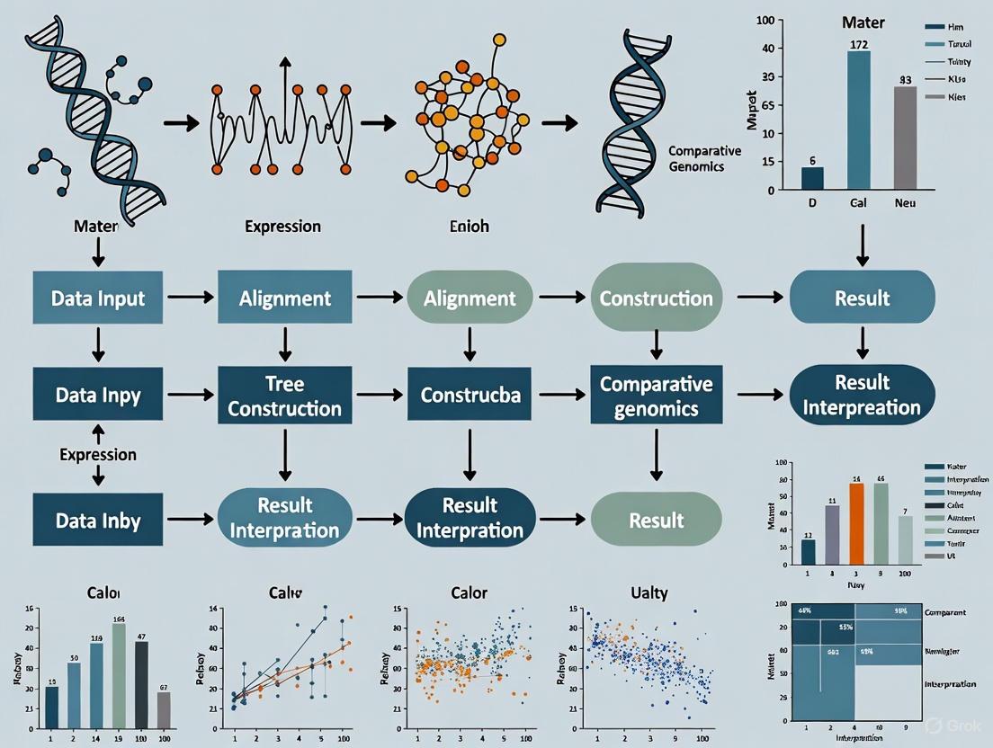
Comparative genomics serves as a cornerstone of modern biological research, enabling scientists to decipher evolutionary relationships, predict gene function, and identify genetic variations through computational analysis of genomic sequences. This field relies on a sophisticated pipeline that transforms raw sequence data into evolutionary insights, with multiple sequence alignment (MSA) and phylogenetic tree construction representing two fundamental computational pillars. The reliability of downstream biological conclusions—from species classification to drug target identification—depends entirely on the accuracy and appropriateness of these computational methods [1].
As genomic databases expand exponentially, the computational challenges in comparative genomics have intensified, driving innovation in algorithm development. Next-generation sequencing technologies now generate trillions of nucleotide bases per run, creating demand for methods that balance scalability, accuracy, and computational efficiency [2]. This guide provides a comprehensive comparison of current methodologies across the comparative genomics workflow, enabling researchers to select optimal strategies for their specific research contexts within drug development and evolutionary studies.
Multiple Sequence Alignment: Methods and Performance Comparison
Multiple sequence alignment establishes the foundational framework for comparative genomics by identifying homologous positions across biological sequences. The MSA process is inherently NP-hard, making heuristic approaches essential for practical applications [1]. Current MSA methods generally fall into three categories: traditional progressive methods, meta-aligners that integrate multiple approaches, and emerging artificial intelligence-based techniques.
Table 1: Performance Comparison of Multiple Sequence Alignment Tools
| Method/Tool | Algorithm Type | Key Features | Accuracy & Performance | Best Use Cases |
|---|---|---|---|---|
| BetaAlign | Deep Learning (Transformer) | Uses NLP techniques trained on simulated alignments; adaptable to specific evolutionary models [3] | Comparable or better than state-of-the-art tools; accuracy depends on training data quality [3] | Large datasets with known evolutionary parameters; phylogenomic studies requiring high precision |
| LexicMap | Hierarchical k-mer indexing | Probe-based seeding with prefix/suffix matching; efficient against million-genome databases [4] | High accuracy with greater speed and lower memory use vs. state-of-the-art methods [4] | Querying genes/plasmids against massive prokaryotic databases; epidemiological studies |
| M-Coffee | Meta-alignment | Consistency-based library from multiple aligners; weighted character pairs [1] | Generally approximates average quality of input alignments [1] | Integrating results from specialized aligners; protein families with challenging regions |
| MAFFT/MUSCLE | Progressive alignment | Heuristic-based; "once a gap, always a gap" principle [1] | Fast but prone to early error propagation [1] | Initial alignment generation; large-scale screening analyses |
Advanced Alignment Strategies: Post-Processing and Realignment
Even the most sophisticated initial alignments often benefit from post-processing refinement to correct errors introduced by heuristic algorithms. Meta-alignment strategies, such as those implemented in M-Coffee and TPMA, integrate multiple independent MSA results to produce consensus alignments that leverage the strengths of different alignment programs [1]. These approaches are particularly valuable when analyzing sequences with regions of high variability or when alignment uncertainty exists.
Realigner methods operate through iterative optimization of existing alignments using horizontal partitioning strategies. These include single-type partitioning (realigning one sequence against a profile), double-type partitioning (aligning two profile groups), and tree-dependent partitioning (dividing alignment based on guide tree topology) [1]. Tools like ReAligner implement these approaches to progressively improve alignment scores until convergence, effectively addressing the "once a gap, always a gap" limitation of progressive methods [1].
Phylogenetic Tree Construction: Methodological Approaches
Phylogenetic trees provide the evolutionary context for comparative genomics, visually representing hypothesized relationships between taxonomic units. The construction of these trees follows a systematic workflow from sequence collection to tree evaluation, with method selection profoundly impacting the resulting topological accuracy.
Phylogenetic Inference Methods: A Comparative Analysis
Table 2: Comparison of Phylogenetic Tree Construction Methods
| Method | Algorithm Principle | Advantages | Limitations | Computational Demand |
|---|---|---|---|---|
| Neighbor-Joining (NJ) | Distance-based clustering using pairwise evolutionary distances [5] | Fast computation; fewer assumptions; suitable for large datasets [5] | Information loss in distance matrix; sensitive to evolutionary rate variation [5] | Low to moderate; efficient for large taxon sets |
| Maximum Parsimony (MP) | Minimizes total number of evolutionary steps [5] | Straightforward principle; no explicit model assumptions [5] | Prone to long-branch attraction; multiple equally parsimonious trees [5] | High for large datasets due to tree space search |
| Maximum Likelihood (ML) | Probability-based; finds tree with highest likelihood under evolutionary model [5] | Explicit model assumptions reduce systematic errors; high accuracy [5] | Computationally intensive; model misspecification risk [5] | Very high; requires heuristic searches for large datasets |
| Bayesian Inference (BI) | Probability-based; estimates posterior probability of trees [5] | Provides natural probability measures; incorporates prior knowledge [5] | Computationally demanding; convergence assessment needed [5] | Extremely high; Markov Chain Monte Carlo sampling |
The selection of phylogenetic inference methods depends on dataset size, evolutionary complexity, and computational resources. Distance-based methods like Neighbor-Joining transform sequence data into pairwise distance matrices before applying clustering algorithms, providing computationally efficient solutions for large datasets [5]. In contrast, character-based methods including Maximum Parsimony, Maximum Likelihood, and Bayesian Inference evaluate individual sequence characters during tree search, typically generating numerous hypothetical trees before identifying optimal topologies according to specific criteria [5].
For large-scale phylogenomic analyses, integrated pipelines like Phyling provide streamlined workflows from genomic data to species trees. Phyling utilizes profile Hidden Markov Models to identify orthologs from BUSCO databases, aligns sequences using tools like Muscle or hmmalign, and supports both consensus (ASTER) and concatenation (IQ-TREE, RAxML-NG) approaches for final tree inference [6]. Such pipelines significantly accelerate phylogenetic analysis while maintaining accuracy comparable to traditional methods.
Integrated Analysis: From Alignment to Tree Assessment
Experimental Protocols for Phylogenomic Workflows
Protocol 1: Standard Phylogenetic Analysis from Genomic Data
Sequence Acquisition and Orthology Determination: Collect protein or coding sequences from samples (minimum of four). For ortholog identification, search sequences against Hidden Markov Model profiles from BUSCO database using hmmsearch (PyHMMER v0.11.0). Exclude samples with multiple hits to the same HMM profile to ensure orthology [6].
Multiple Sequence Alignment: Extract sequences matching HMM profiles and align using hmmalign (default) or Muscle v5.3 for higher quality. Trim alignments with ClipKIT v2.1.1 to retain parsimony-informative sites while removing unreliable regions [6].
Marker Selection and Tree Inference: Construct trees for each marker using FastTree v2.1.1. Evaluate phylogenetic informativeness using treeness over relative composition variability (RCV) score calculated via PhyKIT v2.0.1. Retain top n markers ranked by treeness/RCV scores [6].
Species Tree Construction: Apply either consensus approach (building individual gene trees and inferring species tree using ASTER v1.19) or concatenation approach (combining alignments into supermatrix). For concatenation, determine best-fit substitution model using ModelFinder from IQ-TREE package [6].
Protocol 2: Alignment-Free Viral Classification
Feature Extraction: Transform viral genome sequences into numeric feature vectors using one of six established alignment-free techniques: k-mer counting, Frequency Chaos Game Representation (FCGR), Return Time Distribution (RTD), Spaced Word Frequencies (SWF), Genomic Signal Processing (GSP), or Mash [2].
Classifier Training: Use extracted feature vectors as input for Random Forest classifiers. Train separate models for specific viral pathogens (SARS-CoV-2, dengue, HIV) using known lineage information as classification targets [2].
Validation and Application: Evaluate classifier performance on holdout test sets using accuracy, Macro F1 score, and Matthew's Correlation Coefficient. Apply optimized models to classify new viral sequences without alignment steps [2].
The Scientist's Toolkit: Essential Research Reagents and Software
Table 3: Key Research Reagent Solutions for Comparative Genomics
| Tool/Resource | Type | Function | Application Context |
|---|---|---|---|
| BUSCO Database | Marker gene set | Provides universal single-copy orthologs for orthology assessment [6] | Phylogenomic studies across diverse taxa |
| ClipKIT | Alignment trimming software | Trims multiple sequence alignments to retain parsimony-informative sites [6] | Pre-processing alignments for phylogenetic inference |
| IQ-TREE | Phylogenetic software package | Implements maximum likelihood inference with model selection [6] | Species tree construction from aligned sequences |
| TPMA | Meta-alignment tool | Integrates multiple nucleic acid MSAs using sum-of-pairs scores [1] | Improving alignment accuracy through consensus |
| TOPD/FMTS | Tree comparison software | Calculates Boot-Split Distance between phylogenetic trees [7] | Quantifying topological differences between gene trees |
The comparative genomics workflow represents an integrated system where choices at each stage influence downstream results. Method selection should be guided by research questions, dataset characteristics, and computational resources. For multiple sequence alignment, deep learning approaches like BetaAlign show promise for challenging alignment problems, while efficient tools like LexicMap excel in large-scale database searches. For phylogenetic inference, likelihood-based methods generally provide the highest accuracy when computational resources permit, while distance methods offer practical solutions for massive datasets.
Emerging trends including alignment-free classification and meta-alignment strategies are expanding the methodological toolkit, particularly for applications requiring rapid analysis of large datasets or integration of diverse analytical approaches. As comparative genomics continues to evolve, the optimal application of these methods will remain fundamental to advancing biological discovery and drug development.
Evolutionary distance provides a quantitative framework for measuring genetic divergence between species, serving as a foundational concept in comparative genomics. By quantifying the degree of molecular divergence—through single nucleotide substitutions, insertions, deletions, and structural variations—evolutionary distance enables researchers to select optimal model organisms for studying human biology, disease mechanisms, and evolutionary processes [8]. The strategic selection of species based on evolutionary distance is not merely an academic exercise; it directly impacts the translational potential of biomedical research, where overreliance on traditional "supermodel organisms" has contributed to a 95% failure rate for drug candidates during clinical development [8]. This comparison guide examines current methodologies for quantifying evolutionary distance, evaluates their performance characteristics, and provides a structured framework for selecting species pairs that maximize research insights while acknowledging the limitations of different distance metrics.
The fundamental challenge in evolutionary distance calculation lies in accurately modeling the relationship between observed genetic differences and actual evolutionary divergence time. As sequences diverge, multiple substitutions may occur at the same site, obscuring the true evolutionary history. More sophisticated models account for these hidden changes through various substitution models (Jukes-Cantor, K80, GTR), but each carries specific assumptions about evolutionary processes that may not hold across all lineages or genomic regions [9]. Recent advances in whole-genome sequencing have dramatically expanded the scope of evolutionary comparisons, enabling researchers to move beyond gene-centric analyses to whole-genome comparisons that capture the full complexity of genomic evolution, including structural variations and regulatory element conservation [10] [11].
Methodologies for Quantifying Evolutionary Distance
Alignment-Based Methods
Alignment-based methods constitute the traditional approach for calculating evolutionary distance by directly comparing nucleotide or amino acid sequences. Whole-genome alignment tools like lastZ identify homologous regions between genomes through a seed-and-extend algorithm, providing a foundation for precise nucleotide-level comparison [12]. The key advantage of lastZ lies in its exceptional sensitivity for aligning highly divergent sequences, maintaining alignment coverage even at divergence levels exceeding 40%, where other tools frequently fail [12]. This sensitivity comes at significant computational cost, with mammalian whole-genome alignments requiring approximately 2,700 CPU hours, creating substantial bottlenecks for large-scale analyses [12].
The Average Nucleotide Identity (ANI) approach provides a standardized metric for genomic similarity, traditionally calculated using alignment tools like BLAST or MUMmer [9]. ANI was originally developed as an in-silico replacement for DNA-DNA hybridization (DDH) techniques, with a 95% ANI threshold corresponding to the 70% DDH value used for species delineation [9]. Modern implementations such as OrthoANI and ANIb (available through PyANI) differ in their specific methodologies, with ANIb demonstrating superior accuracy in capturing true evolutionary distances despite being computationally intensive [9]. A significant limitation of traditional ANI calculations is their dependence on "alignable regions," which can result in zero or near-zero estimates for highly divergent genomes where homologous regions represent only a small fraction of the total sequence [9].
Table: Comparison of Alignment-Based Evolutionary Distance Methods
| Method | Algorithm | Optimal Use Case | Sensitivity | Computational Demand |
|---|---|---|---|---|
| lastZ | Seed-filter-extend with gapped extension | Divergent genome pairs (>40% divergence) | Excellent | Extreme (≈2700 CPU hours for mammals) |
| ANIb | BLAST-based average nucleotide identity | Species delineation, closely related genomes | High | High |
| ANIm | MUMmer-based alignment | Rapid comparison of similar genomes | Moderate | Medium |
| KegAlign | GPU-accelerated diagonal partitioning | Large-scale analyses requiring speed | High (lastZ-level) | Moderate (6 hours for human-mouse on GPU) |
Alignment-Free Methods
Alignment-free methods have emerged as efficient alternatives for evolutionary distance estimation, particularly valuable for large-scale comparisons and database searches. These approaches typically employ k-mer-based sketching techniques, such as MinHash implemented in Mash and Dashing, which create compact representations of genomic sequences by storing subsets of their k-mers [9]. By comparing these sketches rather than full sequences, these tools can estimate evolutionary distances several orders of magnitude faster than alignment-based methods while maintaining strong correlation with traditional measures [9].
The KmerFinder tool exemplifies the specialized application of k-mer techniques for taxonomic classification, demonstrating how k-mer profiles can rapidly place unknown samples within evolutionary frameworks [9]. A significant advantage of k-mer-based approaches is their ability to handle incomplete or draft-quality genomes where alignment-based methods struggle with fragmentation and assembly artifacts. However, these methods rely on heuristics and may sacrifice some accuracy for speed, particularly at intermediate evolutionary distances where k-mer composition may not linearly correlate with true evolutionary divergence [9].
Synteny-Based Approaches
Synteny-based approaches represent a paradigm shift in identifying evolutionary relationships beyond sequence similarity. The Interspecies Point Projection (IPP) algorithm identifies orthologous genomic regions based on their relative position between conserved anchor points, independent of sequence conservation [11]. This method leverages syntenic relationships—the conservation of genomic colinearity—to identify functionally conserved regions even when sequences have diverged beyond the detection limits of alignment-based methods.
In comparative analyses between mouse and chicken hearts, IPP demonstrated remarkable utility, identifying five times more conserved regulatory elements than alignment-based approaches [11]. Whereas traditional LiftOver methods identified only 7.4% of enhancers as conserved between these species, IPP revealed that 42% of enhancers showed positional conservation despite sequence divergence [11]. This approach is particularly valuable for studying the evolution of regulatory elements, which often maintain function despite rapid sequence turnover. The method relies on high-quality genome assemblies and annotation of conserved anchor points, typically protein-coding genes with clear orthologous relationships, and benefits from including multiple bridging species to improve projection accuracy [11].
Experimental Protocols for Evolutionary Distance Analysis
Comparative Genomic Analysis Workflow
Diagram 1. Workflow for comprehensive evolutionary distance analysis integrating multiple methodological approaches.
Detailed Protocol: Whole-Genome Alignment with KegAlign
Objective: Perform sensitive pairwise whole-genome alignment for evolutionary distance calculation between mammalian species.
Sample Protocol (Human-Mouse Comparison):
- Data Preparation: Download reference genomes (hg38 human, mm39 mouse) from ENSEMBL or UCSC. Format sequences using
kegalign preprocessto ensure consistent formatting and remove ambiguous bases. - Anchor Point Identification: Identify syntenic anchor points using reciprocal BLAST with E-value threshold of 1e-10 and minimum alignment length of 100 bp. These anchors facilitate the diagonal partitioning strategy.
- GPU Configuration: Configure NVIDIA GPU with multi-instance GPU (MIG) and multi-process service (MPS) enabled to optimize hardware utilization. A minimum of 16GB GPU memory is recommended for mammalian genomes.
- Alignment Execution: Run KegAlign with species-appropriate parameters:
kegalign -t 32 --gpu-batch 8 -x human_mouse.xml hg38.fa mm39.fa -o output.maf. The tool employs diagonal partitioning to minimize tail latency issues common in highly similar genomes. - Post-processing: Filter alignments for minimum length (50 bp) and identity (30%) using
kegalign postprocess. Convert to phylogenetic format if needed for downstream analysis. - Distance Calculation: Calculate evolutionary distance using Jukes-Cantor correction:
d = -3/4 * ln(1 - 4/3 * p), where p is the observed proportion of differing sites in aligned regions [12].
This protocol reduces computational time from approximately 2,700 CPU hours with lastZ to under 6 hours on a single GPU-containing node while maintaining equivalent sensitivity [12].
Detailed Protocol: Synteny-Based Conservation Detection with IPP
Objective: Identify evolutionarily conserved regulatory elements between distantly related species despite sequence divergence.
Sample Protocol (Mouse-Chicken Heart Enhancer Conservation):
- Functional Genomic Data Collection: Generate or obtain chromatin profiling data (ATAC-seq, H3K27ac ChIP-seq) from equivalent developmental stages (mouse E10.5, chicken HH22) [11].
- CRE Identification: Predict cis-regulatory elements using CRUP from histone modifications, integrating with chromatin accessibility and gene expression data to minimize false positives.
- Bridge Species Selection: Curate 14 bridging species from reptilian and mammalian lineages with ancestral vertebrate genomes to serve as evolutionary intermediates.
- Anchor Point Definition: Identify alignable regions between all species pairs using lastZ with minimum match threshold of 0.1. These serve as reference points for interpolation.
- Interspecies Point Projection: Run IPP algorithm to project mouse CRE coordinates to chicken genome through bridged alignments:
ipp --bridges species_list.txt --min_anchors 3 --max_gap 2500 mouse_CREs.bed mouse_chicken.chain[11]. - Classification: Categorize projections as: (1) Directly Conserved (within 300 bp of direct alignment), (2) Indirectly Conserved (projected through bridged alignments with summed distance <2.5 kb), or (3) Non-conserved.
- Functional Validation: Select indirectly conserved enhancers for in vivo reporter assays in transgenic mouse models to confirm functional conservation [11].
This approach identified 42% of mouse heart enhancers as conserved in chicken (compared to 7.4% with alignment-based methods), dramatically expanding the detectable conserved regulome [11].
Research Reagent Solutions for Evolutionary Distance Studies
Table: Essential Research Reagents and Computational Tools for Evolutionary Distance Analysis
| Category | Specific Tools/Resources | Primary Function | Application Context |
|---|---|---|---|
| Genome Alignment | lastZ, KegAlign, MUMmer | Generate base-level genome alignments | Pairwise whole-genome comparison, anchor identification |
| Sequence Similarity | OrthoANI, PyANI, FastANI | Calculate average nucleotide identity | Species delineation, phylogenetic framework construction |
| K-mer Analysis | Mash, Dashing, KmerFinder | Efficient genome sketching and comparison | Large-scale database searches, rapid phylogenetic placement |
| Synteny Analysis | IPP, Cactus, SynMap | Identify conserved genomic organization | Regulatory element evolution, deep evolutionary comparisons |
| Phylogenomics | OrthoFinder, NovelTree, IQ-TREE | Infer gene families and species trees | Evolutionary framework construction, orthology assignment |
| Functional Genomics | CRUP, MACS2, HOMER | Identify cis-regulatory elements | Functional element conservation analysis |
| Data Integration | Airbyte, Displayr, RStudio | Clean, transform, and analyze diverse datasets | Multi-omics data integration, reproducible analysis |
Data Presentation: Performance Comparison of Evolutionary Distance Methods
Table: Quantitative Performance Metrics for Evolutionary Distance Tools
| Method | Human-Mouse Runtime | Hardware Requirements | Sensitivity (Enhancer Detection) | Key Advantage | Primary Limitation |
|---|---|---|---|---|---|
| lastZ | ~2700 CPU hours | High-performance CPU cluster | 10% (alignment-based) | Excellent for highly divergent sequences | Extreme computational demands |
| KegAlign | <6 hours | Single GPU node | Equivalent to lastZ | GPU acceleration without sensitivity loss | Requires specialized hardware |
| Mash (k=21) | Minutes | Standard server | NA (alignment-free) | Extreme efficiency for large datasets | Indirect distance estimation |
| IPP Algorithm | Hours to days (including data generation) | CPU cluster with substantial memory | 42% (synteny-based) | Detects functional conservation beyond sequence similarity | Requires multiple bridging species |
The optimal choice of evolutionary distance methodology depends critically on research objectives, biological questions, and available computational resources. For maximum accuracy in closely related species or when precise nucleotide-level comparison is essential, alignment-based methods like ANIb provide the gold standard despite computational costs [9]. When studying deep evolutionary relationships or regulatory element conservation, synteny-based approaches like IPP reveal conserved elements invisible to sequence-based methods, expanding detectable conservation fivefold between mouse and chicken [11]. For large-scale comparative genomics or database screening, k-mer-based methods offer unparalleled efficiency with minimal sacrifice in accuracy [9].
The integration of GPU acceleration in tools like KegAlign demonstrates how algorithmic innovations can dramatically reduce computational barriers without sacrificing sensitivity [12]. Meanwhile, the recognition that sequence divergence often exceeds functional divergence—particularly for regulatory elements—underscores the importance of complementing traditional alignment methods with synteny-based and functional genomic approaches [11]. By strategically selecting and combining these approaches, researchers can leverage evolutionary distance not merely as a descriptive metric but as a powerful tool for selecting optimal species comparisons that maximize biological insights across the tree of life.
Table of Contents
- Introduction to the Functional Genome
- Benchmarking Genomic Analysis Models
- Experimental Protocols for Model Evaluation
- Pathways in Genomic Element Identification
- The Scientist's Toolkit: Essential Research Reagents
The completion of the Human Genome Project revealed that protein-coding genes comprise a mere 2% of our DNA [13]. The remaining majority, once dismissed as 'junk' DNA, is now understood to be a complex regulatory landscape essential for controlling gene expression [13]. This non-coding genome contains critical functional elements, including promoters, enhancers, insulators, and non-coding RNAs, which orchestrate when and where genes are activated or silenced [13] [14]. Disruptions in these regions are a major contributor to disease; over 90% of genetic variants linked to common conditions lie within these non-coding 'switch' regions [15]. Consequently, accurately identifying these functional elements is a fundamental goal in genomics, driving advances in precision medicine and drug discovery [13] [16].
The field has moved from analyzing isolated segments to understanding the genome as an integrated, three-dimensional structure. DNA is folded intricately inside the nucleus, bringing distant regulatory elements, such as enhancers and promoters, into close physical contact to control gene expression [15]. Mapping these long-range interactions, which can span millions of base pairs, is crucial for a complete understanding of genetic regulation [17]. Recent advances in artificial intelligence (AI) and deep learning have created powerful new models capable of predicting these complex sequence-to-function relationships, necessitating rigorous benchmarking to guide researchers in selecting the right tool for their specific needs [17] [18].
Benchmarking Genomic Analysis Models
To objectively evaluate the performance of modern genomic analysis tools, researchers have developed standardized benchmarks like DNALONGBENCH [17]. This suite tests models on five biologically significant tasks that require understanding dependencies across long DNA sequences—up to 1 million base pairs. The performance of various model types, including specialized "expert" models and more general-purpose "foundation" models, is compared quantitatively.
Table 1: Performance Summary of Model Types on DNALONGBENCH Tasks
| Model Type | Example Models | Key Characteristics | Strengths | Weaknesses |
|---|---|---|---|---|
| Expert Models | ABC, Enformer, Akita, Puffin [17] | Highly specialized, task-specific architecture. | State-of-the-art performance on their designated tasks; superior at capturing long-range dependencies for complex regression (e.g., contact maps) [17]. | Narrow focus; cannot be easily applied to new tasks without retraining. |
| DNA Foundation Models | HyenaDNA, Caduceus [17] | Pre-trained on vast genomic data, then fine-tuned for specific tasks. | Good generalization; reasonable performance on certain classification tasks [17]. | Struggle with complex, multi-channel regression; fine-tuning can be unstable [17]. |
| Lightweight CNNs | 3-layer CNN [17] | Simple convolutional neural networks. | Simplicity and fast training; robust baseline for shorter-range tasks. | Consistently outperformed by expert and foundation models on long-range tasks [17]. |
Table 2: Quantitative Model Performance on Specific Genomic Tasks
| Task | Description | Expert Model (Score) | DNA Foundation Models (Score) | CNN (Score) |
|---|---|---|---|---|
| Enhancer-Target Gene Prediction [17] | Classifies whether an enhancer regulates a specific target gene. | ABC Model (AUROC: 0.892) [17] | Caduceus-PS (AUROC: 0.816) [17] | CNN (AUROC: 0.774) [17] |
| Contact Map Prediction [17] | Predicts 3D chromatin interactions from sequence. | Akita (SCC: 0.856) [17] | Caduceus-PS (SCC: 0.621) [17] | CNN (SCC: 0.521) [17] |
| Transcription Initiation Signal Prediction [17] | Regression task to predict the location and strength of transcription start sites. | Puffin (Avg. Score: 0.733) [17] | Caduceus-PS (Avg. Score: 0.108) [17] | CNN (Avg. Score: 0.042) [17] |
| Regulatory Element Segmentation [19] | Nucleotide-level annotation of elements like exons and promoters. | SegmentNT (Avg. MCC: 0.42 on 10kb sequences) [19] | Nucleotide Transformer (Baseline for SegmentNT) [19] | Not Reported |
Another foundation model, OmniReg-GPT, demonstrates the value of efficient long-sequence training. When benchmarked on shorter regulatory element identification tasks (e.g., promoters, enhancers), it achieved superior Matthews Correlation Coefficient (MCC) scores in 9 out of 13 tasks compared to other foundational models like DNABERT2 and Nucleotide Transformer [14].
Experimental Protocols for Model Evaluation
A critical step in comparing genomic tools is the use of standardized, rigorous experimental protocols. Below is a detailed methodology for a typical benchmarking study, as used in the evaluation of DNALONGBENCH [17].
Protocol 1: Benchmarking Long-Range Genomic Dependencies with DNALONGBENCH
- 1. Objective: To comprehensively evaluate the ability of computational models to capture long-range dependencies in DNA sequence for five key biological tasks.
- 2. Data Curation and Pre-processing:
- Data Sources: Genomic data is collected from public repositories such as ENCODE [17] [19]. For DNALONGBENCH, this includes Hi-C data for 3D genome organization, ChIP-seq and ATAC-seq data for regulatory elements, and RNA-seq data for expression quantitative trait loci (eQTLs) [17].
- Sequence Extraction: Input sequences and their corresponding labels (e.g., contact frequencies, expression levels, element classifications) are extracted in windows of up to 1 million base pairs from the reference genome based on coordinates in BED format files [17].
- Dataset Splitting: Chromosomes are strategically partitioned into training, validation, and test sets (e.g., train on chromosomes 1-16, validate on 17-18, test on 19-22) to ensure no data leakage and robust performance evaluation [17] [19].
- 3. Model Selection and Training:
- Models: Three classes of models are selected:
- Expert Models: State-of-the-art models specifically designed for a single task (e.g., Akita for contact maps, Enformer for eQTLs) [17].
- DNA Foundation Models: General-purpose models pre-trained on large genomic corpora and then fine-tuned on each task (e.g., HyenaDNA, Caduceus) [17].
- Baseline CNN: A lightweight convolutional neural network provides a performance baseline [17].
- Fine-tuning/Training: Expert models are used as published. Foundation models are fine-tuned on each task's training set. The CNN is trained from scratch. Training uses task-appropriate loss functions (e.g., cross-entropy for classification, mean squared error for regression) [17].
- Models: Three classes of models are selected:
- 4. Performance Evaluation:
- Metrics: Models are evaluated on the held-out test set using task-specific metrics.
- Classification (e.g., Enhancer-Target): Area Under the Receiver Operating Characteristic Curve (AUROC) and Area Under the Precision-Recall Curve (AUPR) [17].
- Regression (e.g., Contact Map): Stratum-Adjusted Correlation Coefficient (SCC) and Pearson Correlation [17].
- Nucleotide-Level Segmentation (e.g., Element Annotation): Matthews Correlation Coefficient (MCC), F1-score, and Segment Overlap Score (SOV) [19].
- Analysis: Performance scores are aggregated and compared across model types and tasks to identify strengths and weaknesses.
- Metrics: Models are evaluated on the held-out test set using task-specific metrics.
Protocol 2: Nucleotide-Resolution Genome Annotation with SegmentNT
- 1. Objective: To train a model for annotating multiple genomic elements at single-nucleotide resolution by framing the problem as multilabel semantic segmentation [19].
- 2. Data Preparation:
- 3. Model Architecture and Training:
- Backbone: A pre-trained DNA foundation model (Nucleotide Transformer) serves as the encoder to generate initial sequence representations [19].
- Segmentation Head: A 1D U-Net architecture is attached to the backbone. It downscales and then upscales the representations to make a separate prediction for each element at each nucleotide position [19].
- Loss Function: A focal loss objective is used during training to handle the high class imbalance, as functional elements are sparse in the genome [19].
Pathways in Genomic Element Identification
The following diagram illustrates the logical workflow and key decision points for a researcher choosing a computational strategy to identify functional genomic elements, based on the benchmark data.
The Scientist's Toolkit: Essential Research Reagents
The experiments and models discussed rely on a foundation of wet-lab techniques and computational resources. The following table details key reagents and tools essential for this field.
Table 3: Key Research Reagents and Resources for Genomic Studies
| Category | Reagent / Tool | Function in Research | Example Use-Case |
|---|---|---|---|
| Experimental Assays | ATAC-seq [20] | Identifies regions of open chromatin, indicative of regulatory activity. | Used to validate that conserved non-coding sequences (CNS) are enriched in functionally accessible chromatin [20]. |
| ChIP-seq [20] | Maps the binding sites of specific proteins (e.g., transcription factors, histones) across the genome. | Profiling histone modifications (e.g., H3K9ac, H3K4me3) to characterize the epigenetic state of regulatory elements [20]. | |
| Hi-C [17] | Captures the 3D architecture of the genome by quantifying chromatin interactions. | Generating ground truth data for training and benchmarking models that predict 3D genome organization [17]. | |
| MCC ultra [15] | A high-resolution technique that maps chromatin structure down to a single base pair inside living cells. | Revealing the physical arrangement of gene control switches and how they form "islands" of activity [15]. | |
| Computational Tools & Data | Foundation Models (e.g., Nucleotide Transformer, OmniReg-GPT) [19] [14] | Provide pre-trained, general-purpose representations of DNA sequence that can be fine-tuned for diverse downstream tasks. | Serving as the backbone for SegmentNT for genome annotation or benchmarking for long-range task performance [19] [17]. |
| Benchmark Suites (e.g., DNALONGBENCH) [17] | Standardized datasets and tasks for the objective comparison of different genomic deep learning models. | Enabling rigorous evaluation of model performance on tasks like enhancer-target prediction and contact map modeling [17]. | |
| ENCODE / GENCODE Annotations [19] | Comprehensive, publicly available catalogs of functional elements in the human genome. | Providing the labeled data required to train supervised models like SegmentNT for genome annotation [19]. |
Comparative genomics is the comparison of genetic information within and across species to understand the evolution, structure, and function of genes, proteins, and non-coding regions [21]. This scientific discipline provides powerful tools for systematically exploring biological relationships between species, aiding in understanding gene structure and function, and gaining crucial insights into human disease mechanisms and potential therapeutic targets [21]. The field has accelerated dramatically with advances in DNA sequencing technology, which have generated a flood of genomic data from diverse eukaryotic organisms [22]. The National Institutes of Health (NIH) Comparative Genomics Resource (CGR) is a multi-year project implemented by the National Library of Medicine (NLM) to maximize the impact of eukaryotic research organisms and their genomic data resources to biomedical research [23] [22]. This review provides a comprehensive comparison of CGR against other essential model organism databases, offering performance data and experimental protocols to guide researchers in selecting appropriate resources for their comparative genomics studies.
The NIH CGR is designed as a comprehensive toolkit to facilitate reliable comparative genomics analyses for all eukaryotic organisms through community collaboration and interconnected data resources [23] [24]. CGR aims to maximize the biomedical impact of eukaryotic research organisms by providing high-quality genomic data, improved comparative genomics tools, and scalable analyses that support emerging big data approaches [23]. A key objective is the application of FAIR (Findable, Accessible, Interoperable, Reusable) principles to make genomic data more easily usable with standard bioinformatics platforms and tools [23]. The project is guided by two advisory boards: the NLM Board of Regents CGR working group comprising external biological leaders, and the CGR Executive Steering Committee providing NIH oversight [23].
CGR addresses several critical challenges in contemporary genomics research, including ensuring data quality, enhancing annotation consistency, and improving interoperability between resources [21]. The resource emphasizes connecting NCBI-held genomic content with community-supplied resources such as sample metadata and gene functional information, thereby amplifying the potential for new scientific discoveries [23] [21]. CGR's organism-agnostic approach provides equal access to datasets across the eukaryotic tree of life, enabling researchers to explore biological patterns and generate new hypotheses beyond traditional model organisms [23].
Established Model Organism Databases
Model organism databases (MODs) provide curated, species-specific biological data essential for biomedical research. These resources typically offer comprehensive genetic, genomic, phenotypic, and functional information focused on particular research organisms that serve as models for understanding biological processes relevant to human health. The National Human Genome Research Institute (NHGRI) supports several key model organism databases that represent well-established species with extensive research histories [25].
Table 1: Key Model Organism Databases and Their Research Applications
| Database Name | Research Organism | Primary Research Applications | Key Features |
|---|---|---|---|
| FlyBase [25] | Drosophila melanogaster (Fruit fly) | Genetics, developmental biology, neurobiology | Genetic and genomic data, gene expression patterns, phenotypic data |
| MGI [25] | Mus musculus (House mouse) | Human disease models, mammalian biology | Mouse genome database, gene function, phenotypic alleles |
| RGD [25] | Rattus norvegicus (Brown rat) | Cardiovascular disease, metabolic disorders | Rat genome data, disease portals, quantitative trait loci (QTL) |
| WormBase [25] | Caenorhabditis elegans (Nematode) | Developmental biology, neurobiology, aging | Genome sequence, gene models, genetic maps, functional genomics |
| ZFIN [25] | Danio rerio (Zebrafish) | Developmental biology, toxicology, regeneration | Genetic and genomic data, gene expression, mutant phenotypes |
| SGD [25] | Saccharomyces cerevisiae (Baker's yeast) | Cell biology, genetics, functional genomics | Gene function, metabolic pathways, protein interactions |
These traditional model organisms were selected for biomedical research because they are typically easy to maintain and breed in laboratory settings and possess biological characteristics similar to human systems [22]. However, with advances in comparative genomics, emerging model organisms are increasingly being recognized for their potential to provide unique insights into specific biological processes and human diseases [22].
Performance Comparison: CGR vs. Specialized Model Organism Databases
Table 2: Performance Metrics and Capabilities Comparison Across Genomic Resources
| Feature | NCBI CGR | Specialized MODs | CGR Advantages |
|---|---|---|---|
| Taxonomic Scope | All eukaryotic organisms [23] | Single species or related species [25] | Broader phylogenetic range for discovery |
| Data Integration | Integrates across multiple organisms and connects with community resources [23] [21] | Deep curation within single organism [25] | Enables cross-species comparisons and meta-analyses |
| Tool Availability | Eukaryotic Genome Annotation Pipeline, Foreign Contamination Screen, Comparative Genome Viewer [22] | Organism-specific analysis tools and visualization [25] | Standardized tools applicable across diverse species |
| Data Quality Framework | Contamination screening, consistent annotation [23] [22] | Community-curated gene models and annotations [25] | Systematic quality control across all data |
| Computational Scalability | Support for big data approaches, AI-ready datasets, cloud-ready tools [23] | Varies by resource, typically single-organism focus | Designed for large-scale comparative analyses |
Quantitative assessments of genomic resource utility demonstrate that CGR's primary advantage lies in its cross-species interoperability and scalable infrastructure. For example, CGR facilitates the creation of AI-ready datasets and provides tools that maintain consistent annotation across diverse eukaryotic species, addressing a critical challenge in comparative genomics [23] [22]. While specialized model organism databases typically offer greater depth of curated information for specific organisms, CGR provides superior capabilities for researchers requiring cross-species comparisons or working with non-traditional research organisms.
Experimental Applications and Benchmarking
Key Research Applications of Comparative Genomics
Comparative genomics approaches have enabled significant advances across multiple biomedical research domains. The CGR project has identified several emerging model organisms with particular promise for illuminating specific biological processes relevant to human health [22]:
Pigs (Sus scrofa domesticus) for Xenotransplantation Research: Comparative genomic analyses have identified pigs as optimal donors for organ transplantation due to physiological and genomic similarities to humans. CGR resources facilitate the identification of genetic barriers to transplantation and potential engineering strategies [22].
Bats (Order Chiroptera) for Infectious Disease Studies: Various bat species exhibit unique immune adaptations that allow them to harbor viruses without developing disease. CGR enables comparative analysis of bat immune genes and pathways relevant to understanding viral transmission and host response [21].
Killifish (Nothobranchius furzeri) for Aging Research: These short-lived vertebrates exhibit rapid aging processes. Comparative genomics through CGR helps identify conserved genetic factors influencing longevity and age-related diseases [22].
Thirteen-Lined Ground Squirrels (Ictidomys tridecemlineatus) for Hibernation Studies: These mammals undergo profound metabolic changes during hibernation. CGR tools enable identification of genetic regulators of metabolic depression with potential applications for human metabolic disorders [22].
The CGR platform supports these research applications by providing integrated data and tools for comparing genomic features across species, identifying conserved elements, and analyzing lineage-specific adaptations [23] [21].
Benchmarking Methodologies for Genomic Tools
Rigorous benchmarking is essential for evaluating the performance of computational methods in genomics. Based on comprehensive assessments of benchmarking practices, several key methodological principles have been established [26] [27]:
Purpose and Scope Definition: Clearly define the benchmarking objectives, whether for method development, neutral comparison, or community challenge [27].
Comprehensive Method Selection: Include all relevant methods using predetermined inclusion criteria to avoid selection bias [27].
Diverse Dataset Selection: Utilize both simulated and experimental datasets that represent realistic biological scenarios and varying levels of complexity [27].
Appropriate Evaluation Metrics: Employ multiple performance metrics including accuracy, computational efficiency, scalability, and usability [26] [27].
A recent systematic review of single-cell benchmarking studies analyzed 282 papers and identified critical aspects of benchmarking methodology, including the importance of dataset diversity, method robustness assessment, and downstream evaluation [26]. These principles directly apply to evaluating genomic resources like CGR and model organism databases, where performance can be assessed based on data quality, annotation accuracy, tool interoperability, and user experience.
Diagram 1: Benchmarking workflow for genomic resources following established methodologies [26] [27].
Experimental Protocol for Comparative Genomics Analysis
A standardized protocol for conducting comparative genomics analyses using CGR and model organism databases ensures reproducible and biologically meaningful results:
Research Question Formulation: Clearly define the biological question and select appropriate comparator species based on evolutionary relationships or phenotypic traits.
Data Acquisition and Quality Control:
- Retrieve genome assemblies and annotations from CGR or relevant model organism databases
- Apply quality assessment metrics including completeness, contamination screening, and annotation consistency [22]
- Utilize CGR's Foreign Contamination Screen (FCS) tool to remove contaminated sequences prior to analysis [22]
Comparative Analysis Execution:
- Identify orthologous gene sets using reciprocal best hits or phylogenetic approaches
- Perform multiple sequence alignments of conserved genomic regions
- Utilize CGR's Comparative Genome Viewer (CGV) to visualize structural variations across species [22]
- Conduct evolutionary rate analyses (dN/dS) to identify signatures of selection
Functional Interpretation:
Validation and Follow-up:
- Design experimental validation based on computational predictions
- Utilize model organisms for functional testing of conserved genetic elements
- Iterate between computational and experimental approaches to refine biological models
This protocol leverages the complementary strengths of CGR's cross-species capabilities and the deep curation provided by specialized model organism databases to generate biologically insightful results.
Table 3: Essential Research Reagents and Computational Tools for Comparative Genomics
| Resource Category | Specific Tools/Databases | Function and Application |
|---|---|---|
| Integrated Genomic Platforms | NIH CGR [23] | Provides eukaryotic genome data, annotation tools, and comparative analysis capabilities |
| Model Organism Databases | MGI, FlyBase, WormBase, ZFIN, RGD, SGD [25] | Species-specific genetic and genomic data with community curation |
| Reference Databases | UniProt KnowledgeBase [25] | Curated protein sequence and functional information |
| Pathway Resources | Reactome [25] | Curated resource of core pathways and reactions in human biology |
| Annotation Tools | Eukaryotic Genome Annotation Pipeline [22] | Consistent genome annotation across eukaryotic species |
| Quality Control Tools | Foreign Contamination Screen (FCS) [22] | Detection and removal of contaminated sequences in genome assemblies |
| Visualization Tools | Comparative Genome Viewer (CGV) [22] | Visualization of genomic features and structural variations across species |
| Data Retrieval Systems | NCBI Datasets [22] | Programmatic access to genome-associated data and metadata |
These essential resources provide the foundation for rigorous comparative genomics studies. The CGR project enhances interoperability between these tools, creating a more connected ecosystem for genomic research [23] [21]. For example, CGR facilitates connections between NCBI resources and community databases, enabling researchers to move seamlessly between cross-species comparisons and deep dives into organism-specific biology.
Diagram 2: CGR integration in the biomedical research workflow, showing inputs from various genomic data sources and outputs to key research applications [23] [21].
The NIH Comparative Genomics Resource represents a significant advancement in genomic data integration and analysis capabilities, complementing existing model organism databases by enabling cross-species comparisons and discovery across the eukaryotic tree of life. While specialized model organism databases continue to provide essential depth for particular research organisms, CGR offers unique strengths in taxonomic breadth, tool interoperability, and support for large-scale comparative analyses.
Future developments in comparative genomics will likely focus on enhancing data integration across resources, improving scalability for increasingly large datasets, and developing more sophisticated analytical methods for extracting biological insights from cross-species comparisons [23] [21]. The CGR project is positioned to address these challenges through its ongoing development of improved tools, community engagement initiatives, and commitment to FAIR data principles [23]. As comparative genomics continues to evolve, resources like CGR and specialized model organism databases will play complementary roles in enabling biomedical researchers to translate genomic information into improved understanding of human health and disease.
For researchers embarking on comparative genomics studies, the selection of resources should be guided by specific research questions: specialized model organism databases for depth within established models, and CGR for breadth across diverse eukaryotes and integrated analysis capabilities. Engaging with both types of resources through CGR's connectivity framework provides the most comprehensive approach to addressing complex biological questions through comparative genomics.
Methodological Workflows and Their Transformative Applications in Biomedicine
Genome Sequencing, Assembly, and Annotation Pipelines
Genome analysis pipelines have evolved into sophisticated workflows that integrate diverse sequencing technologies, computational assembly tools, and annotation algorithms. The choice of pipeline components significantly impacts the final output quality, with long-read technologies now enabling telomere-to-telomere assemblies and pangenome references that capture global genetic diversity. This guide objectively compares the performance of leading tools and technologies based on recent experimental benchmarks, providing researchers with evidence-based selection criteria for their genomic investigations.
Sequencing Technologies: Landscape and Performance
Technology Comparison and Selection Criteria
Table 1: Comparison of Modern DNA Sequencing Technologies (2025)
| Technology | Read Length | Accuracy | Key Strengths | Best Applications |
|---|---|---|---|---|
| PacBio HiFi | >15 kb | >99.9% [28] | Ultra-high accuracy, haplotype phasing | Structural variant detection, genome finishing [28] |
| Oxford Nanopore (UL) | >100 kb | ~99% [29] | Ultra-long reads, real-time analysis | Complex SV resolution, base modification detection [30] |
| Illumina NovaSeq X | 200-300 bp | >99.9% [28] | High throughput, low cost | Variant discovery, population sequencing |
| Element AVITI | 300 bp | Q40 [28] | Benchtop flexibility, high accuracy | Targeted sequencing, clinical applications |
| Roche SBX* | N/A | High (CMOS) | Rapid turnaround, Xpandomer chemistry | High-throughput genomics [28] |
| MGI DNBSEQ | Varies | High | Cost-effective, AI-enhanced | Population screening, point-of-care [28] |
*Scheduled for 2026 release [28]
Experimental Evidence and Performance Metrics
Recent large-scale studies demonstrate that technology selection directly impacts assembly quality. Research sequencing 65 diverse human genomes achieved 130 haplotype-resolved assemblies with a median continuity of 130 Mb by combining PacBio HiFi (~47x coverage) with Oxford Nanopore ultra-long reads (~36x coverage) [29]. This hybrid approach enabled:
- Telomere-to-telomere (T2T) status for 39% of chromosomes [29]
- 92% gap closure compared to previous assemblies [29]
- Resolution of 1,852 complex structural variants and 1,246 centromeres [29]
Genome Assembly Tools: Benchmarking and Protocols
Assembly Algorithm Performance Comparison
Table 2: Benchmarking of Genome Assembly Tools (2025 Data)
| Assembler | Contiguity (N50) | Completeness (BUSCO) | Runtime Efficiency | Misassembly Rate | Best Use Cases |
|---|---|---|---|---|---|
| NextDenovo | High | Near-complete [31] | Stable | Low [31] | Large eukaryotic genomes |
| NECAT | High | Near-complete [31] | Efficient | Low [31] | Prokaryotic & eukaryotic |
| Flye | High [32] | Complete | Moderate | Sensitive to input [31] | Balanced accuracy/contiguity |
| Unicycler | Lower than Flye [31] | Complete | Moderate | Low | Hybrid assembly [32] |
| Canu | Moderate (3-5 contigs) [31] | High | Longest runtime [31] | Low | Accuracy-critical projects |
| Verkko | 130 Mb (median) [29] | 99% complete [29] | N/A | Low | Haplotype-resolved diploid |
| hifiasm (ultra-long) | Comparable to Verkko [29] | High [29] | N/A | Low | Complex SV resolution |
Experimental Protocols for Assembly Benchmarking
Methodology from Recent Assembly Studies:
- Data Input Standardization: Assemblers were tested using standardized computational resources with identical preprocessing [31]
- Evaluation Metrics: Contiguity (N50, total length, contig count), GC content, and completeness using Benchmarking Universal Single-Copy Orthologs (BUSCO) [31]
- Quality Control: Integration of Flagger, NucFreq, Merqury, and Inspector for robust error estimates [29]
- Phasing Validation: For diploid assemblies, parental support verification via assembly-to-assembly alignments (median 99.9% support achieved) [29]
Key Finding: Preprocessing strategy significantly impacts output quality. Filtering improved genome fraction and BUSCO completeness, while correction benefited overlap-layout-consensus (OLC) assemblers but occasionally increased misassemblies in graph-based tools [31].
Figure 1: Genome Analysis Pipeline Workflow showing technology and tool integration points
Genome Annotation: Precision and Accuracy Assessment
Annotation Tool Performance Metrics
Evidence from Recent Comparative Studies:
- Error Rates: Automated annotation tools exhibit measurable error rates, with RAST (2.1%) and PROKKA (0.9%) wrongly annotating coding gene sequences [32]
- Error Patterns: Misannotations frequently associate with shorter coding sequences (<150 nt) involving transposases, mobile genetic elements, and hypothetical proteins [32]
- Completeness: Modern eukaryotic genome annotations achieve >99% completeness for known single-copy genes when using integrated approaches [29]
Annotation Methodologies and Protocols
Braker3 Protocol (Evidence-Based):
- Input Requirements: Genome sequence, RNA-seq alignments (BAM format), and curated protein sequences (e.g., UniProt/SwissProt) [33]
- Methodology: Integrates GeneMark-ETP and AUGUSTUS using transcriptomic and protein evidence [33]
- Critical Parameter: RNA-seq alignment must include
--outSAMstrandField intronMotiffor proper intron information [33]
Helixer Protocol (Deep Learning-Based):
- Input Requirements: Only genome sequence required [33]
- Methodology: Cross-species deep learning model predicting gene structures without external evidence [33]
- Lineage Selection: Four predefined models (invertebrate, vertebrate, land plant, fungi) optimized for each lineage [33]
Table 3: Annotation Tool Comparison and Error Analysis
| Annotation Tool | Approach | Evidence Requirements | Error Rate | Strengths | Limitations |
|---|---|---|---|---|---|
| Braker3 | Evidence-based | RNA-seq, protein sequences [33] | Not quantified | High precision with extrinsic support [33] | Dependent on quality of input evidence |
| Helixer | Deep learning | None (ab initio) [33] | Not quantified | Rapid execution, no evidence needed [33] | Limited to four predefined lineages |
| RAST | Automated | None | 2.1% [32] | Comprehensive pipeline | Higher error rate for short CDS |
| PROKKA | Automated | None | 0.9% [32] | Prokaryote-optimized | Higher error rate for short CDS |
Research Reagent Solutions and Essential Materials
Table 4: Essential Research Reagents for Genome Analysis Pipelines
| Reagent/Material | Function | Application Context | Examples/Specifications |
|---|---|---|---|
| PacBio SMRT cells | HiFi read generation | Long-read sequencing | >15 kb reads, >99.9% accuracy [28] |
| Oxford Nanopore flow cells | Ultra-long read generation | Structural variant resolution | PromethION (200 Gb/output) [28] |
| Strand-seq libraries | Global phasing information | Haplotype resolution [29] | Chromosome-specific phasing |
| Hi-C sequencing kits | Chromatin interaction data | Scaffolding, phase separation [29] | Proximity ligation-based |
| Bionano optics chips | Optical mapping | Scaffold validation [29] | Large molecule imaging |
| RNA STAR aligner | Transcriptome alignment | Evidence-based annotation [33] | Requires specific strand parameters |
| UniProt/SwissProt | Curated protein sequences | Protein evidence for annotation [33] | Manually reviewed sequences |
| BUSCO datasets | Completeness assessment | Assembly/annotation QC [31] | Universal single-copy orthologs |
Emerging Trends and Future Directions
The field of genome analysis is rapidly evolving with several significant developments:
Pangenome References: The construction of diverse reference sets from 65 individuals enables capturing essential variation explaining differential disease risk across populations [30]. This approach has increased structural variant detection to 26,115 per individual, dramatically expanding variants available for disease association studies [29].
Complex Variant Resolution: Recent studies have completely resolved previously intractable regions including:
- Major Histocompatibility Complex (MHC) linked to cancer and autoimmune diseases [30]
- SMN1/SMN2 region target for spinal muscular atrophy therapies [30]
- Centromeres with up to 30-fold variation in α-satellite arrays [29]
Methodological Innovations: Current research focuses on overcoming persistent challenges in assembling ultra-long tandem repeats, resolving complex polyploid genomes, and complete metagenome assembly through improved alignment algorithms, AI-driven assembly graph analysis, and enhanced metagenomic binning techniques [34].
Figure 2: Current Challenges and Emerging Solutions in Genome Assembly
Based on current experimental evidence, pipeline selection should be guided by research objectives:
For Complete Eukaryotic Genomes: Hybrid assembly with PacBio HiFi and Oxford Nanopore ultra-long reads using Verkko or hifiasm, followed by evidence-based annotation with Braker3 provides the most comprehensive results [29].
For Prokaryotic Genomes: Long-read assemblers like NextDenovo or Flye offer optimal balance of accuracy and contiguity, with PROKKA providing efficient annotation despite measurable error rates in shorter CDS [32] [31].
For Population Studies: Pangenome graphs incorporating diverse assemblies now enable structural variant association studies at unprecedented scale, significantly advancing equity in genomic medicine applications [30] [29].
The continuous innovation in sequencing technologies and computational methods promises further improvements in resolution, accuracy, and inclusivity of genome analysis pipelines, with emerging capabilities to fully resolve remaining difficult genomic regions including centromeres and highly identical segmental duplications.
Comparative genomics provides fundamental insights into evolutionary biology, functional genetics, and disease mechanisms by analyzing genomic sequences across different species and strains. As sequencing technologies advance, generating unprecedented volumes of genomic data, the computational methods for comparing these genomes have become increasingly sophisticated. This review objectively compares three cornerstone methodologies in modern comparative genomics: whole-genome alignment, ortholog identification, and pangenome analysis. Each approach addresses distinct biological questions while facing unique computational challenges related to scalability, accuracy, and interpretability. We examine recent algorithmic advances that enhance processing efficiency without sacrificing precision, focusing on performance benchmarks from experimental evaluations. The integration of these methodologies enables researchers to trace evolutionary trajectories, infer gene function, and understand the genetic basis of adaptation across the tree of life.
Whole-Genome Alignment Methods
Whole-genome alignment (WGA) establishes base-to-base correspondence between entire genomes, enabling the detection of large-scale structural variations and evolutionary conservation patterns. WGA algorithms can be broadly classified into four categories: suffix tree-based, hash-based, anchor-based, and graph-based methods, each with distinct computational strategies for handling genomic scale and complexity [35].
Suffix tree-based methods, exemplified by the MUMmer suite, utilize data structures that represent all suffixes of a given string to identify maximal unique matches (MUMs) between genomes [35]. MUMmer's algorithm first performs a MUM decomposition to identify subsequences that occur exactly once in both genomes, then filters spurious matches, organizes remaining MUMs by their conserved order, fills gaps between MUMs with local alignment, and finally produces a comprehensive genome alignment [35]. This approach provides exceptional accuracy for identifying conserved regions but faces memory constraints with larger genomes due to suffix tree construction requirements.
Anchor-based methods identify conserved regions ("anchors") between genomes and build alignments around these regions, while hash-based methods use precomputed k-mer tables to efficiently locate potential alignment seeds. Graph-based methods represent genome relationships as graphs, offering flexibility for capturing complex evolutionary events including rearrangements, but requiring substantial computational resources [35].
The choice between WGA algorithms depends heavily on read type applications. Short reads (100-600 bp) benefit from tools like BOWTIE2 and BWA that optimize for high precision in mapping, whereas long reads (extending to thousands of bp) require specialized tools like Minimap2 that can handle higher error rates while resolving complex genomic architectures [35].
Table 1: Performance Characteristics of Major WGA Algorithm Categories
| Algorithm Type | Representative Tools | Strengths | Limitations |
|---|---|---|---|
| Suffix Tree-Based | MUMmer | High accuracy for conserved regions; Efficient MUM identification | Memory-intensive for large genomes |
| Hash-Based | BWA, BOWTIE2 | Optimized for short reads; High precision for small variants | Struggles with complex genomic regions |
| Anchor-Based | Minimap2 | Effective for long reads; Handles structural variants | Higher error rate tolerance needed |
| Graph-Based | SibeliaZ, BubbZ | Captures complex evolutionary events | Computationally demanding |
Figure 1: Classification of whole-genome alignment methodologies showing four computational approaches for comparing complete genomes.
Ortholog Identification Approaches
Orthologs are genes diverging after a speciation event, making their accurate identification crucial for functional annotation transfer and evolutionary studies. Orthology inference methods face substantial computational challenges with the expanding repertoire of sequenced genomes, necessitating scalable solutions that maintain precision.
NCBI Orthologs Methodology
The NCBI Orthologs resource implements a high-precision pipeline integrating multiple evidence types to identify one-to-one orthologous relationships across eukaryotic genomes. This approach combines protein sequence similarity, nucleotide alignment conservation, and microsynteny information to resolve complex evolutionary relationships [36]. The pipeline processes genomes individually, ensuring scalability across the expanding RefSeq database.
The method begins with all-against-all protein comparisons using DIAMOND (BLASTP-like alignment scores), selecting the best protein isoform pairs based on a modified Jaccard index that normalizes alignment scores against potential maximum similarity [36]. For candidate pairs, the pipeline evaluates nucleotide-level conservation by aligning concatenated exonic sequences with flanking regions using discontiguous-megablast, again applying a modified Jaccard index. Finally, microsynteny conservation is assessed by counting homologous gene pairs within a 20-locus window surrounding the candidate genes [36]. The integration of these metrics enables the algorithm to identify true orthologs amidst complex gene families, particularly when microsynteny evidence is present.
FastOMA Algorithm
FastOMA addresses critical scalability limitations in orthology inference through a complete algorithmic redesign of the established Orthologous Matrix (OMA) approach. It achieves linear time complexity through k-mer-based homology clustering, taxonomy-guided subsampling, and parallel computing architecture [37]. This enables processing of 2,086 eukaryotic proteomes in under 24 hours using 300 CPU cores - a dramatic improvement over original OMA (50 genomes in same timeframe) and outperforming other contemporary tools like OrthoFinder and SonicParanoid that exhibit quadratic scaling [37].
The algorithm employs a two-stage process: first, identifying root hierarchical orthologous groups (HOGs) via OMAmer placement and Linclust clustering; second, inferring nested HOG structures through leaf-to-root species tree traversal [37]. Benchmarking on Quest for Orthologs references demonstrates FastOMA maintains high precision (0.955 on SwissTree) with moderate recall, positioning it on the Pareto frontier of orthology inference methods [37]. The method also incorporates handling of alternative splicing isoforms and fragmented gene models, further enhancing its practical applicability to diverse genomic datasets.
Table 2: Orthology Inference Tool Performance Benchmarks
| Method | Precision (SwissTree) | Recall (SwissTree) | Time Complexity | Scalability (Genomes in 24h) |
|---|---|---|---|---|
| FastOMA | 0.955 | 0.69 | Linear | 2,086 |
| OMA | 0.945 | 0.65 | Quadratic | 50 |
| OrthoFinder | 0.925 | 0.75 | Quadratic | ~500 |
| SonicParanoid | 0.910 | 0.72 | Quadratic | ~600 |
| NCBI Orthologs | Not reported | Not reported | Near-linear | Not reported |
Figure 2: Ortholog identification workflows comparing the scalable FastOMA approach with the evidence-integration strategy of NCBI Orthologs.
Pangenome Analysis Frameworks
Pangenome analysis characterizes the total gene repertoire within a taxonomic group, distinguishing core genes (shared by all individuals) from accessory genes (variable presence). This approach reveals evolutionary dynamics, adaptation mechanisms, and genetic diversity patterns across populations.
PGAP2 Toolkit
PGAP2 represents a significant advancement in prokaryotic pangenome analysis, integrating quality control, ortholog identification, and visualization in a unified toolkit. Designed to process thousands of genomes, it employs a dual-level regional restriction strategy for precise ortholog inference [38]. The workflow begins with format-flexible input processing (GFF3, GBFF, FASTA), followed by automated quality control that identifies outlier strains based on average nucleotide identity (ANI < 95%) or unique gene content [38].
Ortholog identification in PGAP2 utilizes fine-grained feature analysis within constrained genomic regions. The system constructs two network representations: a gene identity network (edges represent similarity) and a gene synteny network (edges represent gene adjacency) [38]. Through iterative regional refinement, PGAP2 evaluates clusters using gene diversity, connectivity, and bidirectional best hit criteria while employing conserved gene neighborhoods to ensure acyclic graph structures. This approach specifically addresses challenges in clustering mobile genetic elements and paralogs that complicate simpler methods.
Validation on simulated datasets demonstrates PGAP2's superior accuracy in ortholog/paralog distinction compared to existing tools, particularly under conditions of high genomic diversity [38]. The toolkit additionally introduces four quantitative parameters derived from inter- and intra-cluster distances, enabling statistical characterization of homology clusters beyond qualitative descriptions. Application to 2,794 Streptococcus suis strains illustrates PGAP2's practical utility in revealing population-specific genetic adaptations in a zoonotic pathogen [38].
Table 3: Pangenome Analysis Method Categories and Capabilities
| Method Category | Representative Tools | Typical Application Scale | Ortholog Determination Approach |
|---|---|---|---|
| Reference-Based | eggNOG, COG | Dozens of genomes | Database homology searching |
| Graph-Based | PGAP2 | Thousands of genomes | Identity/synteny network clustering |
| Phylogeny-Based | OrthoFinder, OMA | Hundreds of genomes | Phylogenetic tree reconciliation |
Experimental Protocols and Benchmarking
Orthology Benchmarking Standards
Orthology inference tools are typically evaluated using the Quest for Orthologs (QfO) benchmark suite, which includes reference datasets like SwissTree containing curated gene phylogenies with validated orthologous relationships [37]. Performance is measured by precision (fraction of predicted orthologs that are true orthologs) and recall (fraction of true orthologs successfully detected). FastOMA achieved a precision of 0.955 and recall of 0.69 on this benchmark, outperforming most state-of-the-art methods on precision while maintaining moderate recall [37].
The generalized species tree benchmark evaluates how well inferred gene trees match expected species phylogenies using normalized Robinson-Foulds distances. FastOMA achieved a distance of 0.225 at the Eukaryota level, indicating high topological concordance with reference evolutionary histories [37].
Pangenome Validation Methods
PGAP2 validation employs both simulated datasets with known orthology/paralogy relationships and gold-standard curated genomes. Performance metrics include clustering accuracy, robustness to evolutionary distance variation, and scalability with increasing genome numbers [38]. On simulated data, PGAP2 maintained stable performance across different ortholog/paralog thresholds, demonstrating particular strength in distinguishing recent gene duplications - a challenging scenario for many alternative methods [38].
Research Reagent Solutions
Table 4: Essential Computational Tools for Comparative Genomics
| Tool/Resource | Function | Application Context |
|---|---|---|
| DIAMOND | Protein sequence similarity search | NCBI Orthologs pipeline for initial homology detection |
| OMAmer | k-mer-based protein placement | FastOMA root HOG identification |
| Linclust | Highly scalable sequence clustering | FastOMA clustering of unplaced sequences |
| Discontiguous Megablast | Nucleotide alignment of divergent sequences | NCBI Orthologs exon-based conservation analysis |
| PGAP2 | Pangenome analysis and visualization | Prokaryotic pangenome construction and quantification |
| MUMmer | Whole-genome alignment using suffix trees | Global genome comparison and alignment |
| Minimap2 | Long-read alignment and comparison | WGA of Oxford Nanopore/PacBio data |
Integrated Workflow and Future Directions
The integration of whole-genome alignment, ortholog identification, and pangenome analysis creates a powerful framework for comparative genomics. WGA provides the structural context for understanding genome evolution, orthology inference enables functional comparisons across taxa, and pangenome analysis reveals population-level diversity patterns. Together, these approaches facilitate comprehensive studies of gene family evolution, adaptive mechanisms, and phylogenetic relationships.
Future methodological development will likely focus on enhanced scalability to accommodate exponentially growing genomic datasets, with approaches like FastOMA's linear-time algorithms setting new standards. Integration of additional data types, particularly structural protein information and three-dimensional chromatin architecture, promises to improve orthology resolution at deeper evolutionary levels [37]. For pangenome analysis, quantitative characterization of gene clusters - as implemented in PGAP2 - represents a shift from qualitative to statistical frameworks for understanding gene evolutionary dynamics [38].
As these methodologies continue to mature, their convergence will enable increasingly comprehensive reconstructions of evolutionary history, functional constraint, and adaptive mechanisms across the tree of life. The development of standardized benchmarks, such as those provided by the Quest for Orthologs initiative, ensures objective performance assessment and method refinement, ultimately advancing the field of comparative genomics.
Comparative genomics, the comparison of genetic information across and within species, serves as a powerful tool for understanding evolution, gene function, and disease mechanisms [21]. By analyzing genomic data from diverse organisms, researchers can identify essential biological elements that have been conserved through evolutionary history or uniquely adapted in specific lineages. This approach has become particularly valuable for identifying novel drug targets, especially those targeting pathogens or processes absent from human biology [21] [39]. The fundamental premise is that genes essential for pathogen survival but absent in humans represent ideal therapeutic targets, as inhibiting them would potentially disable the pathogen with minimal side effects on the human host.
The completion of high-quality genomic sequences from diverse species has dramatically accelerated this field. Recent breakthroughs in sequencing technology have enabled the production of complete, telomere-to-telomere human genomes and similar high-quality assemblies for other organisms [30] [29]. These resources provide unprecedented views of previously inaccessible genomic regions, such as centromeres and areas rich in complex structural variations, opening new avenues for comparative analysis and target discovery [30]. This article examines the methodologies, experimental approaches, and reagent solutions enabling researchers to systematically identify essential non-human genes as potential drug targets.
Key Methodologies in Comparative Genomics
Genomic Sequencing and Assembly
The foundation of any comparative genomics study is the generation of complete and accurate genome sequences. Modern approaches combine multiple sequencing technologies to overcome the limitations of any single method. The Human Genome Structural Variation Consortium (HGSVC), for instance, has pioneered methods that integrate PacBio HiFi reads for high base-level accuracy and Oxford Nanopore Technologies (ONT) ultra-long reads for superior continuity across repetitive regions [29]. This multi-platform approach, complemented by Hi-C sequencing and Strand-seq for phasing, has enabled the assembly of 130 haplotype-resolved human genomes with a median continuity of 130 Mb, closing 92% of previous assembly gaps [29].
For drug target identification, the critical step is the comparative analysis of these assemblies to pinpoint genes essential for a pathogen's viability that are absent in the human genome. This involves several computational approaches:
- Phylogenetic Analysis: Controlling for evolutionary relationships is crucial, as species, genomes, and genes cannot be treated as independent data points in statistical tests [40]. Phylogeny-based comparative methods account for shared ancestry, preventing spurious associations and improving the identification of truly divergent genes [40].
- Ortholog Identification: Software tools are used to identify orthologs—genes in different species that evolved from a common ancestral gene. The absence of an ortholog in humans for an essential pathogen gene flags it as a potential target.
- Pan-genome Analysis: Constructing a pan-genome that captures the genetic diversity of a pathogen species helps distinguish core genes (present in all strains) from accessory genes. Core essential genes represent the most reliable targets, as they are likely fundamental to the pathogen's biology.
Table 1: Key Sequencing Technologies for Comparative Genomics
| Technology | Key Feature | Application in Target Discovery |
|---|---|---|
| PacBio HiFi Sequencing | Long reads (∼18 kb) with high accuracy (>99.9%) | Resolving complex genomic regions with high confidence [29] |
| Oxford Nanopore (ULTRA) | Ultra-long reads (>100 kb) | Spanning large repetitive regions (e.g., centromeres, segmental duplications) [29] |
| Hi-C Sequencing | Captures chromatin interactions | Phasing haplotypes and scaffolding assemblies [29] |
| Strand-seq | Single-cell template strand sequencing | Phasing genetic variants without parent-child trios [29] |
Functional Validation through Perturbation Omics
Identifying a gene absent in humans is only the first step. The critical follow-up is to determine if that gene is essential for the pathogen's survival or virulence. Perturbation omics provides a powerful framework for this functional validation by introducing systematic perturbations and measuring global molecular responses [41].
A leading method for functional screening is pooled, image-based screening coupled with CRISPR/Cas9 gene knockout. This approach was harnessed by scientists at the Whitehead Institute and Broad Institute to systematically evaluate the functions of over 5,000 essential human genes [42]. The method involves creating a library of CRISPR guides targeting the genes of interest, introducing them into a population of cells, and then using high-content imaging to analyze the phenotypic consequences of each knockout. Automated image analysis quantifies hundreds of cellular parameters (e.g., nucleus size and shape, DNA damage response, cytoskeleton organization), generating a unique "phenotypic fingerprint" for each gene knockout [42]. This allows researchers to infer gene function and identify those critical for cellular processes like cell division, the failure of which would be lethal to a pathogen.
Figure 1: A workflow for identifying and validating essential non-human genes for drug targeting, combining perturbation omics and AI analysis.
Artificial intelligence (AI) significantly enhances this process. Neural networks, graph neural networks (GNNs), and causal inference models can analyze the complex, high-dimensional data from perturbation screens to predict gene essentiality and identify functional relationships between genes [41]. For example, AI can cluster genes with similar phenotypic fingerprints, suggesting they operate in the same biological pathway or protein complex [42].
Experimental Protocols for Target Identification
Protocol: Pooled Image-Based CRISPR Screening for Essential Genes
This protocol is adapted from the landmark study by Funk et al. that mapped the phenotypic landscape of essential human genes [42].
Objective: To systematically identify and characterize genes essential for pathogen survival using a pooled, image-based CRISPR screening platform.
Materials:
- Culturable pathogen cells or a relevant eukaryotic model (e.g., yeast, Plasmodium falciparum).
- A CRISPR/Cas9 system optimized for the target organism.
- A library of guide RNAs (gRNAs) designed to target all putative protein-coding genes in the pathogen's genome.
- A high-content imaging system (e.g., confocal microscope with automated stage).
- Fixation and staining reagents for DNA, cytoskeletal components, and other relevant cellular markers.
- Computational infrastructure for large-scale image storage and analysis.
Method:
- Library Transduction: Transduce the population of pathogen cells with the pooled gRNA library at a low Multiplicity of Infection (MOI) to ensure most cells receive only a single gRNA.
- Selection and Expansion: Apply appropriate selection pressure (e.g., antibiotics) to select for cells that have successfully integrated a gRNA. Allow the cell population to expand for several generations.
- Cell Fixation and Staining: At a predetermined endpoint, fix cells and stain them with fluorescent dyes targeting key cellular components. The study by Funk et al. used markers for DNA, DNA damage response, actin, and tubulin [42].
- High-Throughput Imaging: Image millions of cells in an automated fashion using a high-content microscope.
- Image Analysis and Feature Extraction: Use image analysis software (e.g., CellProfiler) to segment individual cells and extract quantitative data for hundreds of morphological features (size, shape, intensity, texture) for each cell. This creates a rich phenotypic profile for each gRNA.
- Phenotypic Clustering: Employ computational clustering algorithms to group gRNAs (and hence their target genes) based on the similarity of their phenotypic fingerprints. Genes that cluster together are likely to be involved in related biological processes.
- Essentiality Scoring: Genes whose knockout leads to cell death or a severe, non-viable phenotype are classified as essential. The specific phenotypic fingerprints can also reveal the biological function of the essential gene (e.g., defects in mitosis, transcription, or metabolism).
Protocol: In Silico Comparative Genomics for Target Prioritization
Objective: To computationally identify genes present and essential in a pathogen but absent in the human host.
Materials:
- High-quality, annotated genome sequences for the pathogen of interest and Homo sapiens (e.g., T2T-CHM13v2.0 or GRCh38) [29].
- Genomic data from multiple pathogen strains to define the core genome.
- Ortholog prediction software (e.g., OrthoFinder, eggNOG).
- Functional annotation databases (e.g., Gene Ontology, KEGG Pathways).
- Essentiality data from public databases (e.g., DEG) or from internal mutagenesis screens.
Method:
- Define the Core Genome: Compare genome sequences from multiple strains of the pathogen to identify the set of genes conserved across all strains (the core genome). These genes are more likely to encode fundamental functions.
- Identify Human-Pathogen Orthologs: Perform a whole-genome comparison between the pathogen's core genome and the human genome to identify orthologous gene pairs.
- Filter for Absent Genes: Create a list of pathogen core genes that lack a clear ortholog in the human genome. These are candidate targets for selective inhibition.
- Integrate Essentiality Data: Cross-reference the list of absent genes with experimental data on gene essentiality. This can come from transposon mutagenesis screens, CRISPR knockout studies, or RNAi screens in the pathogen. Prioritize genes that are both absent in humans and essential for the pathogen's growth/survival in vitro or in vivo.
- Assess 'Druggability': Analyze the prioritized list of genes using bioinformatics tools to predict which encode proteins with characteristics of druggable targets (e.g., enzymes with active sites, receptors with ligand-binding domains, and not highly similar to any human protein). Structural biology AI models, such as AlphaFold, can predict protein structures to systematically annotate potential binding sites [41].
Research Reagent Solutions Toolkit
Successful execution of comparative genomics and functional screening relies on a suite of specialized reagents and platforms. The following table details key solutions used in the featured experiments and the broader field.
Table 2: Essential Research Reagents and Platforms for Target Discovery
| Reagent / Platform | Function | Application Context |
|---|---|---|
| CRISPR/Cas9 Gene Knockout System | Precise disruption of gene function to test essentiality. | Pooled phenotypic screens to determine gene function [42]. |
| PacBio HiFi & ONT Ultra-Long Reads | Generating complete, contiguous genome assemblies. | Resolving complex structural variants and repetitive regions for accurate comparative analysis [30] [29]. |
| CETSA (Cellular Thermal Shift Assay) | Validating direct drug-target engagement in intact cells. | Confirming that a drug candidate binds to its intended target protein within a physiological cellular environment [43]. |
| eProtein Discovery System (Nuclera) | Automated protein production from DNA design to purified protein. | Rapidly expressing and purifying potential target proteins for structural studies and in vitro assays [44]. |
| MO:BOT Platform (mo:re) | Automating 3D cell culture and organoid screening. | Generating reproducible, human-relevant disease models for more predictive target validation [44]. |
| Verkko & hifiasm (ultra-long) | Automated software for assembling complete genomes. | Generating the haplotype-resolved assemblies that form the foundation of the pangenome reference [29]. |
The integration of complete genomic sequences, advanced functional screening technologies, and sophisticated AI-driven analysis is revolutionizing the identification of essential non-human genes as drug targets. The methods detailed here—from telomere-to-telomere sequencing and phylogenetic comparisons to pooled CRISPR imaging and AI-enhanced causal inference—provide a robust framework for target discovery. These approaches are shifting the drug discovery paradigm from a reliance on known biology to a systematic, data-driven exploration of genomic differences, promising a new generation of therapeutics that selectively target pathogens while minimizing harm to the human host. As these technologies continue to mature and become more accessible, they hold the potential to significantly accelerate the development of novel antibiotics, antifungals, and anti-parasitic drugs, directly addressing critical unmet medical needs such as antimicrobial resistance [21].
Combating Zoonotic Diseases and Antimicrobial Resistance (AMR)
Zoonotic diseases, which are transmitted between animals and humans, constitute approximately 60% of all known infectious diseases and account for 75% of emerging infectious diseases [45]. The coronavirus pandemic has underscored that zoonotic infections have historically caused numerous outbreaks and millions of deaths over centuries, with significant pandemic potential [46]. Concurrently, antimicrobial resistance (AMR) has emerged as a "silent pandemic," projected to cause 10 million deaths annually by 2050 if left unaddressed, thereby undermining decades of progress in infectious disease control [47] [48]. These twin challenges intersect at the human-animal-environment interface, where zoonotic pathogen transmission creates opportunities for resistance genes to transfer between bacterial populations, complicating treatment outcomes and threatening global health security.
The One Health approach, which integrates human, animal, and environmental health, has become essential for addressing these complex challenges [46] [45]. This framework recognizes that the health of humans, domestic and wild animals, plants, and the wider environment are closely linked and interdependent. Effective implementation of One Health strategies enhances zoonotic surveillance and facilitates cross-sectoral collaboration, though significant operational challenges persist, including limited resources, inadequate infrastructure, and fragmented data systems [45]. This review examines how comparative genomics methods provide powerful tools for understanding and combating these interconnected health threats within a One Health framework.
Comparative Analysis of Major Zoonotic Pathogens
Viral Zoonoses: Reservoir Hosts and Transmission Dynamics
Zoonotic viruses demonstrate remarkable diversity in their reservoir hosts, transmission mechanisms, and pathogenic potential. Table 1 summarizes the key characteristics of significant zoonotic viral pathogens, highlighting their comparative attributes across multiple parameters.
Table 1: Comparative Characteristics of Major Zoonotic Viral Pathogens
| Zoonotic Infection | Causative Agent | Reservoir Host(s) | Primary Transmission Route to Humans | Human-to-Human Transmission | Case Fatality Rate |
|---|---|---|---|---|---|
| Ebola/Marburg Hemorrhagic Fever | Ebola virus, Marburg virus | Fruit bats [46] | Contact with body fluids of infected animals [46] | Yes [46] | 25-90% |
| MERS | MERS-CoV | Bats, dromedary camels [49] | Direct contact with infected camels [49] | Limited | ~35% |
| SARS-CoV-1 | SARS-CoV-1 | Bats, palm civets [49] | Contact with infected animals [49] | Yes | ~9.6% |
| COVID-19 | SARS-CoV-2 | Bats (likely) [49] | Respiratory droplets | Yes | Variable (1-3%) |
| Nipah Virus Infection | Nipah virus | Bats (fruit bats, flying-foxes) [46] | Contact with body fluids or respiratory secretions of infected animals, consumption of contaminated date palm sap [46] | Yes [46] | 40-75% |
| Lassa Fever | Lassa virus | Rodents (multimammate mouse) [46] | Direct exposure to rodent excreta, bodily fluids or indirect exposure via contaminated surfaces and food [46] | Yes [46] | 15-20% |
| Crimean-Congo Hemorrhagic Fever | CCHF virus | Cattle, goat, sheep, hare, wild boars [46] | Tick bite or direct contact with blood or secretions of infected animal [46] | Yes [46] | 10-40% |
Genomic analyses reveal that despite their classification within the same viral family, significant genetic differences exist between major zoonotic coronaviruses. SARS-CoV-2 shares approximately 79% of its genome with SARS-CoV-1 and about 50% with MERS-CoV [49]. The shared receptor protein, ACE2, exhibits the most striking genetic similarities between SARS-CoV-1 and SARS-CoV-2, though significant differences exist in the S-gene sequence, including three short insertions in the N-terminal domain and changes in crucial residues in the receptor-binding motif [49].
Bacterial Zoonoses and Antimicrobial Resistance Profiles
The emergence and spread of antimicrobial resistance in zoonotic bacterial pathogens represent a critical challenge at the human-animal interface. Table 2 presents the resistance profiles and genomic characteristics of clinically significant bacterial pathogens with zoonotic potential.
Table 2: Antimicrobial Resistance Profiles and Genomic Features of Key Bacterial Pathogens
| Pathogen | Infection Types | Key Resistance Mechanisms | High-Risk Clones/Lineages | One Health Reservoirs |
|---|---|---|---|---|
| Escherichia coli | Urinary tract infections, bloodstream infections, gastrointestinal infections | ESBL production, carbapenemase genes (blaNDM, blaKPC), plasmid-borne tet(X3)/tet(X4) tigecycline resistance genes [48] [50] | ST131, ST410, ST167 [48] [50] | Humans, swine, poultry, environment [50] |
| Salmonella enterica | Gastrointestinal infections, bloodstream infections | Multidrug resistance, robust biofilm formation [48] | pESI-like megaplasmids in S. Schwarzengrund [48] | Cattle, swine, poultry [48] |
| Klebsiella pneumoniae | Pneumonia, bloodstream infections, urinary tract infections | Carbapenem resistance (blaKPC, blaNDM, blaOXA-48), extended-spectrum β-lactamases [47] | CRKP lineages | Humans, healthcare environments |
| Staphylococcus aureus | Skin infections, pneumonia, bloodstream infections | mecA gene encoding PBP2a with low affinity for β-lactams [47] | MRSA | Humans, livestock |
| Pseudomonas aeruginosa | Healthcare-associated infections, cystic fibrosis infections | Efflux pumps, porin mutations, β-lactamase production [47] | Persisting clones in cystic fibrosis patients [48] | Humans, environment |
Surveillance data from the WHO Global Antimicrobial Resistance and Use Surveillance System (GLASS), which compiled data from 23 million bacteriologically confirmed cases across 104 countries in 2023, demonstrates the alarming global scale of AMR [51]. Treatment failure rates for infections caused by resistant pathogens such as Klebsiella pneumoniae and Acinetobacter baumannii exceed 50% in some regions, with limited therapeutic options available [47]. The mobility of resistance determinants between bacterial species, often facilitated by plasmids and other mobile genetic elements, accelerates the dissemination of resistance genes across human, animal, and environmental compartments [48].
Genomic Methodologies for Zoonotic Disease and AMR Surveillance
Experimental Workflow for Integrated Pathogen Surveillance
The following diagram illustrates a comprehensive genomic surveillance workflow for zoonotic diseases and AMR within a One Health framework:
Detailed Methodological Protocols
Protocol for Cross-Species Viral Susceptibility Testing
In vitro infection assays using pseudotyped viruses provide a standardized approach for comparing viral host ranges across diverse species while maintaining biosafety [52]. The experimental methodology encompasses the following key steps:
Cell Culture Preparation: Primary cell cultures are isolated from multiple tissues (kidney, lung, brain, spleen, and heart) of healthy young adult males of each species to reduce the effects of sex, age, and immunity. Tissues are minced into tiny pieces using dissecting scissors and subjected to enzyme digestion using 0.25% EDTA-trypsin at 37°C for 30 minutes. The resulting cell solution is centrifuged at 250 g for 5 minutes at 4°C, after which pellet cells are collected, resuspended, counted, and seeded into Petri dishes [52].
Pseudotyped Virus Production: Human codon-optimized spike (S) genes of target viruses (SARS-CoV-2, SARS-CoV, MERS-CoV) are synthesized and cloned into a pcDNA3.1 vector. These constructed plasmids (pcDNA3.1-SARS-S, pcDNA3.1-SARS2-S, pcDNA3.1-MERS-S) are used to generate pseudotyped viruses alongside appropriate packaging plasmids in a producer cell line such as HEK-293T. The pseudotyped viruses incorporate reporter genes (e.g., eGFP) to enable infection quantification [52].
Infection Assay and Quantification: Cell cultures are exposed to standardized doses of pseudotyped viruses. After 48-72 hours, transduction rates are measured via flow cytometry for fluorescent reporters or luminescence readings for luciferase-based systems. Susceptibility is calculated as the percentage of transduced cells relative to positive controls. Each assay should include appropriate controls (empty vector, VSV-G pseudotype) and be performed with multiple technical and biological replicates [52].
Site-Directed Mutagenesis: To evaluate how specific mutations affect host range, site-directed mutagenesis is performed on S protein genes using overlap extension PCR or commercial mutagenesis kits. Mutant pseudotypes are then tested across the same panel of cell cultures to identify mutations that alter tropism [52].
Protocol for Genomic Surveillance of AMR in One Health Contexts
Whole-genome sequencing of bacterial isolates from multiple reservoirs enables tracking of AMR dissemination across human, animal, and environmental compartments:
Bacterial Isolation and Identification: Fecal, environmental, or clinical samples are collected using standardized protocols. For swine sampling, fecal samples are collected from individual animals after morning feeding and placed in sterile bags at 4°C for subsequent processing. Escherichia coli and other target bacteria are isolated using selective media, with presumptive colonies confirmed through MALDI-TOF mass spectrometry or PCR-based identification [50].
Whole-Genome Sequencing and Assembly: Genomic DNA is extracted using commercial kits with quality verification through spectrophotometry. Libraries are prepared with fragmentation to appropriate insert sizes and sequenced using Illumina short-read platforms (2×150 bp). For resolution of complex genomic regions, Oxford Nanopore long-read sequencing may be incorporated for hybrid assembly. De novo assembly is performed using tools such as SPAdes, with assembly quality assessed through metrics including N50, contig counts, and completeness [48] [50].
AMR Gene and Mobile Genetic Element Analysis: Assembled genomes are annotated using Prokka or similar tools. AMR genes are identified using the Comprehensive Antibiotic Resistance Database (CARD) with ABRicate or similar tools, applying threshold criteria of ≥90% identity and ≥80% coverage. Plasmid replicons are identified using PlasmidFinder, and virulence factors are detected using the Virulence Factor Database. Mobile genetic elements including insertion sequences and transposons are annotated using ISfinder and additional specialized databases [48] [50].
Phylogenetic and Comparative Genomic Analysis: Core genome multilocus sequence typing (cgMLST) or single nucleotide polymorphism (SNP)-based phylogenetic trees are constructed to elucidate genetic relationships between isolates from different reservoirs. Population structure is analyzed using tools such as RhierBAPS, and recombination is assessed through Gubbins. Statistical analysis of AMR gene associations with mobile genetic elements is performed using correlation tests and network analysis [50].
The Scientist's Toolkit: Essential Research Reagents and Platforms
Implementation of the methodologies described above requires specific research reagents and platforms essential for robust zoonotic disease and AMR research:
Table 3: Essential Research Reagents and Platforms for Zoonotic Disease and AMR Research
| Reagent/Platform Category | Specific Examples | Research Application | Key Considerations |
|---|---|---|---|
| Cell Culture Systems | Primary cell cultures from diverse mammalian species; Immortalized cell lines (Vero E6, Huh-7, A549) [52] | In vitro susceptibility testing, viral replication studies | Species representation, physiological relevance, authentication |
| Sequencing Platforms | Illumina (short-read), Oxford Nanopore (long-read), PacBio (long-read) [48] [50] | Whole genome sequencing, metagenomic analysis | Read length, accuracy, cost, throughput requirements |
| Bioinformatic Tools | CARD, PlasmidFinder, Virulence Factor Database, SPAdes, Prokka [48] [50] | AMR gene detection, plasmid typing, virulence profiling | Database curation, update frequency, accuracy metrics |
| Cloning Systems | pcDNA3.1 vector, site-directed mutagenesis kits [52] | Pseudotyped virus production, mutation functional analysis | Expression efficiency, cloning fidelity, scalability |
| Antimicrobial Agents | Standardized antibiotic panels for MIC testing [47] [51] | Phenotypic resistance confirmation, breakpoint determination | Stability, purity, concentration verification |
| One Health Data Integration Platforms | GLASS, Africa CDC assessment tools, JEE protocols [45] [51] | Multisectoral data integration, capacity assessment | Interoperability, standardization, data security |
Comparative Performance of Genomic Surveillance Approaches
Different genomic approaches offer distinct advantages and limitations for zoonotic disease and AMR surveillance, as summarized in the diagram below:
Whole-genome sequencing currently represents the gold standard for comprehensive AMR surveillance, enabling high-resolution analysis of resistance mechanisms, mobile genetic elements, and strain relatedness [48] [50]. Metagenomic approaches offer culture-independent analysis of complex samples but face challenges in sensitivity and data complexity. The selection of appropriate genomic methods depends on research objectives, available resources, and the specific questions being addressed in zoonotic disease and AMR research.
The converging threats of zoonotic diseases and antimicrobial resistance demand integrated approaches that leverage advanced genomic tools within a One Health framework. Comparative genomics enables researchers to dissect the molecular mechanisms underlying pathogen emergence and resistance dissemination across human, animal, and environmental compartments. The methodologies and tools detailed in this review provide a foundation for robust surveillance systems capable of informing evidence-based interventions.
Despite significant advances, critical challenges remain in implementing comprehensive genomic surveillance globally. Economic constraints, technical capacity limitations, and fragmented institutional frameworks hinder effective implementation, particularly in low- and middle-income countries where zoonotic threats often emerge [45]. Future efforts must focus on strengthening laboratory infrastructure, promoting data sharing standards, and developing cost-effective sequencing solutions that can be deployed at scale.
The ongoing evolution of zoonotic pathogens and antimicrobial resistance mechanisms necessitates continuous innovation in surveillance methodologies. Emerging technologies including CRISPR-based diagnostics, nanopore sequencing, and artificial intelligence-driven analysis platforms hold promise for more rapid and precise characterization of these intersecting threats. By integrating these technological advances with collaborative One Health partnerships, the global community can enhance preparedness and response capabilities for the complex health challenges at the human-animal-environment interface.
Addressing Computational and Analytical Challenges in Genomic Studies
Navigating Data Quality, Contamination, and Annotation Inconsistencies
In comparative genomics, the reliability of biological insights is fundamentally dependent on the quality and integrity of the underlying data. Researchers face significant challenges in ensuring data remains accurate, uncontaminated, and consistently annotated across different tools and platforms. As genomic datasets expand in scale and complexity, systematic approaches for monitoring data quality metrics, detecting contamination events, and resolving annotation discrepancies become increasingly critical for producing valid, reproducible research. This guide examines the core principles and methodologies for addressing these challenges, providing a structured framework for evaluating bioinformatics tools and data quality in genomic studies.
Data Quality Framework for Genomic Research
High-quality data is the foundation of robust genomic analysis. Data quality is assessed across several key dimensions, each providing specific, measurable indicators of data health [53] [54] [55].
Table 1: Core Data Quality Dimensions and Metrics for Genomic Data
| Dimension | Definition | Example Metrics | Genomic Application |
|---|---|---|---|
| Completeness | Degree to which all required data is present [54] | Percentage of missing values per dataset; Ratio of populated fields to total required fields [55] | Missing genomic positional information or annotation fields |
| Accuracy | How closely data reflects real-world entities or biological truth [53] [56] | Percentage of records matching authoritative sources; Number of data entry or format errors [55] | Variant calls matching validated experimental results |
| Consistency | Uniformity of data across systems, formats, and processes [53] [54] | Percentage of conflicting values across systems; Count of mismatched values for shared fields [55] | Concordance of variant annotations across different tools |
| Validity | Conformance to defined rules, formats, or business logic [54] [56] | Percentage of values outside accepted ranges; Ratio of records failing validation rules [55] | Adherence to HGVS nomenclature standards for variants |
| Timeliness | How current and up-to-date data is relative to when it's used [53] [56] | Data latency; Percentage of records updated within SLA timeframes [55] | Currency of genome assembly versions and annotations |
| Uniqueness | Assurance that each record exists only once within a dataset [53] [54] | Duplicate record rate; Percentage of unique keys or identifiers [55] | Non-redundant genomic sequences in a collection |
These dimensions are evaluated through specific data quality metrics—quantifiable measures that track how well data meets defined standards over time, typically expressed as percentages, ratios, or scores [54]. For genomic data, implementation involves automated validation checks at ingestion, cross-referencing against authoritative databases, and continuous monitoring for anomalies across these dimensions.
Data Quality Framework Relationships
Data Contamination in Genomic Analysis
Data contamination occurs when elements from external sources improperly mix with primary datasets, compromising analytical integrity. In genomics, this manifests through several mechanisms with distinct implications for research validity.
- Cross-Species Contamination: Introduction of genetic material from different species during sample processing or sequencing, leading to erroneous variant calls and misinterpreted findings [57].
- Annotation Transfer Errors: Automated function prediction through sequence similarity can propagate mis-annotations across databases, creating circular references where incorrect annotations gain false credibility through repetition [57].
- Benchmark Contamination: When data used for training genomic prediction models overlaps with evaluation datasets, creating artificially inflated performance metrics that don't reflect real-world applicability [58].
The consequences of undetected contamination include distorted phylogenetic analyses, incorrect functional assignments, invalidated therapeutic targets, and ultimately reduced reproducibility in genomic studies.
Detection and Mitigation Strategies
Multiple methodologies exist for identifying and addressing contamination in genomic data:
- Matching-Based Methods: Systematic scanning for identical or highly similar sequences between test and reference datasets using information retrieval approaches to identify duplicated content [58].
- Phylogenetic Anomaly Detection: Identification of evolutionarily implausible patterns, such as eukaryotic-specific protein domains appearing in bacterial genomes, which indicate likely contamination or mis-assignment [57].
- Guessing Analysis: Testing models on improbable questions about specific genomic elements; correct answers suggest prior exposure to contaminated data rather than genuine predictive capability [58].
Mitigation approaches include implementing stringent experimental controls, applying computational filtering techniques, utilizing dynamic benchmarks with temporally separated training and test data, and establishing robust provenance tracking for all genomic annotations [58].
Annotation Inconsistencies in Genomic Tools
Variant annotation is a critical step in genomic analysis, providing functional context to genetic variants. However, different annotation tools can produce inconsistent results, directly impacting clinical interpretations and research conclusions.
Experimental Comparison of Annotation Tools
A comprehensive 2025 study evaluated three widely used annotation tools—ANNOVAR, SnpEff, and Variant Effect Predictor (VEP)—using 164,549 high-quality variants from ClinVar [59]. The analysis assessed consistency in HGVS nomenclature and coding impact predictions, with significant discrepancies identified.
Table 2: Annotation Concordance Across Bioinformatics Tools
| Tool | HGVSc Match Rate | HGVSp Match Rate | Coding Impact Concordance | Notable Strengths | Key Limitations |
|---|---|---|---|---|---|
| ANNOVAR | Moderate | Moderate | 55.9% (LoF accuracy) | Flexible annotation sources | Highest rate of incorrect PVS1 interpretations |
| SnpEff | Highest (0.988) | High | 66.5% (LoF accuracy) | Excellent HGVSc syntax matching | Moderate PVS1 misinterpretation rate |
| VEP | High | Highest (0.977) | 67.3% (LoF accuracy) | Superior HGVSp syntax matching | Still significant PVS1 errors |
The study revealed substantial discrepancies in loss-of-function (LoF) variant categorization, with incorrect PVS1 (very strong pathogenicity criterion) interpretations affecting 55.9-67.3% of variants across tools [59]. These inconsistencies directly impacted final pathogenicity classifications, potentially leading to both false positive and false negative clinical reports.
Multiple technical factors contribute to annotation inconsistencies:
- Transcript Selection: The same variant may receive different functional annotations depending on which transcript isoform is used as reference, particularly challenging for genes with multiple transcripts [59].
- Strand Alignment Differences: VCF format enforces left-alignment (genome reference direction), while HGVS nomenclature uses right-alignment based on the 3' rule (transcript direction), creating representation discrepancies, especially in repetitive regions [59].
- Syntax Representation: HGVS nomenclature allows both preferred and non-preferred syntax for the same variant (e.g., expressing a duplication as an insertion), leading to tool-specific representation choices that affect string-matching comparisons [59].
Annotation Inconsistency Sources
Best Practices for Quality Assurance
Implementing systematic quality control processes is essential for maintaining data integrity throughout genomic research workflows.
Quality Control Protocols
- Multi-Tool Validation: Annotate variants using at least two complementary tools and resolve discrepancies through manual review, prioritizing MANE (Matched Annotation from NCBI and EMBL-EBI) transcripts as standardized references [59].
- Data Provenance Tracking: Maintain detailed records of data sources, processing steps, and transformations to enable contamination tracing and impact assessment when issues are identified [57] [58].
- Threshold-Based Alerting: Implement automated monitoring of key data quality metrics with configurable thresholds to trigger alerts when quality deteriorates beyond acceptable levels [54] [55].
The Researcher's Toolkit for Genomic Quality Control
Table 3: Essential Research Reagents and Computational Tools
| Tool/Resource | Category | Primary Function | Application Context |
|---|---|---|---|
| ANNOVAR | Variant Annotation | Functional interpretation of genetic variants | Linking variants to phenotypic consequences |
| SnpEff | Variant Annotation | Genomic variant effect prediction | Rapid annotation of coding impact |
| VEP | Variant Annotation | Effect prediction with regulatory context | Comprehensive variant annotation |
| MANE Transcripts | Reference Standard | Curated transcript set for annotation consistency | Standardizing clinical variant interpretation |
| CheckM | Quality Control | Assess genome completeness and contamination | Metagenomic assembly validation |
| ClinVar | Reference Database | Public archive of variant interpretations | Clinical variant classification benchmarking |
| HGVS Standards | Nomenclature Guideline | Standardized variant description syntax | Consistent variant representation |
Navigating data quality, contamination, and annotation inconsistencies requires a systematic, multi-layered approach throughout the genomic research lifecycle. By implementing rigorous quality metrics, employing contamination detection methods, utilizing standardized annotation protocols across multiple tools, and maintaining comprehensive provenance tracking, researchers can significantly enhance the reliability and reproducibility of genomic findings. As comparative genomics continues to evolve with increasingly complex datasets and analytical methods, these foundational practices will remain essential for generating biologically meaningful and clinically actionable insights from genomic data.
In the field of comparative genomics, the selection of bioinformatics software is a foundational decision that directly determines the accuracy, reproducibility, and biological relevance of research outcomes. These tools form the essential pipeline for transforming raw sequencing data into actionable biological insights, enabling applications ranging from personalized medicine and drug discovery to evolutionary biology and agricultural improvement [60]. The bioinformatics landscape in 2025 features a diverse ecosystem of specialized software, each with distinct strengths, computational requirements, and optimal use cases [61] [62]. For researchers, scientists, and drug development professionals, navigating this complex tool landscape requires a clear understanding of both algorithmic principles and empirical performance data derived from rigorous benchmarking studies.
This guide provides a structured framework for selecting bioinformatics software by integrating objective performance comparisons, detailed experimental methodologies, and practical implementation workflows. By synthesizing evidence from large-scale multi-center studies and direct tool comparisons, we aim to equip researchers with the criteria necessary to match software capabilities to specific research objectives within the broader context of comparative genomics methods review.
The table below summarizes the key features, strengths, and limitations of major bioinformatics tools commonly used in genomic research.
Table 1: Overview of Major Bioinformatics Tools and Their Primary Applications
| Tool | Primary Category | Best For | Key Strengths | Notable Limitations |
|---|---|---|---|---|
| BLAST [61] [63] | Sequence Alignment | Sequence similarity searches | Widely adopted, comprehensive databases, user-friendly web interface | Limited for large-scale NGS analysis, basic visualization |
| Bioconductor [61] [62] | Genomic Analysis | Omics data analysis using R | Extensive package ecosystem, high flexibility, strong statistical capabilities | Steep learning curve (requires R programming) |
| Galaxy [61] [62] | Workflow Platform | Accessible, reproducible workflow management | No-code web interface, excellent reproducibility, tool integration | Performance depends on server resources, limited advanced customization |
| Cytoscape [61] [62] | Network Analysis | Biological network visualization and analysis | Powerful visualization, highly extensible via plugins | Can be resource-intensive with large networks |
| GATK [62] | Variant Discovery | Variant calling in NGS data | High accuracy variant detection, well-documented best practices | Computationally intensive, requires bioinformatics expertise |
| Clustal Omega [61] [64] | Multiple Sequence Alignment | Multiple sequence alignment of proteins/DNA | Fast and scalable for large datasets, accurate progressive alignment | Limited for highly divergent sequences, basic visualization |
| HISAT2 [65] [66] | Read Alignment | RNA-seq read alignment (splice-aware) | Fast runtime, efficient memory usage, handles SNPs | Lower mapping rates on complex/draft genomes [67] |
| STAR [65] [66] | Read Alignment | RNA-seq read alignment (splice-aware) | High accuracy, handles complex genomes, fast mapping speed | Higher memory requirements than HISAT2 [67] |
| QIIME 2 [61] | Microbiome Analysis | Microbiome data analysis | Specialized for microbiome studies, reproducible workflows | Niche focus (primarily for microbiome data) |
| Rosetta [61] | Protein Modeling | Protein structure prediction and design | Leading accuracy in protein modeling, versatile applications | Computationally intensive, complex setup |
Performance Benchmarking: Experimental Data and Results
Comparative Performance of Short-Read Aligners
Large-scale empirical comparisons provide critical insights into the real-world performance of bioinformatics tools. A systematic evaluation of short-read sequence aligners using RNA-seq data from 48 geographically distinct samples of grapevine powdery mildew fungus offers valuable performance metrics for researchers [65] [66].
Table 2: Performance Comparison of Short-Read Aligners Based on Experimental Data
| Aligner | Alignment Rate | Performance on Long Transcripts (>500 bp) | Runtime Efficiency | Key Application Notes |
|---|---|---|---|---|
| BWA | High performance | Moderate | Moderate | Excellent overall performance in alignment rate and gene coverage [65] [66] |
| HISAT2 | High performance | Excellent | ~3x faster than next fastest aligner | Supersedes TopHat2; efficient for transcriptome alignment [65] [66] |
| STAR | High performance | Excellent | Moderate | Excellent for longer transcripts; handles complex genomes well [65] [66] [67] |
| Bowtie2 | Good performance | Moderate | Moderate | Reliable performance but outperformed by specialized tools [65] [66] |
| TopHat2 | Lower performance | Not specified | Not specified | Largely superseded by newer aligners like HISAT2 [65] [66] |
Multi-Center RNA-Seq Benchmarking Study
A landmark 2024 study published in Nature Communications conducted an extensive real-world RNA-seq benchmarking across 45 laboratories using Quartet and MAQC reference materials, generating over 120 billion reads from 1080 libraries [68]. This study provides unprecedented insights into the performance variations across experimental protocols and bioinformatics pipelines.
The study revealed that inter-laboratory variations were significantly more pronounced when detecting subtle differential expression (as with the Quartet samples) compared to large biological differences (as with the MAQC samples) [68]. Key experimental factors contributing to performance variation included mRNA enrichment protocols and library strandedness, while all bioinformatics steps—from alignment through quantification to differential analysis—represented major sources of variation [68]. Based on these comprehensive assessments, the study provided best practice recommendations for experimental designs, strategies for filtering low-expression genes, and optimal gene annotation and analysis pipelines [68].
Figure 1: Multi-Center RNA-Seq Benchmarking Study Design and Key Findings
HISAT2 vs. STAR: A Community Perspective
Practical experiences from the research community provide complementary insights to formal benchmarking studies. On the Biostars bioinformatics forum, users have reported that STAR generally achieves higher mapping rates (often >90-95% for unique mappings) compared to HISAT2, particularly for complex or draft genomes [67]. However, HISAT2 consistently demonstrates advantages in computational efficiency, using fewer resources than STAR [67]. HISAT2 also offers specialized functionality for handling known SNPs when the aligner is configured with appropriate variant databases [67].
Experimental Protocols: Methodologies for Tool Evaluation
Reference Materials and Ground Truth Data
Robust benchmarking of bioinformatics tools requires well-characterized reference materials with established "ground truth." The Quartet project reference materials—derived from immortalized B-lymphoblastoid cell lines from a Chinese quartet family—provide precisely controlled samples with known biological relationships [68]. These materials enable the evaluation of a tool's ability to detect subtle differential expression, which is particularly relevant for clinical applications where biological differences between sample groups may be minimal [68].
The Microarray Quality Control (MAQC) consortium reference samples, consisting of large biological differences between cancer cell lines (MAQC A) and brain tissues (MAQC B), provide complementary reference materials with known expression profiles [68]. Additionally, synthetic RNA spike-in controls, such as those from the External RNA Control Consortium (ERCC), offer precisely defined ratios of known transcripts that serve as internal controls for technical performance assessment [68].
Performance Metrics and Evaluation Framework
Comprehensive tool evaluation incorporates multiple orthogonal metrics that capture different aspects of performance:
- Alignment Accuracy: Typically assessed through alignment rate and the proportion of reads properly aligned to coding regions [65] [66].
- Expression Measurement Accuracy: Evaluated using correlation with orthogonal validation data (e.g., TaqMan assays) and spike-in control recovery rates [68].
- Differential Expression Detection: Measured through precision and recall for identifying known differentially expressed genes [68].
- Computational Efficiency: Assessed via runtime and memory consumption, particularly important for large-scale studies [65] [66] [67].
- Reproducibility: Quantified through inter-laboratory consistency in results when using identical reference materials [68].
Table 3: Essential Research Reagents and Reference Materials for Bioinformatics Benchmarking
| Resource Type | Specific Examples | Primary Function in Evaluation | Key Characteristics |
|---|---|---|---|
| Reference Materials | Quartet Project samples [68] | Evaluating subtle differential expression detection | Four related cell lines with small biological differences |
| Reference Materials | MAQC samples (A/B) [68] | Evaluating large differential expression detection | Two sample types with large biological differences |
| Spike-in Controls | ERCC RNA Spike-in Mix [68] | Technical performance assessment | 92 synthetic RNAs with defined concentrations |
| Annotation Databases | GENCODE, RefSeq, Ensembl [68] | Standardized genome annotation and quantification | Curated gene models and annotations |
| Validation Data | TaqMan qPCR datasets [68] | Orthogonal validation of expression measurements | Gold-standard quantitative measurements |
Implementation Workflows: From Raw Data to Biological Insights
RNA-Seq Analysis Pipeline
A typical RNA-seq analysis involves multiple processing steps, each with several tool options. The diagram below illustrates a standard workflow with common tool choices at each stage:
Figure 2: Standard RNA-Seq Analysis Workflow with Tool Options
Best Practice Recommendations for Tool Selection
Based on the comprehensive benchmarking studies and community experience, researchers should consider the following best practices when selecting bioinformatics tools:
Match the Tool to Your Biological Question: Specific tools excel in particular applications. HISAT2 works well for standard RNA-seq analyses with limited computational resources, while STAR demonstrates advantages for complex genomes or when maximum alignment sensitivity is required [65] [66] [67].
Consider Your Computational Resources: Tools vary significantly in their memory and processing requirements. HISAT2 uses approximately 3-fold less runtime than other aligners, making it suitable for resource-constrained environments [65] [66].
Prioritize Reproducibility: Platforms like Galaxy facilitate reproducible analyses through workflow sharing and complete provenance tracking, which is particularly valuable for collaborative projects and clinical applications [61] [62].
Validate Findings with Multiple Approaches: Given the significant variations in performance across tools and pipelines, particularly for detecting subtle differential expression, orthogonal validation using different algorithms or experimental methods strengthens research findings [68].
Leverage Established Benchmarking Data: Consult recent large-scale benchmarking studies to understand typical performance characteristics of tools for your specific data type and organism [65] [66] [68].
Selecting appropriate bioinformatics software requires careful consideration of multiple factors, including the specific research question, data characteristics, computational resources, and required accuracy levels. Empirical benchmarking data reveals that while many modern tools perform adequately for standard analyses, significant differences emerge in challenging scenarios such as detecting subtle differential expression or working with complex genomes.
The bioinformatics software landscape continues to evolve rapidly, with emerging trends including the integration of artificial intelligence and machine learning approaches, improved cloud-based solutions for scalable computation, and enhanced focus on reproducibility and interoperability standards. By grounding tool selection in empirical evidence and following established best practices, researchers can maximize the reliability and biological relevance of their genomic analyses, ultimately accelerating scientific discovery and translational applications.
Optimizing Species Selection for Specific Biological Questions
Selecting the appropriate species for biological research is a critical step that directly determines the success, relevance, and translational potential of a study. In comparative genomics and drug development, this choice balances phylogenetic considerations, functional genomics, and practical experimental constraints. This guide provides an objective comparison of selection strategies, supported by experimental data and methodological protocols, to help researchers align their species choice with specific biological questions.
The Critical Role of Species Selection in Research
The foundational principle of species selection is that the chosen model must be biologically relevant to the hypothesis being tested. An inappropriate choice can lead to misleading conclusions, wasted resources, and failed translational efforts.
In comparative genomics, the selection of species for comparison is paramount. The ideal evolutionary distance is a balance: too close, and functional sequences are obscured by overwhelming background conservation; too distant, and they are hidden by excessive random divergence [69]. Research on the gray fox (Urocyon cinereoargenteus) quantitatively demonstrates that using a genetically distant reference genome, such as the domestic dog, instead of a species-specific genome resulted in a 30–60% underestimation of population size and generated false signals of population decline and spurious signs of natural selection [70]. This underscores that the choice of reference genome, a form of species selection for analysis, can directly alter conservation outcomes.
In pharmaceutical safety assessment, regulatory guidelines require testing in animal species that are relevant for predicting human risk. For New Chemical Entities (NCEs), key factors include similarity of metabolic profiles, bioavailability, and species sensitivity. For biologics, the paramount factor is pharmacological relevance, determined by the presence of the intended human target epitope and a similar pharmacological response [71] [72]. A review of 172 drug candidates found that the use of non-human primates (NHPs) for monoclonal antibodies was most often justified by target cross-reactivity and pharmacological relevance, whereas the selection of rats and dogs was frequently based on the availability of extensive historical background data and regulatory expectation [72].
Key Methodologies for Informed Species Selection
A robust species selection strategy relies on specific experimental protocols to empirically determine relevance.
Experimental Protocols for Selection
1. Protocol for Pharmacological Relevance (Target Binding) This protocol is essential for selecting species for biologics (e.g., monoclonal antibodies) or target-specific small molecules.
- Objective: To identify which test species express the target of interest with sufficient homology to the human target to allow binding and elicit a similar pharmacological response.
- Materials: Cultured cells or tissue homogenates from human and candidate test species (e.g., mouse, rat, NHP, dog).
- Procedure:
- In Vitro Binding Assays: Perform surface plasmon resonance (SPR) or similar kinetic binding assays to quantify the affinity of the therapeutic agent for the target from different species.
- Cell-Based Activity Assays: Treat cells expressing the human or animal ortholog of the target with the therapeutic agent. Measure a downstream pharmacological response (e.g., cAMP production, cell proliferation, or reporter gene activation).
- Immunohistochemistry: Use the therapeutic agent or anti-target antibodies to stain tissue sections from human and candidate species to confirm target distribution and expression patterns are comparable.
- Data Interpretation: A species is considered pharmacologically relevant if the binding affinity (KD) is within a pre-defined range (e.g., within one order of magnitude) of the human target affinity and if it elicits a similar functional response in cell-based assays [72] [73].
2. Protocol for Comparative Genomic Analysis This protocol is used to identify functionally conserved genomic elements or to select evolutionarily informative species for comparison.
- Objective: To identify conserved non-coding elements (e.g., enhancers), lineage-specific accelerated regions, or genes under selection.
- Materials: Whole-genome sequence data from a clade of species relevant to the biological question.
- Procedure:
- Genome Alignment: Use whole-genome alignment tools (e.g., MULTIZ, LASTZ) to generate a multiple sequence alignment for the genomic region of interest across several species.
- Identification of Conserved Elements: Apply conservation-scoring programs like
phastConsfrom the PHAST package to identify sequences that have evolved more slowly than the neutral background rate [74]. - Detection of Accelerated Evolution: Use programs like
phyloPto scan conserved elements for signatures of accelerated substitution rates in specific lineages (e.g., mammalian or avian basal lineages) [74]. - Functional Annotation: Overlap the identified regions with chromatin marks (e.g., H3K27ac for enhancers) and validate putative functional elements in vivo (e.g., using transgenic zebrafish or mouse assays).
- Data Interpretation: Genomic regions showing high conservation across deep evolutionary time are likely functionally important. Lineage-specific acceleration in these regions (e.g., Mammalian Accelerated Regions - MARs) may be linked to the evolution of clade-specific traits [74].
The following workflow integrates these protocols for a systematic approach to species selection, applicable to both biomedical and evolutionary studies.
Quantitative Comparison of Research Organisms
The table below summarizes key metrics and optimal use cases for commonly used species in biomedical and genomic research, based on compiled industry data and genomic studies.
| Species | Common Research Context | Key Quantitative Metric | Primary Justification |
|---|---|---|---|
| Rat | Small Molecule Toxicology [72] | ~97% use in small molecule programs [72] | Extensive historical background data, regulatory expectation [72] |
| Dog (Beagle) | Small Molecule Toxicology [72] | Common non-rodent species [72] | Extensive historical data, physiological similarity for CVS [72] |
| Non-Human Primate (NHP) | Biologics (mAbs) Toxicology [72], Comparative Genomics [74] | ~96% use for mAbs; ~65% as single species [72] | Target cross-reactivity, pharmacological relevance, PK similarity [72] |
| Mouse | Comparative Genomics, Model Organism | 30–40% of mAbs if pharmacologically relevant [72] | Genetic tractability, vast repertoire of genetic tools [72] |
| Minipig | Small Molecule Toxicology (Alternative) | Considered for some small molecules & biologics [72] | Ethical (3Rs) alternative to dog for some endpoints [72] |
| Mimulus guttatus (Yellow Monkeyflower) | Evolutionary Genomics [75] | Up to 7.4% SNP divergence between complexes [75] | Exceptional genetic diversity for studying genome evolution [75] |
| Gray Fox | Conservation Genomics [70] | 26-32% more variants detected with correct genome [70] | Species-specific reference genome critical for accurate analysis [70] |
A second table highlights critical considerations and potential pitfalls identified through empirical studies.
| Species/Context | Critical Consideration/Pitfall | Supporting Data / Consequence |
|---|---|---|
| Any (Comparative Genomics) | Using a non-specific reference genome [70] | Population size estimates 30-60% too low; false signals of selection [70] |
| Biologics Programs | Limited to species with target reactivity [72] [73] | 65% of mAb programs use only one (NHP) species due to specificity [72] |
| Evolutionary Studies | Annotation heterogeneity across genomes [76] | Apparent "lineage-specific genes" inflated by up to 15-fold [76] |
| Cross-Species Genomics | Optimal evolutionary distance is crucial [69] | Too close: functional regions obscured. Too far: regions hidden by drift [69] |
| Mimulus guttatus | High diversity complicates resequencing [75] | Pairwise differences ~3.2% within a single population; large unalignable regions [75] |
Successful species selection and subsequent research depend on key reagents and databases.
| Tool / Resource | Function / Purpose | Example Use Case |
|---|---|---|
| Species-Specific Reference Genome | Master sequence for aligning and analyzing DNA from individuals [70] | Serves as the baseline for variant calling and population genetics studies; critical for accuracy [70] |
| Whole-Genome Alignment Tools (e.g., MULTIZ) | Aligns homologous genomic regions across multiple species [69] [74] | Enables identification of evolutionarily conserved non-coding sequences [69] |
| Conservation/Acceleration Software (e.g., phastCons, phyloP) | Identifies sequences evolving slower (conserved) or faster (accelerated) than neutral expectation [74] | Used to find Mammalian or Avian Accelerated Regions (MARs/AvARs) linked to lineage-specific traits [74] |
| In Vitro Binding Assay Kits (e.g., SPR) | Quantifies binding affinity (KD) of a drug to its target from different species [72] | Determines pharmacological relevance for species selection in toxicology studies [72] |
| Phylogenetic Comparative Methods | Statistical framework accounting for shared evolutionary history in cross-species comparisons [77] | Prevents spurious correlations in comparative genomics analyses [77] |
| NCBI Comparative Genomics Resource (CGR) | Centralized platform for eukaryotic genomic data, tools, and analysis [21] | Supports comparative genomics across a wide range of species for biomedical discovery [21] |
In conclusion, optimizing species selection is a multifaceted process that requires careful consideration of genetic, physiological, and practical factors. By applying the methodologies and data-driven comparisons outlined in this guide, researchers can make informed decisions that enhance the validity and impact of their work.
Integrating Machine Learning for Enhanced Prediction of Gene Function and Resistance
The rapid expansion of genomic data has far outpaced the capacity for experimental characterization of gene function, creating a critical bottleneck in biomedical and agricultural research [78]. This annotation inequality hinders progress in drug development and crop improvement, particularly in the context of emerging antimicrobial resistance and plant diseases that threaten global food security [79] [80]. Computational prediction methods have traditionally relied on sequence similarity to infer function, but this approach fails for proteins without characterized homologs and compounds existing annotation biases [78].
Machine learning (ML) now offers powerful alternatives that can integrate diverse data types and identify complex patterns beyond simple sequence homology. This review provides a comprehensive comparison of ML approaches for predicting gene function and resistance mechanisms, evaluating their performance, underlying methodologies, and suitability for different research contexts. We focus specifically on applications in antimicrobial resistance (AMR) gene identification and plant resistance (R) gene prediction, two areas with significant implications for human health and agricultural sustainability.
By synthesizing experimental data from recent benchmarking studies, we aim to guide researchers and drug development professionals in selecting appropriate computational tools for their specific needs. Our analysis reveals that while ML methods generally outperform traditional approaches, their relative performance depends heavily on data availability, genetic architecture, and the specific prediction task.
Comparative Performance of Machine Learning Approaches
Performance Metrics Across Methodologies
Table 1: Performance comparison of machine learning methods for genomic prediction
| Method Category | Specific Method | Application Context | Performance Metrics | Reference |
|---|---|---|---|---|
| Deep Learning | PRGminer | Plant resistance gene identification | Phase I accuracy: 95.72% (independent testing), MCC: 0.91; Phase II accuracy: 97.21% | [81] |
| Ensemble Methods | EvoWeaver (Logistic Regression) | Gene functional associations | AUC: 0.94 (Complexes benchmark), AUC: 0.91 (Modules benchmark) | [78] |
| Traditional ML | XGBoost | Antimicrobial resistance prediction | Performance varies by annotation tool and antibiotic class | [82] |
| Neural Networks | Neural Networks | Arabidopsis thaliana trait prediction | Most accurate and robust for high heritability traits | [83] |
| Linear Models | gBLUP/Elastic Net | Arabidopsis thaliana trait prediction/AMR prediction | Competitive performance, strong baseline | [83] [82] |
Task-Dependent Performance Variations
The performance of ML methods varies significantly based on the specific prediction task and genetic architecture of the target traits. In plant genomics, deep learning models like PRGminer demonstrate exceptional accuracy in classifying resistance genes, achieving 95.72% accuracy in independent testing for initial identification and 97.21% accuracy for classifying R-genes into specific categories [81]. The model utilizes dipeptide composition features from protein sequences, suggesting that this representation effectively captures essential patterns for resistance gene identification.
For predicting gene functional associations, ensemble methods that combine multiple coevolutionary signals show superior performance. EvoWeaver integrates 12 different algorithms across four categories—phylogenetic profiling, phylogenetic structure, gene organization, and sequence-level methods—achieving an AUC of 0.94 for identifying protein complexes and 0.91 for detecting pathway modules [78]. This comprehensive approach outperforms individual coevolutionary analysis methods by amplifying weaker signals through their combination.
In genomic prediction of quantitative traits, neural networks statistically outperform linear models for traits with high heritability, while linear models like gBLUP remain competitive, particularly when sample sizes are limited [83]. The superiority of neural networks appears most pronounced for traits where non-additive genetic effects contribute substantially to phenotypic variation, though linear models can capture some of these effects through their representation in additive variance.
Experimental Protocols and Methodologies
Benchmarking Frameworks and Data Splitting Strategies
Robust evaluation of ML methods requires careful experimental design to avoid overoptimistic performance estimates. The PEREGGRN benchmarking platform implements a non-standard data splitting strategy where no perturbation condition occurs in both training and test sets, providing a more realistic assessment of model performance on unseen genetic interventions [84]. This approach prevents illusory success where models simply learn to predict that knocked-down genes will produce fewer transcripts.
For genomic prediction tasks, nested cross-validation is essential to avoid information leak and provide unbiased performance estimates [83]. This involves splitting data k times, with each split creating independent training and validation sets, plus an additional inner cross-validation for hyperparameter tuning. Without this rigorous approach, performance metrics can be significantly inflated.
Feature Engineering and Data Representation
The representation of biological data significantly impacts ML model performance. For protein function prediction, profile-based descriptors including Position Scoring Matrices (PSSM) and custom Hidden Markov Models (HMM) extracted from non-cytoplasmic domains have been identified as the most impactful features for classifying xylose transport capacity [85]. These features capture evolutionary patterns and structural information beyond simple sequence homology.
In plant resistance gene identification, dipeptide composition has been shown to outperform other sequence representations, achieving Matthews correlation coefficients of 0.91 in independent testing [81]. This representation effectively captures compositional biases without requiring alignment to reference sequences, making it particularly valuable for identifying divergent resistance genes.
For genomic prediction, the standard approach utilizes genomic relationship matrices derived from single-nucleotide polymorphisms (SNPs), though several studies are exploring the integration of additional omics layers [79] [83]. The conversion of genomic data into numerical representations suitable for ML algorithms remains an active area of research, with significant implications for model performance.
Signaling Pathways and Workflow Visualization
PRGminer Deep Learning Workflow for Plant Resistance Gene Identification
Table 2: Key components of the PRGminer resistance gene identification system
| Component | Function | Implementation Details |
|---|---|---|
| Input Representation | Protein sequence encoding | Dipeptide composition feature extraction |
| Architecture | Deep neural network | Multiple layers for feature extraction from raw sequences |
| Phase I | R-gene vs non-R-gene classification | Binary classification with exclusion of non-R-genes |
| Phase II | R-gene categorization | Multi-class classification into 8 resistance gene types |
| Output | Annotated resistance genes | Classification with confidence scores |
EvoWeaver Multi-Signal Integration for Functional Association Prediction
Research Reagent Solutions and Essential Materials
Table 3: Essential research reagents and computational resources
| Resource Name | Type | Primary Function | Application Context |
|---|---|---|---|
| CARD (Comprehensive Antibiotic Resistance Database) | Manually curated database | Reference database of AMR genes and mechanisms | Antimicrobial resistance prediction [80] |
| AMRFinderPlus | Annotation tool | Identifies AMR genes, mutations, and stress response elements | Bacterial AMR gene detection [82] [80] |
| PRGminer | Deep learning tool | Plant resistance gene identification and classification | Plant R-gene discovery [81] |
| EvoWeaver | Ensemble method platform | Integrates 12 coevolutionary signals for functional association | Gene function prediction [78] |
| GGRN/PEREGGRN | Benchmarking platform | Expression forecasting and perturbation response evaluation | Method comparison and benchmarking [84] |
| ResFinder/PointFinder | Specialized database | Identifies acquired AMR genes and chromosomal mutations | Bacterial AMR detection [80] |
Discussion and Future Directions
The integration of machine learning for gene function and resistance prediction represents a paradigm shift from similarity-based approaches to pattern-based predictive modeling. Our comparison reveals that while deep learning and ensemble methods generally achieve superior performance for specific well-defined tasks, their implementation requires substantial computational resources and expertise [81] [78]. Linear models remain competitive, particularly when data are limited or traits are primarily influenced by additive genetic effects [83].
A critical challenge in the field is the incompleteness of gold standard datasets for training and evaluation. Even in well-characterized model organisms, approximately 20% of genes lack functional annotations below root-level categories, and the majority have only single annotations, suggesting substantial incomplete annotation [86]. This sparsity adversely affects performance evaluation, with different methods being differentially underestimated, leading to potentially misleading comparisons [86].
Future methodology development should focus on multi-omics integration, combining genomic, transcriptomic, epigenomic, proteomic, and metabolomic data to provide a more comprehensive understanding of biological systems [79]. Machine learning approaches are particularly well-suited to handling these heterogeneous, high-dimensional datasets and capturing nonlinear relationships prevalent in biological systems. The emerging paradigm of "Breeding 4.0" proposes integrating multi-omics data with artificial intelligence to enable data-driven decisions in breeding pipelines, with similar applications possible in biomedical contexts [79].
As the field advances, robust benchmarking platforms like PEREGGRN will be essential for neutral evaluation of method performance across diverse biological contexts [84]. Standardized evaluation metrics and data splitting strategies that properly assess performance on unseen perturbations will enable more meaningful comparisons and accelerate method development.
For researchers and drug development professionals, method selection should be guided by specific use cases: deep learning approaches like PRGminer for plant resistance gene identification, ensemble methods like EvoWeaver for gene functional association prediction, and specialized annotation tools like AMRFinderPlus integrated with machine learning classifiers for antimicrobial resistance profiling. As these computational tools continue to mature, they promise to significantly accelerate gene function discovery and resistance mechanism characterization, with profound implications for therapeutic development and crop improvement.
Validation Frameworks, Benchmarking, and Real-World Impact
The Zoonomia Project represents the most comprehensive comparative genomics resource for mammals ever developed, enabling systematic analysis of genomic elements through cross-species comparison. By aligning and comparing the genomes of 240 placental mammal species, representing over 80% of mammalian families, this project establishes a new benchmark for identifying functional genomic elements and understanding mammalian evolution [87]. The project's scale—spanning approximately 100 million years of evolution—provides unprecedented power to distinguish conserved, functionally important genomic regions from neutral sequences [88] [89].
This project addresses a fundamental challenge in genomics: while humans possess a large genome, the function of most of it remains unknown [88] [89]. Zoonomia's approach leverages evolutionary constraint to identify functionally important regions, demonstrating how comparative genomics can illuminate both genome evolution and human disease mechanisms [88]. The resource has already generated numerous insights across diverse fields, from human medicine to conservation biology [90].
Project Methodology and Technical Framework
Genome Selection and Sequencing
The Zoonomia Project employed a systematic approach to genome selection, ensuring representation across the mammalian phylogenetic tree. The project team analyzed DNA samples collected from more than 50 institutions worldwide, with significant contributions from the San Diego Wildlife Alliance that provided genomes from threatened and endangered species [88] [89]. This strategic selection enables comparative analyses across diverse mammalian lineages and ecological adaptations.
Table: Zoonomia Project Dataset Composition
| Component | Scale | Evolutionary Timespan | Taxonomic Coverage |
|---|---|---|---|
| Mammalian species | 240 species | ~100 million years | >80% of mammalian families |
| Research collaboration | >150 researchers across 7 time zones | N/A | International consortium |
| Data sources | >50 institutions worldwide | N/A | Includes threatened/endangered species |
Genome Alignment and Conservation Scoring
The technical foundation of Zoonomia involves sophisticated computational methods for aligning sequences and measuring evolutionary constraint:
Whole-genome alignment: The project performed multiple sequence alignments across all 240 species, a massive computational task that required specialized algorithms and infrastructure [87].
Conservation scoring: Researchers used phyloP scores at single-base resolution to quantify evolutionary constraint across the alignment [91]. These scores range from -20 to 8.9, with:
- Negative values indicating accelerated evolution
- Scores near 0 suggesting neutral evolution
- Positive values signifying constrained evolution [91]
Statistical significance threshold: A false discovery rate (FDR) of 5% was established, with sites possessing phyloP scores ≥2.27 considered significantly conserved [91].
Performance Comparison: Zoonomia Versus Alternative Genomic Approaches
Zoonomia represents a quantum leap in scale compared to previous comparative genomics resources. Where earlier efforts typically compared dozens of species, Zoonomia's 240-mammal dataset provides substantially greater statistical power for identifying constrained elements and tracing evolutionary trajectories.
Table: Comparative Analysis of Genomic Approaches for Identifying Functional Elements
| Method | Number of Species | Evolutionary Timespan | Identified Functional Genome | Key Limitations |
|---|---|---|---|---|
| Zoonomia Project | 240 mammalian species | ~100 million years | ~10% of human genome under constraint | Limited to placental mammals |
| Traditional model organism comparisons | Typically <10 species | Variable | ~1-2% protein-coding regions | Limited phylogenetic scope |
| GWAS studies | Human populations only | ~100,000 years | Disease-associated variants | Cannot distinguish causal elements |
| Zoonomia's precursor projects | Dozens of species | Limited spans | Partial constraint maps | Incomplete taxonomic sampling |
Conservation Metrics and Functional Genome Annotation
Zoonomia's analysis revealed that approximately 10% of the human genome is highly conserved across mammalian species [88] [87]. This represents a ten-fold increase over the approximately 1% that codes for proteins, highlighting the extensive functional non-coding genome. Key findings include:
- 4,500 elements are almost perfectly conserved across >98% of species studied [88]
- 20.8% of four-fold degenerate (4d) sites show significant conservation (phyloP ≥2.27) despite their synonymity [91]
- Conservation patterns differ by functional category: 74.1% of non-degenerate sites show significant conservation compared to 29.4% of three-fold and 36.6% of two-fold degenerate sites [91]
The project demonstrated that most conserved regions play roles in embryonic development and regulation of RNA expression, while more rapidly evolving regions typically shape an animal's interaction with its environment through immune responses or skin development [88].
Experimental Applications and Validation Protocols
Disease Variant Prioritization Framework
Zoonomia enabled development of a systematic protocol for identifying disease-causing genetic variants:
Constraint-based filtering: Researchers identified variants occurring in evolutionarily conserved positions (phyloP ≥2.27) [91]
Cross-species validation: Variants were examined across the mammalian alignment to assess functional conservation
Experimental validation: For medulloblastoma, researchers identified mutations in conserved positions that cause brain tumors to grow faster or resist treatment [87]
Mechanistic follow-up: Specific deletions were linked to neuronal function through experimental analysis [88]
This approach demonstrated that variants in evolutionarily constrained regions are more likely to be causally involved in disease than variants in non-conserved regions [88].
Trait Evolution Analysis
The project developed methodologies for linking genomic changes to unusual mammalian traits:
For each specialized trait (e.g., hibernation, exceptional olfactory ability), researchers:
- Identified lineage-specific adaptations through phylogenetic analysis
- Detected accelerated evolution in relevant genomic regions
- Applied machine learning to identify regulatory elements associated with traits like brain size [88] [87]
- Validated findings through experimental follow-up where feasible
Conservation Genomics Applications
Zoonomia established protocols for using genomic data to inform conservation efforts:
- Genetic diversity assessment: Quantified genetic variation across species
- Extinction risk prediction: Found that species with fewer genetic changes at conserved sites face greater extinction risk [88]
- Population history reconstruction: Determined that species with smaller historical populations are at higher extinction risk today [88] [87]
Table: Essential Zoonomia Project Resources for Researchers
| Resource | Type | Function | Access |
|---|---|---|---|
| 240-species whole genome alignment | Data resource | Core comparative genomics analyses | Available through Zoonomia website |
| Base-wise phyloP conservation scores | Analysis resource | Quantifying evolutionary constraint at single-base resolution | Downloadable from project site |
| Mammalian phylogenetic tree | Reference resource | Evolutionary relationships among 240 species | Provided with alignment |
| Variant call files | Data resource | Species-specific genetic variation | Available for download |
| Machine learning classifiers | Analytical tool | Identifying genomic regions associated with specific traits | Methods described in publications |
Comparative Performance Assessment
Validation Against Established Biological Knowledge
The Zoonomia resource was validated through multiple approaches confirming its biological relevance:
- Rediscovery of known drug targets: The constraint maps successfully identified genes encoding targets of licensed drugs, validating the approach for pharmaceutical applications [92]
- Explanation of unusual traits: The data provided genetic explanations for extraordinary mammalian capabilities, including hibernation and superior sensory abilities [88]
- Disease variant prioritization: Demonstrated superior identification of causal disease variants compared to methods without evolutionary constraint information [88]
Advantages Over Alternative Approaches
Zoonomia provides distinct advantages for genomic medicine and evolutionary biology:
- Functional genome annotation: Identifies constrained elements with far greater precision than model organism comparisons alone
- Variant interpretation: Enables prioritization of deleterious variants in both coding and non-coding regions
- Trait discovery: Facilitates identification of genetic bases for unusual mammalian phenotypes
- Conservation assessment: Provides metrics for evaluating species vulnerability and conservation priorities
The project has already demonstrated practical impact, with studies identifying genetic factors in cancer, neurological disorders, and unusual adaptations across the mammalian tree of life [88] [87]. The resource continues to grow as new species are added and analytical methods are refined, promising ongoing insights into genome function and evolution.
The rise of invasive fungal infections poses a significant global health threat, contributing to over 1.5 million deaths annually and presenting a formidable challenge to medical science [93]. The identification of novel antifungal drug targets is increasingly urgent due to the growing emergence of multidrug-resistant pathogens such as Candida auris and azole-resistant Aspergillus fumigatus [94] [95]. This review explores how modern comparative genomics and innovative delivery technologies are validating new antifungal targets, moving beyond the limitations of the current therapeutic arsenal which comprises only four main drug families [95]. We will objectively compare the performance of these emerging strategies against conventional approaches, providing a detailed analysis of the experimental data supporting their efficacy.
Comparative Genomics in Antifungal Target Discovery
Comparative genomics has emerged as a powerful methodology for identifying potential antifungal targets by analyzing genetic differences across fungal pathogens, their non-pathogenic relatives, and isolates with varying susceptibility profiles.
Core Principles and Workflows
The process involves large-scale genomic comparisons to identify genes essential for fungal viability, virulence, or resistance that are absent in human hosts. Advanced sequencing technologies have enabled the assembly of comprehensive genomic databases, with repositories like the Genome Taxonomy Database (GTDB) expanding from 402,709 bacterial and archaeal genomes in April 2023 to 732,475 by April 2025, demonstrating the explosive growth of available data [96]. This expansion provides an unprecedented resource for identifying fungal-specific targets.
The standard workflow begins with DNA extraction from pure cultures, followed by library preparation, sequencing, and quality control. Subsequent genome assembly can be performed via de novo assembly or reference-based alignment, with the former using algorithms like de Bruijn graphs to reconstruct longer DNA fragments (contigs) without a reference genome [96]. Following assembly, genomic annotation ascribes biological information to identified sequences, enabling researchers to pinpoint potential drug targets.
Key Genomic Analyses for Target Identification
Comparative genomics enables several analytical approaches crucial for antifungal target discovery:
- Pangenome Analysis: Differentiates between core genes (shared by all individuals within a species) and accessory genes that may provide selective advantages like virulence or antifungal resistance [96].
- Phylogenetic Analysis: Organizes biological diversity to understand the evolutionary origins and trajectories of resistance mechanisms [96].
- Variant Analysis: Identifies single nucleotide polymorphisms (SNPs) and insertions/deletions (indels) associated with drug resistance or increased virulence [96].
- Orthology Annotation: Predicts bacterial protein sequences and identifies conserved essential pathways across fungal pathogens [96].
These approaches have revealed that human-associated fungi employ distinct genomic adaptation strategies, including gene acquisition in Pseudomonadota and genome reduction in Actinomycetota and certain Bacillota, providing insights into potential therapeutic targets [10].
Table: Comparative Genomics Approaches for Antifungal Target Identification
| Analytical Method | Key Objective | Output for Target Validation | Limitations |
|---|---|---|---|
| Pangenome Analysis | Define core vs. accessory genome | Identifies essential genes conserved across pathogen populations | May miss conditionally essential genes |
| Variant Analysis (SNPs/Indels) | Correlate genetic changes with resistance | Pinpoints specific mutations conferring antifungal resistance | Requires large sample sizes for statistical power |
| Phylogenetic Studies | Trace evolutionary relationships | Reveals historical development of resistance mechanisms | Computational complexity increases with dataset size |
| Machine Learning Integration | Predict resistance from genomic data | Builds models classifying susceptibility from genetic markers | Dependent on quality and size of training datasets |
Experimental Validation of Synergistic Cell Wall Targets
Rationale for Dual-Targeting Strategy
The fungal cell wall presents an ideal therapeutic target due to its essential structural role and absence in human hosts. While current echinocandins target β-(1,3)-D-glucan synthesis, resistance mechanisms and limited spectrum have driven the search for complementary targets. A promising approach involves the simultaneous disruption of both β-(1,3)-glucan and chitin biosynthesis, two essential cell wall components [97]. This synergistic strategy was recently validated through an innovative platform combining nanotechnology with antisense oligonucleotides (ASOs).
Nanoconstruct-Mediated Target Validation
Researchers hypothesized that dual targeting of FKS1 (encoding β-1,3-glucan synthase) and CHS3 (encoding chitin synthase) could synergistically inhibit fungal growth [97]. To test this hypothesis, they developed a library of fungal-targeted nanoconstructs (FTNx) designed for efficient delivery of antisense oligonucleotides to fungal cells.
The experimental workflow involved:
- Library Construction: Creating cationic gold nanoconstructs (5 nm core) with varying secondary polymeric cations including chitosan (CS), polyethyleneimine (PEI), poly(allylamine) (PAA), and protamine (PTN) [97].
- Formulation Characterization: Measuring hydrodynamic diameters (48-158 nm range) and zeta potentials (+19.3 mV to +68.4 mV for most formulations) using dynamic light scattering [97].
- Uptake Optimization: Screening formulations for preferential fungal cell internalization over mammalian cells, with chitosan-based nanoconstructs (CSlow) showing punctuate intracellular staining patterns indicating successful endocytosis [97].
- In Vitro Efficacy Testing: Evaluating antifungal activity against Candida albicans and selectivity versus mammalian NIH-3T3 fibroblasts [97].
- In Vivo Validation: Assessing efficacy in mouse models of disseminated candidiasis, measuring fungal burden reduction and survival rates [97].
The lead FTNx formulation demonstrated remarkable specificity, with minimal uptake in mammalian cells (NIH-3T3 fibroblasts) while achieving potent intracellular delivery in fungal cells [97]. This targeted approach resulted in significant antifungal effects both in vitro and in vivo, with treated mice showing diminished fungal growth and enhanced survival rates [97].
Diagram Title: FTNx Experimental Workflow
Table: Key Research Reagent Solutions for Target Validation
| Reagent/Category | Specific Examples | Function in Experimental Process |
|---|---|---|
| Nanoconstruct Components | Cationic gold nanoparticles (5nm core), Chitosan (CSlow), Polyethyleneimine (PEI) | Forms delivery vehicle for antisense oligonucleotides |
| Antisense Oligonucleotides (ASOs) | FKS1-targeting fso, CHS3-targeting fso | Specifically inhibits expression of essential cell wall genes |
| Characterization Tools | Dynamic Light Scattering (DLS), Zeta Potential Measurement | Determines particle size, distribution, and surface charge |
| Cell Culture Models | Candida albicans strains, NIH-3T3 fibroblasts | Provides in vitro systems for efficacy and selectivity testing |
| In Vivo Models | Mouse disseminated candidiasis model | Evaluates therapeutic efficacy in whole organism context |
Performance Comparison of Antifungal Targeting Strategies
Comparative Efficacy Metrics
The FTNx platform represents a significant advancement over conventional antifungal approaches. Quantitative comparison reveals distinct performance characteristics across different targeting strategies.
Table: Performance Comparison of Antifungal Targeting Approaches
| Targeting Strategy | Mechanism of Action | Efficacy Metrics | Resistance Potential | Key Limitations |
|---|---|---|---|---|
| FTNx Dual-Targeting | ASO-mediated inhibition of FKS1 & CHS3 | >80% fungal burden reduction in murine models; enhanced survival [97] | Low (synergistic target inhibition) | Complex formulation requirements |
| Conventional Azoles | Inhibition of ergosterol biosynthesis | Fungistatic against yeasts; 30-40% treatment failure in resistant strains [93] [95] | High (single-target mechanism) | Drug interactions; hepatotoxicity |
| Echinocandins | Inhibition of β-(1,3)-D-glucan synthesis | Fungicidal against Candida; first-line for invasive candidiasis [93] [95] | Moderate (emerging resistance) | Limited spectrum; poor oral bioavailability |
| Polyenes | Membrane disruption via ergosterol binding | Concentration-dependent killing; broad-spectrum activity [93] | Low | Significant nephrotoxicity |
| Medicinal Plant Phytochemicals | Multiple mechanisms including membrane disruption | Variable efficacy; synergistic with conventional antifungals [98] | Not fully established | Standardization challenges; limited clinical data |
Advantages of Multi-Target Approaches
The dual-targeting strategy employed by FTNx demonstrates several advantages over conventional single-target antifungals. By simultaneously disrupting both β-(1,3)-glucan and chitin synthesis, this approach creates synergistic stress on the fungal cell wall that is difficult to overcome through conventional resistance mechanisms [97]. This is particularly relevant given that current antifungal drugs are hampered by toxicity, limited spectra, and the emergence of resistance, with some fungi like Fusarium solani exhibiting intrinsic resistance to multiple drug classes [94].
The specificity of targeted approaches like FTNx also addresses the fundamental challenge in antifungal development: the eukaryotic nature of fungal cells, which shares many biochemical pathways with human hosts [95]. By utilizing antisense oligonucleotides with precise sequence complementarity to fungal genes, and combining this with fungal-specific delivery systems, such platforms achieve selectivity that eludes many conventional small-molecule antifungals.
Diagram Title: Dual-Target Mechanism of FTNx
Future Directions and Implementation Considerations
The validation of synergistic targets like FKS1 and CHS3 through advanced delivery platforms opens new avenues for antifungal development. Several implementation considerations will determine the translational potential of these approaches.
First, the scalability and manufacturing consistency of complex nanoconstructs must be addressed for clinical translation. While the research-grade FTNx demonstrated excellent efficacy, Good Manufacturing Practice (GMP) production presents engineering challenges that require further development.
Second, regulatory pathways for combination-targeting agents need clarification. Current antifungal approval processes typically focus on single agents with defined mechanisms, while multi-target approaches may require adapted regulatory frameworks that acknowledge their synergistic mechanisms.
Third, diagnostic compatibility is essential for targeted therapies. The optimal deployment of target-specific antifungals will require companion diagnostics capable of rapidly identifying not just fungal species, but specific resistance markers and target gene sequences to guide therapy selection.
Finally, the economic feasibility of targeted approaches must be considered, particularly for deployment in resource-limited settings where the burden of fungal disease is often highest [95]. Platform technologies like FTNx that can be adapted to target different fungal pathogens through modification of their oligonucleotide payloads may offer economies of scale that make targeted approaches more accessible globally.
The successful validation of synergistic antifungal targets through advanced delivery platforms represents a paradigm shift in antifungal development. The FTNx approach, combining dual targeting of essential cell wall biosynthesis genes with fungal-specific delivery, demonstrates superior performance compared to conventional single-target agents across multiple metrics, including efficacy, specificity, and resistance potential. While implementation challenges remain, these targeted strategies offer a promising path forward against the growing threat of drug-resistant fungal infections. As comparative genomics continues to identify new target opportunities, and delivery technologies advance, the antifungal arsenal appears poised for meaningful expansion, potentially reversing the current trend of rising antifungal resistance.
In the field of comparative genomics, the accurate identification of functional genomic elements is paramount for advancing biological discovery and drug development. The performance of genomic tools is primarily quantified by three critical metrics: sensitivity, the ability to correctly identify true functional elements; specificity, the ability to correctly reject non-functional regions; and scalability, the capacity to maintain or improve performance as data volume and complexity increase. This guide provides an objective comparison of contemporary genomic tool performance, underpinned by experimental data and structured within a broader thesis on comparative genomics methods.
Performance Metrics and Experimental Protocols
Core Performance Metrics
The evaluation of genomic tools relies on a standard set of metrics derived from binary classification outcomes (True Positives, False Positives, True Negatives, False Negatives).
- Sensitivity (Recall): Proportion of true functional elements correctly identified. Calculated as TP / (TP + FN).
- Specificity: Proportion of true non-functional elements correctly identified. Calculated as TN / (TN + FP).
- Precision: Proportion of identified elements that are truly functional. Calculated as TP / (TP + FP).
- F1 Score: Harmonic mean of precision and sensitivity, providing a single metric for balanced assessment.
- Area Under the Receiver Operating Characteristic Curve (AUROC): Measures the overall ability to distinguish between functional and non-functional elements across all classification thresholds.
- Area Under the Precision-Recall Curve (AUPR): Particularly informative for imbalanced datasets where non-functional regions far outnumber functional ones.
Benchmarking Experimental Protocols
Robust benchmarking requires standardized datasets and data splitting strategies to ensure realistic performance evaluation.
1. Benchmarking for Gene Identification
- Objective: To evaluate the power of discriminative metrics for distinguishing protein-coding exons from non-coding regions [99].
- Dataset: A benchmark set of 10,722 known protein-coding exons from Drosophila melanogaster and 39,181 random intergenic regions of identical length and strand distribution [99].
- Alignment: Genomic regions are extracted from whole-genome alignments (e.g., using MULTIZ or MAVID) of multiple related species (e.g., 12 Drosophila genomes) [99].
- Metrics Tested: A variety of single-species (e.g., codon bias, Fourier transform), pairwise comparative (e.g., KA/KS, Codon Substitution Frequencies), and multi-species comparative metrics (e.g., dN/dS test, multi-species CSF) [99].
- Evaluation: The discriminatory power of each metric is measured by its ability to correctly classify the known exons against the non-coding background, assessing how performance scales with phylogenetic distance and the number of species compared [99].
2. Benchmarking for Expression Forecasting
- Objective: To assess the accuracy of machine learning methods in predicting gene expression changes resulting from novel genetic perturbations [84].
- Dataset & Platform: Utilization of benchmarking platforms like PEREGGRN, which aggregates multiple large-scale perturbation transcriptomics datasets (e.g., from Perturb-seq assays) [84].
- Critical Data Splitting: A key methodological step is a non-standard data split where no specific perturbation condition is allowed to occur in both the training and test sets. This ensures evaluation reflects real-world predictive power for novel interventions [84].
- Evaluation Metrics: A suite of metrics is employed, including standard metrics like Mean Absolute Error (MAE) and Spearman correlation, metrics focused on the top differentially expressed genes, and accuracy in predicting cell type changes following perturbation [84].
3. Benchmarking for Long-Range DNA Prediction
- Objective: To evaluate the capability of deep learning models to capture dependencies in DNA sequences spanning up to 1 million base pairs [17].
- Dataset & Tasks: Benchmarks like DNALONGBENCH are used, which cover five biologically significant long-range tasks: enhancer-target gene interaction, expression quantitative trait loci (eQTL), 3D genome organization, regulatory sequence activity, and transcription initiation signals [17].
- Models Compared: Performance is typically compared across several model types [17]:
- Lightweight Convolutional Neural Networks (CNNs)
- Task-specific Expert Models (e.g., Enformer, Akita)
- Fine-tuned DNA Foundation Models (e.g., HyenaDNA, Caduceus)
- Evaluation: Models are assessed using task-appropriate metrics (e.g., AUROC, AUPR for classification; stratum-adjusted correlation coefficient for contact map prediction) to determine their effectiveness in capturing long-range genomic interactions [17].
Tool Performance Comparison Tables
Table 1: Performance of Discriminative Metrics in Gene Identification (12 Drosophila Genomes)
This table summarizes the performance of different classes of metrics in discriminating protein-coding exons from non-coding regions, based on a large-scale benchmark in Drosophila melanogaster [99].
| Metric Category | Example Metrics | Key Findings | Performance Scalability |
|---|---|---|---|
| Single-Species | Codon Bias, Fourier Transform, ICMs, Z Curve | Effective for basic gene identification, but outperformed by comparative methods, especially for shorter exons (≤240 nt) [99]. | Limited; relies on signals within a single genome. |
| Pairwise Comparative | KA/KS, Codon Substitution Frequencies (CSF), Reading Frame Conservation (RFC) | Robustly outperforms single-species metrics. Effectiveness is maintained across a broad range of phylogenetic distances [99]. | Plateaus at larger phylogenetic distances. |
| Multi-Species Comparative | dN/dS test, Multi-species CSF, Multi-species RFC | Achieves the highest discriminatory power. Combines independent features from single-species and comparative metrics for superior performance [99]. | Continued improvement with each additional species (up to 12 tested) with no apparent saturation [99]. |
Table 2: Performance of Model Types on DNALONGBENCH Long-Range Tasks
This table compares the performance of different model architectures across a suite of five long-range DNA prediction tasks, demonstrating that expert models generally achieve the highest scores [17].
| Model Type | Example Models | Enhancer-Target (AUROC) | eQTL (AUROC) | Contact Map (SCC) | Reg. Sequence Activity (Avg Score) | Transcription Initiation (Avg Score) |
|---|---|---|---|---|---|---|
| CNN | Lightweight CNN | - | - | - | - | 0.042 [17] |
| DNA Foundation | HyenaDNA, Caduceus | Reasonable performance in certain tasks [17] | - | - | - | 0.132 [17] |
| Expert Model | ABC, Enformer, Akita, Puffin | Highest scores [17] | Highest scores [17] | Highest scores [17] | Highest scores [17] | 0.733 [17] |
| Key Insight | Expert models show a greater advantage in complex regression tasks (e.g., contact maps) than in some classification tasks [17]. | The contact map prediction task is notably challenging for all models [17]. |
Table 3: Optimizing Sensitivity and Specificity in Genomic Selection
This table presents results from a study on genomic selection in plant breeding, showing how tuning classification thresholds to balance Sensitivity and Specificity can enhance the identification of top-performing cultivars [100].
| Model/Method | Description | F1 Score Improvement vs. Baseline | Key Performance Insight |
|---|---|---|---|
| RC | Bayesian Best Linear Unbiased Predictor (GBLUP) | Baseline | Standard regression model. |
| B | Threshold Bayesian Probit Binary (TGBLUP) | - | Uses a fixed threshold of 0.5. |
| BO | TGBLUP with Optimal Threshold | +9.62% over RC [100] | Optimizes threshold to balance Sensitivity and Specificity, leading to better performance. |
| RO | Regression Optimal | +17.63% over RC [100] | Combines a regression model with an optimized threshold, achieving the highest F1 score and Sensitivity [100]. |
Visualizing Workflows and Relationships
Diagram 1: Comparative Genomics Benchmarking Workflow
Diagram 2: Sensitivity-Specificity Trade-off in Classification
The Scientist's Toolkit: Research Reagent Solutions
This table details key resources and tools essential for conducting rigorous performance assessments in comparative genomics.
| Tool / Resource | Function & Application |
|---|---|
| Whole-Genome Aligners (MULTIZ, MAVID) | Generates multiple sequence alignments from different species, forming the foundational data for comparative metrics [99]. |
| Benchmarking Platforms (PEREGGRN) | Provides standardized, curated collections of perturbation datasets and software engines for neutral evaluation of expression forecasting methods [84]. |
| Specialized Benchmark Suites (DNALONGBENCH) | Offers a comprehensive set of biologically meaningful long-range DNA prediction tasks for evaluating model performance on dependencies spanning up to 1 million base pairs [17]. |
| Visualization Tools (VISTA, PipMaker) | Converts raw orthologous sequence data into visually interpretable plots to identify conserved coding and non-coding sequences between species [101]. |
| Discriminative Metrics (CSF, RFC, dN/dS) | Algorithms that produce scores indicating the likelihood of a genomic region being protein-coding, based on evolutionary signatures [99]. |
| Expert Models (Enformer, Akita) | State-of-the-art, specialized deep learning models designed for specific genomic prediction tasks, often serving as performance benchmarks [17]. |
The shift from one-size-fits-all medicine to precision healthcare is fundamentally powered by advances in genomic technologies. The accurate and comprehensive analysis of genetic information now directly influences diagnostic capabilities, therapeutic development, and clinical decision-making. In this rapidly evolving landscape, selecting the optimal genomic method is paramount. Different technologies and bioinformatics tools offer distinct advantages and limitations in terms of resolution, accuracy, cost, and applicability [102] [103]. This guide provides a structured comparison of current genomic methods, focusing on their performance metrics across key impact areas—scientific discovery, clinical application, and industrial scale-up. We objectively evaluate these alternatives using supporting experimental data to equip researchers, scientists, and drug development professionals with the information needed to align their methodological choices with specific project goals.
Performance Comparison of Genomic Technologies
DNA Sequencing Technologies
The evolution of DNA sequencing technologies has provided researchers with a suite of options, each with distinct performance characteristics suitable for different applications. The table below summarizes the key features of prominent sequencing technologies.
Table 1: Comparison of DNA Sequencing Technology Generations
| Technology Generation | Examples | Key Technology | Read Length | Key Advantages | Key Limitations |
|---|---|---|---|---|---|
| First-Generation | Sanger Sequencing | Chain-termination | Long (~700-1000 bp) | High accuracy, gold standard | Low-throughput, high cost, labor-intensive [103] |
| Second-Generation (NGS) | Illumina, Ion Torrent | Sequencing by Synthesis (SBS) | Short (50-600 bp) | High throughput, low cost per base, massively parallel [103] | Requires amplification (potential bias), shorter reads [103] |
| Third-Generation | PacBio SMRT, Oxford Nanopore | Single-molecule real-time sequencing | Very Long (10 kb to >100 kb) | No amplification bias, long reads, real-time data access [103] | Higher error rates (though improving), relatively expensive [103] |
DNA Methylation Detection Methods
DNA methylation is a critical epigenetic mark, and its accurate profiling is essential for understanding gene regulation in development and disease. A 2025 systematic study compared four major genome-wide methylation profiling methods—Whole-Genome Bisulfite Sequencing (WGBS), Illumina MethylationEPIC (EPIC) microarray, Enzymatic Methyl-Sequencing (EM-seq), and Oxford Nanopore Technologies (ONT) sequencing—across three human genome samples (tissue, cell line, and whole blood) [104]. The following table synthesizes the key comparative findings.
Table 2: Performance Comparison of DNA Methylation Detection Methods [104]
| Method | Technology Principle | Resolution | Genomic Coverage & Strengths | Limitations |
|---|---|---|---|---|
| WGBS | Bisulfite Conversion | Single-base | Nearly every CpG site (~80% of all CpGs); considered a default for absolute methylation levels [104] | DNA degradation/fragmentation; incomplete conversion can cause false positives [104] |
| EPIC Microarray | Bisulfite Conversion + Hybridization | Pre-designed CpG sites (~850,000-935,000) | Cost-effective for large sample numbers; standardized, easy data processing [104] | Limited to pre-selected CpG sites; cannot discover novel sites [104] |
| EM-seq | Enzymatic Conversion (TET2, APOBEC) | Single-base | High concordance with WGBS; superior uniformity of coverage; preserves DNA integrity; lower DNA input [104] | Relatively newer method with less established community protocols [104] |
| ONT Sequencing | Direct Electrical Detection | Single-base (from long reads) | Captures long-range methylation patterns; accesses challenging genomic regions; identifies unique loci [104] | Lower agreement with WGBS/EM-seq; requires high DNA input (~1 µg); higher error rates [104] |
The study concluded that EM-seq and ONT are robust alternatives to WGBS and EPIC, offering unique advantages: EM-seq delivers consistent and uniform coverage, while ONT excels in long-range methylation profiling and access to challenging genomic regions [104].
AI-Powered Genomic Analysis Tools
The complexity and volume of genomic data have made Artificial Intelligence (AI) and Machine Learning (ML) indispensable for interpretation. The following table compares some of the prominent AI-driven tools available.
Table 3: Comparison of Key AI-Powered Genetic Analysis Tools [102] [105]
| Tool | Primary Application | Core AI Technology | Pros | Cons |
|---|---|---|---|---|
| DeepVariant | Variant Calling | Deep Learning (Convolutional Neural Networks) | High accuracy in identifying SNPs and small indels; open-source [102] [105] | High computational demands; limited for complex structural variants [105] |
| Bioconductor | High-throughput Genomic Analysis | R-based statistical modeling and ML | Highly extensible with thousands of packages; strong community support; free [105] | Requires R programming expertise; steep learning curve [105] |
| Galaxy | Accessible Genomic Workflows | AI-driven tools with a web interface | Beginner-friendly, no-coding-required platform; highly customizable workflows [105] | Limited advanced features for experts; public servers can be slow [105] |
| Rosetta | Protein Structure Prediction | Deep Learning | Highly accurate for protein folding and structure prediction; scalable for drug discovery [105] | Computationally intensive; steep learning curve; licensing fees for commercial use [105] |
Experimental Protocols for Method Validation
Protocol: Comparative Evaluation of DNA Methylation Methods
The following workflow details the methodology used in the 2025 comparative study of DNA methylation detection methods [104].
Title: DNA Methylation Method Comparison Workflow
Detailed Methodology [104]:
Sample Collection and DNA Extraction:
- Samples: Three human samples are used: colorectal cancer tissue (fresh frozen), MCF-7 breast cancer cell line, and whole blood from a healthy volunteer.
- Ethics: Approval from an institutional ethics committee and informed consent are mandatory for human samples.
- Extraction: DNA is extracted using commercial kits (e.g., Nanobind Tissue Big DNA Kit, DNeasy Blood & Tissue Kit) or the salting-out method for blood.
- Quality Control: DNA purity is assessed via NanoDrop (260/280 and 260/230 ratios), and quantity is measured using a fluorometer (e.g., Qubit).
Method-Specific Library Preparation and Processing:
- WGBS: DNA is subjected to bisulfite conversion using a kit like the EZ DNA Methylation Kit, which deaminates unmethylated cytosines to uracils, before library prep and sequencing.
- EPIC Array: 500 ng of DNA is bisulfite-converted and hybridized to the Infinium MethylationEPIC BeadChip.
- EM-seq: DNA is treated with the TET2 enzyme to oxidize 5-methylcytosine (5mC) and protect 5-hydroxymethylcytosine (5hmC), followed by APOBEC deamination of unmodified cytosines. This preserves DNA integrity.
- ONT: DNA is prepared for sequencing without conversion, as methylation is detected directly via changes in electrical current as DNA passes through nanopores.
Data Analysis and Comparison:
- Processing: Raw data from each method is processed through standardized bioinformatic pipelines (e.g.,
minfipackage for EPIC array data to obtain β-values). - Metrics for Comparison: The methods are systematically compared based on:
- Resolution: Single-base vs. pre-defined sites.
- Genomic Coverage: Proportion and location of CpG sites covered.
- Concordance: Agreement of methylation calls between methods (e.g., EM-seq vs. WGBS).
- Identification of Unique Sites: Number of CpG sites detected exclusively by one method.
- Practicality: Cost, time, and DNA input requirements.
- Processing: Raw data from each method is processed through standardized bioinformatic pipelines (e.g.,
Protocol: Benchmarking AI Variant Callers
Validating the performance of AI-based tools like DeepVariant requires a robust benchmarking pipeline.
Title: AI Variant Caller Benchmarking Workflow
Detailed Methodology:
Reference Dataset:
- Use a reference sample with a well-characterized "ground truth" variant set, such as those from the Genome in a Bottle (GIAB) Consortium.
Sequencing Data Generation:
- Generate whole-genome sequencing data for the reference sample using one or more platforms (e.g., Illumina NovaSeq for short-reads, PacBio or ONT for long-reads) to produce BAM or FASTQ files [105].
Variant Calling:
Performance Metrics Calculation:
- Compare the variant calls from each tool against the GIAB ground truth.
- Calculate standard performance metrics:
- Precision: Proportion of identified variants that are true variants (minimizing false positives).
- Recall (Sensitivity): Proportion of true variants that are correctly identified (minimizing false negatives).
- F1-Score: The harmonic mean of precision and recall, providing a single metric for overall accuracy.
Successful genomic research relies on a foundation of high-quality reagents, datasets, and software tools. The following table catalogues key resources for the field.
Table 4: Essential Reagents and Resources for Genomic Research
| Item / Resource | Function / Application | Examples / Specifications |
|---|---|---|
| High-Quality DNA Extraction Kits | To obtain pure, high-molecular-weight DNA for sequencing and arrays. | Nanobind Tissue Big DNA Kit, DNeasy Blood & Tissue Kit, salting-out method [104]. |
| Bisulfite Conversion Kit | For converting unmethylated cytosine to uracil in WGBS and EPIC protocols. | EZ DNA Methylation Kit (Zymo Research) [104]. |
| NGS Library Prep Kits | For preparing sequencing libraries from DNA or RNA for various platforms. | Platform-specific kits from Illumina, PacBio, and Oxford Nanopore. |
| Infinium MethylationEPIC BeadChip | Microarray for cost-effective, large-scale methylation profiling of >900,000 sites. | Illumina MethylationEPIC v1.0 or v2.0 [104]. |
| Public Genomic Data Repositories | Provide large-scale, annotated genomic datasets for analysis and validation. | The Cancer Genome Atlas (TCGA), Genomic Data Commons (GDC), Gene Expression Omnibus (GEO) [103]. |
| Bioinformatics Analysis Portals | Web-based platforms for interactive exploration and analysis of genomic data. | cBioPortal, UCSC Genome Browser [103]. |
| AI/ML Analysis Software | Tools for advanced analysis, including variant calling and pattern recognition. | DeepVariant, Bioconductor, Rosetta [105]. |
Conclusion
Comparative genomics has matured into an indispensable multidisciplinary field, providing a powerful lens through which to decipher evolutionary biology, functional genetics, and the mechanisms of disease. The integration of robust foundational principles with advanced methodological workflows—from pangenome analysis to machine learning—is consistently yielding actionable insights for human health. This is exemplified by the successful identification of novel drug targets against fungal pathogens and the tracking of antibiotic resistance. Future progress hinges on overcoming challenges of data standardization, interoperability, and the development of more accessible computational tools. As sequencing technologies continue to advance and datasets expand, comparative genomics is poised to deepen our understanding of complex diseases, accelerate therapeutic discovery, and play a pivotal role in personalized medicine, ultimately fulfilling its promise as a cornerstone of modern biomedical research.
References
- 1. Advances in post-processing methods for multiple ... [https://www.frontiersin.org/journals/genetics/articles/10.3389/fgene.2025.1722317/full]
- 2. Alignment-free viral sequence classification at scale [https://bmcgenomics.biomedcentral.com/articles/10.1186/s12864-025-11554-5]
- 3. a deep learning approach for multiple sequence alignment [https://pubmed.ncbi.nlm.nih.gov/39775454/]
- 4. Efficient sequence alignment against millions of prokaryotic ... [https://www.nature.com/articles/s41587-025-02812-8]
- 5. Common Methods for Phylogenetic Tree Construction and ... [https://www.mdpi.com/2306-5354/11/5/480]
- 6. Phyling: phylogenetic inference from annotated genomes [https://pmc.ncbi.nlm.nih.gov/articles/PMC12324385/]
- 7. GENOME-WIDE COMPARATIVE ANALYSIS OF ... [https://pmc.ncbi.nlm.nih.gov/articles/PMC3842619/]
- 8. Leveraging evolution to identify novel organismal models ... [https://research.arcadiascience.com/pub/result-evolutionary-organismal-selection]
- 9. EvANI benchmarking workflow for evolutionary estimation... distance [https://pmc.ncbi.nlm.nih.gov/articles/PMC12159288/]
- 10. Frontiers | Comparative reveals key adaptive mechanisms... genomics [https://www.frontiersin.org/journals/microbiology/articles/10.3389/fmicb.2025.1543610/full]
- 11. Conservation of regulatory elements with highly... | Nature Genetics [https://www.nature.com/articles/s41588-025-02202-5?error=cookies_not_supported&code=a57b3011-4163-43ab-b38a-88dd86edc1ae]
- 12. KegAlign: optimizing pairwise alignments with diagonal partitioning [https://genomebiology.biomedcentral.com/articles/10.1186/s13059-025-03830-0]
- 13. Unveiling the regulatory potential of the non-coding genome [https://pmc.ncbi.nlm.nih.gov/articles/PMC12355918/]
- 14. Omnireg-gpt: a high-efficiency foundation model for ... [https://www.nature.com/articles/s41467-025-65066-7]
- 15. Scientists capture genome's structure in unprecedented ... [https://www.cam.ac.uk/research/news/scientists-capture-genomes-structure-in-unprecedented-detail]
- 16. Advancing genome-based precision medicine: a review on ... [https://pmc.ncbi.nlm.nih.gov/articles/PMC12265892/]
- 17. a benchmark suite for long-range DNA prediction tasks [https://www.nature.com/articles/s41467-025-65077-4]
- 18. The transformative role of Artificial Intelligence in genomics [https://www.sciencedirect.com/science/article/abs/pii/S2452014425001876]
- 19. Annotating the genome at single-nucleotide resolution with ... [https://pmc.ncbi.nlm.nih.gov/articles/PMC12615259/]
- 20. insights into gene regulation and phenotypic variation in maize [https://bmcgenomics.biomedcentral.com/articles/10.1186/s12864-025-11221-9]
- 21. The NIH Comparative Genomics Resource: addressing the ... [https://bmcgenomics.biomedcentral.com/articles/10.1186/s12864-023-09643-4]
- 22. NIH Comparative Genomics Resource project - NCBI Insights [https://ncbiinsights.ncbi.nlm.nih.gov/2023/01/05/cgr-model-organisms-human-health/]
- 23. About CGR - NCBI - NLM - NIH [https://www.ncbi.nlm.nih.gov/comparative-genomics-resource/about/]
- 24. NIH Comparative Genomics Resource (CGR) - NCBI - NLM [https://www.ncbi.nlm.nih.gov/comparative-genomics-resource]
- 25. Model Organism Databases Supported by NHGRI [https://www.genome.gov/10001837/model-organism-databases]
- 26. The current landscape and emerging challenges of ... [https://pmc.ncbi.nlm.nih.gov/articles/PMC12495992/]
- 27. Essential guidelines for computational method benchmarking [https://pmc.ncbi.nlm.nih.gov/articles/PMC6584985/]
- 28. DNA Sequencing: How to Choose the Right Technology [https://frontlinegenomics.com/dna-sequencing-how-to-choose-the-right-technology/]
- 29. Complex genetic variation in nearly complete human ... [https://www.nature.com/articles/s41586-025-09140-6]
- 30. The most complete view of the human genome yet sets ... [https://www.jax.org/news-and-insights/2025/july/the-most-complete-view-of-the-human-genome-yet-sets-new-standard-for-use-in-precision-medicine]
- 31. Research Article Benchmarking long-read assembly tools ... [https://www.sciencedirect.com/science/article/pii/S2215017X2500058X]
- 32. Comparison of genomic assembly and annotation based ... [https://www.sciencedirect.com/science/article/pii/S0923250825000804]
- 33. Comparison of two annotation tools - Helixer and Braker3 [https://training.galaxyproject.org/training-material/topics/genome-annotation/tutorials/comparison-braker-helixer-annotation/tutorial.html]
- 34. Recent advances and challenges in de novo genome ... [https://www.maxapress.com/article/id/68882108fa6c58586441813c]
- 35. Whole-Genome Alignment: Methods, Challenges, and ... [https://www.mdpi.com/2076-3417/14/11/4837]
- 36. NCBI Orthologs: Public Resource and Scalable Method for ... [https://link.springer.com/article/10.1007/s00239-025-10268-2]
- 37. Orthology inference at scale with FastOMA | Nature Methods [https://www.nature.com/articles/s41592-024-02552-8]
- 38. PGAP2: A comprehensive toolkit for prokaryotic pan-genome ... [https://pmc.ncbi.nlm.nih.gov/articles/PMC12603064/]
- 39. Comparative Genomics - an overview | ScienceDirect Topics [https://www.sciencedirect.com/topics/neuroscience/comparative-genomics]
- 40. A phylogenetic approach to comparative genomics [https://pubmed.ncbi.nlm.nih.gov/39779997/]
- 41. Artificial intelligence for drug target identification [https://www.sciencedirect.com/science/article/pii/S0065774325000028]
- 42. Scientists unveil the functional landscape of essential genes [https://www.broadinstitute.org/news/scientists-unveil-functional-landscape-essential-genes]
- 43. Top 5 Drug Discovery Trends 2025 Driving Breakthroughs [https://www.pelagobio.com/cetsa-drug-discovery-resources/blog/drug-discovery-trends-2025/]
- 44. Inside ELRIG's Drug Discovery 2025 [https://www.drugtargetreview.com/article/190237/inside-elrigs-drug-discovery-2025/]
- 45. Performance of the One Health platform in zoonotic ... [https://www.frontiersin.org/journals/public-health/articles/10.3389/fpubh.2025.1634641/full]
- 46. Advancements and Challenges in Addressing Zoonotic Viral ... [https://pmc.ncbi.nlm.nih.gov/articles/PMC11946417/]
- 47. The Global Challenge of Antimicrobial Resistance [https://pmc.ncbi.nlm.nih.gov/articles/PMC12284435/]
- 48. Genomic Characterization of Antimicrobial Resistance and ... [https://pmc.ncbi.nlm.nih.gov/articles/PMC12466589/]
- 49. Comparative highlights on MERS-CoV, SARS-CoV-1, SARS ... [https://pmc.ncbi.nlm.nih.gov/articles/PMC9727256/]
- 50. Genomic insights into temporal trends of antimicrobial ... [https://www.sciencedirect.com/science/article/abs/pii/S0304389425033953]
- 51. Global antibiotic resistance surveillance report 2025 [https://www.who.int/publications/i/item/9789240116337]
- 52. Comparative susceptibility of SARS-CoV-2, ... [https://www.nature.com/articles/s41396-023-01368-2]
- 53. 12 Data Quality Metrics to Measure Data Quality in 2025 [https://lakefs.io/data-quality/data-quality-metrics/]
- 54. 9 Key Data Quality Metrics You Need to Know in 2025 [https://atlan.com/data-quality-metrics/]
- 55. Data Quality Metrics: How to Measure Data Accurately [https://www.alation.com/blog/data-quality-metrics/]
- 56. Mastering Data Quality Metrics: A Comprehensive Guide to ... [https://www.arkondata.com/en/post/data-quality-metrics-a-comprehensive-guide]
- 57. Annotation inconsistencies beyond sequence similarity ... [https://pmc.ncbi.nlm.nih.gov/articles/PMC4653902/]
- 58. An Overview of Data Contamination: The Causes, Risks ... [https://www.holisticai.com/blog/overview-of-data-contamination]
- 59. discrepancies in variant nomenclature and syntax for ClinVar ... [https://humgenomics.biomedcentral.com/articles/10.1186/s40246-025-00778-x]
- 60. The translational impact of bioinformatics on traditional wet ... [https://pubmed.ncbi.nlm.nih.gov/40175046/]
- 61. Top 10 Bioinformatics Tools in 2025: Features, Pros, Cons ... [https://www.scmgalaxy.com/tutorials/top-10-bioinformatics-tools-in-2025-features-pros-cons-comparison/]
- 62. Top 10 Bioinformatics Tools in 2025: Features, Pros, Cons ... [https://www.devopsschool.com/blog/top-10-bioinformatics-tools-in-2025-features-pros-cons-comparison/]
- 63. Top Tools for Bioinformatics Analysts: From BLAST to Galaxy [https://dromicsedu.com/blog-details/top-tools-for-bioinformatics-analysts-from-blast-to-galaxy]
- 64. Top 10 Bioinformatics Tools in 2025: Features, Pros, Cons ... [https://www.cotocus.com/blog/top-10-bioinformatics-tools-in-2025-features-pros-cons-comparison/]
- 65. Comparison of Short-Read Sequence Aligners Indicates ... [https://www.frontiersin.org/journals/plant-science/articles/10.3389/fpls.2021.657240/full]
- 66. Comparison of Short-Read Sequence Aligners Indicates ... [https://pmc.ncbi.nlm.nih.gov/articles/PMC8087178/]
- 67. HISAT2 V.S. STAR [https://www.biostars.org/p/288726/]
- 68. A real-world multi-center RNA-seq benchmarking study ... [https://www.nature.com/articles/s41467-024-50420-y]
- 69. Comparative genomic analysis as a tool for biological ... [https://pmc.ncbi.nlm.nih.gov/articles/PMC1664741/]
- 70. Want to save an endangered species? Start with the right DNA ... [https://dornsife.usc.edu/news/stories/genomic-reference-dna-affects-genetic-research-results/]
- 71. Species selection for nonclinical safety assessment of drug ... [https://www.sciencedirect.com/science/article/pii/S0273230021001707]
- 72. Justification for species selection for pharmaceutical ... [https://pmc.ncbi.nlm.nih.gov/articles/PMC7786171/]
- 73. Preclinical Species Selection [https://www.itrlab.com/preclinical-species-selection/]
- 74. Comparative genomics sheds light on mammalian and ... [https://www.nature.com/articles/s41467-025-64134-2]
- 75. Comparative Analyses of Four Reference Genomes ... [https://pubmed.ncbi.nlm.nih.gov/40693537/]
- 76. Mixing genome annotation methods in a comparative ... [https://www.sciencedirect.com/science/article/pii/S0960982222007217]
- 77. A phylogenetic approach to comparative genomics [https://www.nature.com/articles/s41576-024-00803-0]
- 78. EvoWeaver: large-scale prediction of gene functional ... [https://www.nature.com/articles/s41467-025-59175-6]
- 79. Integrating multi-omics and machine learning for disease ... [https://pmc.ncbi.nlm.nih.gov/articles/PMC12204941/]
- 80. Identification of antibiotic resistance genes from whole ... [https://onehealthadv.biomedcentral.com/articles/10.1186/s44280-025-00079-x]
- 81. PRGminer: harnessing deep learning for the prediction of ... [https://www.frontiersin.org/journals/plant-science/articles/10.3389/fpls.2025.1411525/full]
- 82. Comparative assessment of annotation tools reveals ... [https://www.nature.com/articles/s41598-025-24333-9]
- 83. Comparison of machine learning methods for genomic ... [https://journals.plos.org/plosone/article?id=10.1371/journal.pone.0308962]
- 84. A comparison of computational methods for expression ... [https://genomebiology.biomedcentral.com/articles/10.1186/s13059-025-03840-y]
- 85. Machine learning and comparative genomics approaches for ... [https://biotechnologyforbiofuels.biomedcentral.com/articles/10.1186/s13068-022-02153-7]
- 86. an experimental benchmark for protein function prediction - PMC [https://pmc.ncbi.nlm.nih.gov/articles/PMC2735660/]
- 87. Zoonomia Project Compares Genomes from 240 Mammal ... [https://www.genengnews.com/topics/omics/zoonomia-project-compares-genomes-from-240-mammal-species/]
- 88. Genomes from 240 mammalian species reveal what makes ... [https://www.broadinstitute.org/news/zoonomia]
- 89. Genomes from 240 mammalian species reveal what makes ... [https://www.nsf.gov/news/genomes-240-mammalian-species-reveal-what-makes]
- 90. The Project [https://zoonomiaproject.org/the-project/]
- 91. Interpreting mammalian synonymous site conservation in ... [https://www.nature.com/articles/s41467-025-57179-w]
- 92. Human Genomics and Drug Development - PMC [https://pmc.ncbi.nlm.nih.gov/articles/PMC8805642/]
- 93. Antifungal Agents in the 21st Century: Advances, Challenges ... [https://pmc.ncbi.nlm.nih.gov/articles/PMC12386373/]
- 94. New antifungal strategies and drug development against ... [https://pmc.ncbi.nlm.nih.gov/articles/PMC12507853/]
- 95. Antifungal Drugs for the Treatment of Invasive Fungal ... [https://www.mdpi.com/1424-8247/18/8/1220]
- 96. Comparative genomics: a key tool in bacterial research [https://efor-group.com/en/comparative-genomics-bacterial-research/]
- 97. Effective treatment of systemic candidiasis by synergistic ... [https://www.nature.com/articles/s41467-025-60684-7]
- 98. The Potential of Medicinal Plants in Antifungal Drug ... [https://e-jmi.org/archive/detail/163?is_paper=y]
- 99. Performance and Scalability of Discriminative Metrics for ... [https://pmc.ncbi.nlm.nih.gov/articles/PMC2291194/]
- 100. Balancing Sensitivity and Specificity Enhances Top and ... [https://www.mdpi.com/2223-7747/14/3/308]
- 101. Comparative genomic tools and databases [https://pmc.ncbi.nlm.nih.gov/articles/PMC152942/]
- 102. 2025 and Beyond: The Future of Genomic Data Analysis ... [https://blog.crownbio.com/2025-and-beyond-the-future-of-genomic-data-analysis-and-innovations-in-genomics-services]
- 103. A Comprehensive Review of Bioinformatics Tools for Genomic ... [https://pmc.ncbi.nlm.nih.gov/articles/PMC11353282/]
- 104. Comparison of current methods for genome-wide DNA ... [https://pmc.ncbi.nlm.nih.gov/articles/PMC12376410/]
- 105. Top 10 AI Genetic Analysis Tools in 2025: Features, Pros ... [https://www.devopsschool.com/blog/top-10-ai-genetic-analysis-tools-in-2025-features-pros-cons-comparison/]