From FASTQ to Insights: Your Complete Beginner's Guide to RNA-Seq Data Analysis in 2024
This comprehensive tutorial guides researchers, scientists, and drug development professionals through the complete RNA-Seq analysis workflow.
From FASTQ to Insights: Your Complete Beginner's Guide to RNA-Seq Data Analysis in 2024
Abstract
This comprehensive tutorial guides researchers, scientists, and drug development professionals through the complete RNA-Seq analysis workflow. Designed for beginners, it demystifies the foundational concepts of transcriptomics, provides a step-by-step methodological pipeline from raw data (FASTQ) to biological interpretation (Differential Expression, Pathway Analysis), addresses common troubleshooting and optimization challenges, and covers essential validation and comparative best practices. The article bridges the gap between theoretical knowledge and practical application, empowering you to confidently analyze your own RNA-Seq datasets and derive meaningful, publication-ready results for biomedical and clinical research.
RNA-Seq Demystified: Core Concepts, Applications, and Experimental Design for Beginners
RNA Sequencing (RNA-Seq) is a high-throughput, next-generation sequencing (NGS) technology used to profile the transcriptome—the complete set of RNA transcripts present in a biological sample at a specific point in time. Framed within a thesis on RNA-Seq data analysis for beginner researchers, this overview provides a foundational understanding of the methodology, its applications, and the initial steps required to generate and analyze data. This technology has revolutionized our ability to quantify gene expression, discover novel transcripts, and analyze alternative splicing, providing critical insights in basic research, biomarker discovery, and drug development.
Core Principles and Quantitative Workflow
RNA-Seq converts a population of RNA into a library of complementary DNA (cDNA) fragments, which are then sequenced, aligned to a reference genome, and quantified. The core quantitative output is a matrix of read counts mapped to genomic features (e.g., genes, transcripts). The following table summarizes the key quantitative metrics and considerations at each primary stage.
Table 1: Key Quantitative Metrics in a Standard RNA-Seq Workflow
| Workflow Stage | Key Metric | Typical Value/Range | Purpose/Impact |
|---|---|---|---|
| Sample QC | RNA Integrity Number (RIN) | ≥ 8.0 (optimal for most apps) | Measures RNA degradation; critical for library quality. |
| Library Prep | Input Total RNA | 10 ng - 1 µg | Depends on protocol (e.g., full-length vs. 3' enrichment). |
| Sequencing | Read Depth (per sample) | 20 - 50 million reads (bulk RNA-Seq) | Balances cost with detection sensitivity for expressed genes. |
| Sequencing | Read Length | 75 bp - 150 bp (single/paired-end) | Longer reads improve alignment, isoform resolution. |
| Data Output | Mapping Rate | 70% - 90% (species/genome quality dependent) | Percentage of reads aligned to reference; QC for experiment. |
| Analysis | Detected Genes | 10,000 - 15,000 (mammalian cells) | Number of genes with non-zero counts; indicates coverage. |
Detailed Experimental Protocol: Standard mRNA-Seq Library Preparation (Poly-A Selection)
This protocol describes a common method for constructing sequencing libraries from protein-coding mRNA.
Protocol Title: mRNA-Seq Library Preparation Using Poly-A Selection and Strand-Specific Synthesis
Objective: To generate a strand-specific, Illumina-compatible cDNA sequencing library from total RNA, enriched for polyadenylated mRNA transcripts.
Principle: Total RNA is purified using oligo(dT) beads to capture polyadenylated RNA. The enriched mRNA is then fragmented, reverse-transcribed into cDNA using random primers and dUTP incorporation for strand marking, and prepared for sequencing with adapter ligation and PCR amplification.
Materials & Reagents: See "The Scientist's Toolkit" section.
Procedure:
RNA Quality Control and Quantification:
- Assess RNA integrity using an Agilent Bioanalyzer or TapeStation. A RINe value of ≥ 8.0 is recommended.
- Precisely quantify RNA using a fluorometric method (e.g., Qubit RNA HS Assay).
Poly-A mRNA Selection:
- Dilute 100 ng - 1 µg of total RNA in nuclease-free water to 50 µL.
- Add 50 µL of oligo(dT) bead suspension (e.g., NEBNext Poly(A) mRNA Magnetic Isolation Module).
- Incubate at 65°C for 5 minutes, then at room temperature for 5 minutes to allow poly-A binding.
- Place tube on a magnetic stand, discard supernatant.
- Wash beads twice with 200 µL of Wash Buffer.
- Elute mRNA from beads using 10.5 µL of Elution Buffer at 80°C for 2 minutes. Immediately transfer supernatant to a new tube.
mRNA Fragmentation and Priming:
- Add 3.5 µL of Fragmentation Buffer to the eluted mRNA.
- Incubate at 94°C for 5-15 minutes (time optimization required for desired fragment size, e.g., ~200 bp).
- Immediately place on ice and add 3.5 µL of Fragmentation Stop Solution.
- Place on a magnetic stand, transfer the fragmented mRNA supernatant to a new tube.
First-Strand cDNA Synthesis:
- Add 1 µL of random hexamer primer (75 µM) and 1 µL of dNTP Mix (10 mM each) to the fragmented RNA.
- Incubate at 65°C for 5 minutes, then place on ice.
- Add 4 µL of First-Strand Synthesis Reaction Buffer and 1 µL of DTT (100 mM).
- Add 1 µL of Reverse Transcriptase (e.g., SuperScript II) and 1 µL of Actinomycin D (optional, inhibits DNA-dependent synthesis).
- Incubate at 25°C for 10 minutes, then 42°C for 50 minutes. Heat inactivate at 70°C for 15 minutes.
Second-Strand cDNA Synthesis (dUTP Incorporation):
- Add 31 µL of nuclease-free water, 10 µL of Second-Strand Synthesis Reaction Buffer, 3 µL of dNTP Mix (10 mM dATP, dCTP, dGTP; 5 mM dTTP, 25 mM dUTP), 1 µL of E. coli DNA Polymerase I, and 1 µL of RNase H to the first-strand reaction.
- Incubate at 16°C for 60 minutes.
- Purify the double-stranded cDNA using a SPRI bead cleanup (1.8x ratio). Elute in 17 µL of 0.1x TE Buffer.
End Repair, dA-Tailing, and Adapter Ligation:
- To the purified cDNA, add 2.5 µL of End Prep Reaction Buffer and 1.5 µL of End Prep Enzyme Mix.
- Incubate at 20°C for 30 minutes, then 65°C for 30 minutes.
- Add 1.5 µL of Ligation Enhancer, 2.5 µL of Ligation Buffer, 1 µL of diluted Unique Dual Index (UDI) Adapters, and 1 µL of DNA Ligase.
- Incubate at 20°C for 15 minutes.
- Add 3 µL of USER Enzyme to the ligation mix and incubate at 37°C for 15 minutes (cleaves the uracil strand, enabling strand specificity).
Library Amplification and Final Cleanup:
- Add 25 µL of PCR Master Mix and 5 µL of PCR Primer Mix to the USER-treated ligation.
- Amplify using the following cycler program:
- 98°C for 30 seconds
- (98°C for 10 seconds, 65°C for 30 seconds, 72°C for 30 seconds) x 10-15 cycles
- 72°C for 5 minutes
- Perform a final SPRI bead cleanup (0.9x ratio) to select desired library size (e.g., ~300 bp). Elute in 20 µL of 10 mM Tris-HCl, pH 8.5.
- Quantify the final library using a fluorometric assay (e.g., Qubit dsDNA HS Assay) and assess size distribution (e.g., Bioanalyzer High Sensitivity DNA kit).
Sequencing:
- Pool libraries at equimolar concentrations.
- Sequence on an Illumina platform (e.g., NovaSeq 6000) to achieve the desired read depth (see Table 1) with paired-end 150 bp reads recommended for optimal alignment and isoform analysis.
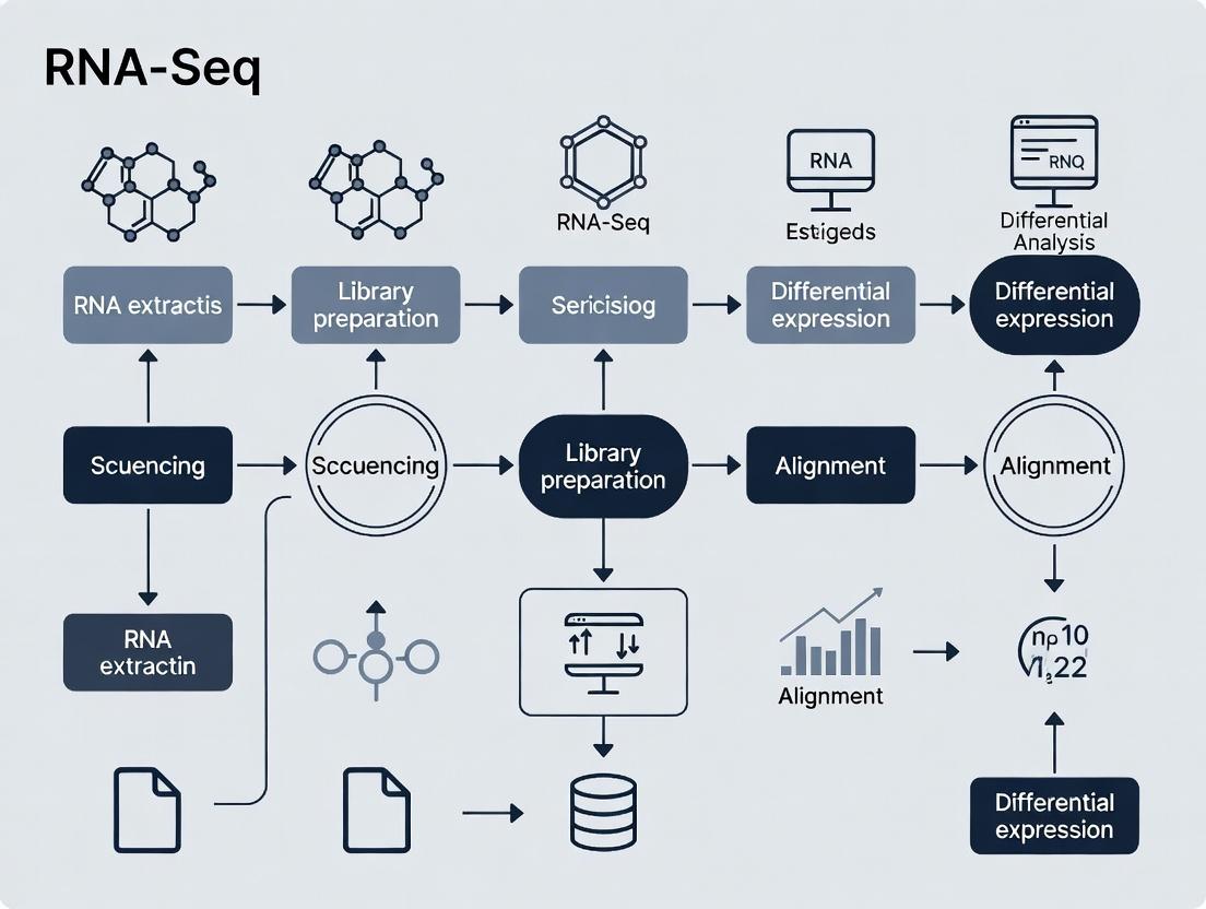
RNA-Seq Experimental and Analysis Workflow
RNA-Seq End-to-End Process from Sample to Insight
Core Data Analysis Pathway for Differential Expression
Key Steps in Differential Expression Analysis Workflow
The Scientist's Toolkit: Essential Research Reagents & Materials
Table 2: Key Reagent Solutions for Standard mRNA-Seq Library Prep
| Item | Function | Example Product (Supplier) |
|---|---|---|
| Total RNA Isolation Kit | Extracts high-quality, DNA-free total RNA from various sample types. | RNeasy Mini Kit (Qiagen), TRIzol Reagent (Thermo Fisher). |
| RNA Integrity Assessor | Provides quantitative assessment of RNA degradation (RIN/RINe). | RNA 6000 Nano Kit for Bioanalyzer (Agilent). |
| Poly-A Selection Beads | Magnetic beads coated with oligo(dT) to isolate polyadenylated mRNA. | NEBNext Poly(A) mRNA Magnetic Isolation Module (NEB), Dynabeads mRNA DIRECT Purification Kit (Thermo Fisher). |
| RNA Fragmentation Buffer | Chemically fragments mRNA into optimal sizes for sequencing. | Provided in NEBNext Ultra II RNA Library Prep Kit (NEB). |
| Reverse Transcriptase | Enzyme that synthesizes first-strand cDNA from RNA template. | SuperScript II or IV Reverse Transcriptase (Thermo Fisher). |
| Second-Strand Synthesis Mix (with dUTP) | Synthesizes second cDNA strand while incorporating dUTP for strand marking. | NEBNext Second Strand Synthesis Module (NEB). |
| DNA Cleanup Beads (SPRI) | Magnetic beads for size selection and purification of cDNA/library fragments. | AMPure XP Beads (Beckman Coulter), Sera-Mag Select Beads (Cytiva). |
| Sequencing Adapters (UDI) | Short, barcoded DNA oligos ligated to fragments for sequencing and multiplexing. | IDT for Illumina RNA UD Indexes (Integrated DNA Technologies). |
| High-Fidelity DNA Polymerase | Amplifies the final adapter-ligated library with minimal bias. | KAPA HiFi HotStart ReadyMix (Roche), Q5 High-Fidelity DNA Polymerase (NEB). |
| Library Quantification Kit | Accurately measures concentration of final dsDNA library. | Qubit dsDNA HS Assay Kit (Thermo Fisher). |
Application Notes
Within the thesis context of an RNA-Seq data analysis tutorial for beginners, the transition from raw sequencing data to biomedical applications is critical. The primary applications are biomarker discovery for diagnostics and prognostics, and drug target identification for therapeutic development. These rely on robust differential expression analysis, pathway enrichment, and network biology approaches.
Biomarker Discovery
RNA-Seq enables the identification of differentially expressed genes (DEGs), non-coding RNAs, and splice variants between diseased and healthy samples. These molecular signatures serve as potential biomarkers for early detection, patient stratification, and monitoring treatment response. Single-cell RNA-Seq (scRNA-seq) further refines this by identifying cell-type-specific biomarkers.
Drug Target Identification
By analyzing transcriptional changes in disease states, researchers can pinpoint key driver genes and dysregulated pathways. Validated targets are genes/proteins whose modulation is expected to have a therapeutic effect. CRISPR screening integrated with RNA-Seq (e.g., Perturb-seq) directly links genetic perturbations to transcriptional outcomes, accelerating target validation.
Table 1: Common RNA-Seq Analysis Outputs for Biomedical Applications
| Analysis Type | Typical Output Metrics | Relevance to Application |
|---|---|---|
| Differential Expression | Log2 Fold Change, p-value, Adjusted p-value (FDR) | Identifies potential biomarker genes or therapeutic targets. |
| Pathway Enrichment | Enrichment Score (NES), p-value, FDR q-value | Reveals dysregulated biological processes for targetable pathways. |
| scRNA-Seq Clustering | Number of distinct cell clusters, Cluster markers | Discovers cell-type-specific biomarkers and targets. |
| Survival Analysis (Cohort Data) | Hazard Ratio (HR), Log-rank p-value | Validates biomarker association with clinical outcomes. |
Table 2: Example Public Data Resources for Integration
| Resource Name | Data Type | Primary Use in Applications |
|---|---|---|
| The Cancer Genome Atlas (TCGA) | Bulk RNA-Seq, Clinical Data | Pan-cancer biomarker and target discovery. |
| Genotype-Tissue Expression (GTEx) | Healthy Tissue RNA-Seq | Defines normal expression baseline. |
| GEO / ArrayExpress | Curated Study Datasets | Independent validation of findings. |
| DepMap (Cancer Dependency Map) | CRISPR Screens, Expression | Prioritizes essential genes as drug targets. |
Experimental Protocols
Protocol 1: Bulk RNA-Seq Workflow for Biomarker Discovery
Objective: To identify a gene expression signature distinguishing disease from control samples.
- Sample Preparation & Sequencing: Extract total RNA from tissue/blood (n≥3 per group). Assess RNA integrity (RIN > 8). Prepare libraries (e.g., poly-A selection) and sequence on an Illumina platform to a depth of 20-40 million paired-end reads per sample.
- Bioinformatics Analysis (Beginner Tutorial Context):
- Quality Control: Use
FastQCto assess read quality. Trim adapters/low-quality bases withTrimmomatic. - Alignment: Map reads to a reference genome (e.g., GRCh38) using a splice-aware aligner like
HISAT2orSTAR. - Quantification: Generate gene-level read counts using
featureCounts. - Differential Expression: Import count matrix into R/Bioconductor. Use
DESeq2to perform statistical testing. Genes with |log2FC| > 1 and FDR-adjusted p-value < 0.05 are considered significant DEGs.
- Quality Control: Use
- Downstream Analysis: Perform pathway over-representation analysis on DEGs using
clusterProfilerand the KEGG database. Correlate top DEG expression with patient survival data using TCGA data and thesurvivalR package.
Protocol 2: In Silico Drug Target Prioritization Pipeline
Objective: To filter candidate genes from DEG analysis into high-confidence drug targets.
- Initial Candidate List: Start with significant DEGs from Protocol 1.
- Essentiality Filter: Cross-reference with DepMap data. Remove genes whose CRISPR knockout does not significantly reduce cell viability in relevant cancer cell lines (Common Essential genes may be poor drug targets).
- Druggability Assessment: Query the
Open Targets PlatformandDGIdbdatabases to identify which candidate genes have known drug compounds, predicted small molecule binders, or are members of druggable protein families (e.g., kinases, GPCRs). - Literature & Clinical Trial Validation: Search PubMed and
ClinicalTrials.govto determine if the gene/protein is already under investigation, to avoid redundancy. - Final Prioritization: Rank candidates by a composite score incorporating expression fold-change, essentiality score, and druggability evidence.
Visualizations
RNA-Seq to Applications Workflow
Target Prioritization Logic
The Scientist's Toolkit: Research Reagent Solutions
Table 3: Essential Reagents & Kits for RNA-Seq Based Applications
| Item | Function | Example Vendor/Product |
|---|---|---|
| Total RNA Isolation Kit | Extracts high-integrity RNA from diverse biological samples (tissue, cells, biofluids). Essential for input material quality. | Qiagen RNeasy, Thermo Fisher PureLink RNA Mini Kit. |
| Poly-A Selection Beads | Enriches for polyadenylated mRNA, removing ribosomal RNA. Standard for mRNA-seq library prep. | NEBNext Poly(A) mRNA Magnetic Isolation Module. |
| Stranded mRNA Library Prep Kit | Converts mRNA into sequencer-ready, strand-preserving DNA libraries. | Illumina Stranded mRNA Prep, Takara Bio SMART-Seq. |
| 10x Genomics Single Cell Kit | Enables barcoding and partitioning of single cells for scRNA-seq applications. | 10x Genomics Chromium Single Cell 3' Gene Expression. |
| CRISPR Guide RNA Library | Pooled guides for genome-wide knockout screens. Integrated with RNA-Seq (Perturb-seq). | Synthego CRISPR Libraries, Broad Institute GPP. |
| Reverse Transcription Master Mix | Converts RNA to cDNA for downstream quantification (e.g., qPCR) required for biomarker validation. | Bio-Rad iScript, Applied Biosystems High-Capacity Kit. |
Within the framework of a thesis on RNA-Seq data analysis for beginners, mastering core terminology is the critical first step. This tutorial deconstructs four fundamental concepts—Reads, Alignments, Transcripts, and Count Tables—that form the scaffold of any RNA-Seq experiment, from wet-lab preparation to computational analysis. Their precise understanding is non-negotiable for researchers, scientists, and drug development professionals aiming to derive biologically meaningful insights from gene expression data.
Core Terminology Explained
Reads
Definition: A "read" is a short DNA sequence generated by a high-throughput sequencing instrument. In RNA-Seq, these are complimentary DNA (cDNA) sequences derived from fragmented RNA molecules. Role in Pipeline: Reads are the primary raw data output. Their quality and quantity directly influence all downstream analyses. Key Metrics:
- Read Length: Typically 50-300 base pairs (bp).
- Read Depth/Count: Total number of reads per sample, often in millions.
- Quality Score (Q-score): Phred-scaled probability of a base call being incorrect (e.g., Q30 = 99.9% accuracy).
Table 1: Common NGS Read Specifications (2023-2024)
| Platform | Typical Read Length (bp) | Output per Flow Cell (approx.) | Common RNA-Seq Application |
|---|---|---|---|
| Illumina NovaSeq X | 150-300 | 8-16 Tb | Bulk RNA-Seq, large cohorts |
| Illumina NextSeq 2000 | 50-300 | 120-600 Gb | Standard bulk RNA-Seq |
| PacBio Sequel II/IIe | HiFi reads: 10-25 kb | 30-50 Gb | Full-length isoform sequencing |
| Oxford Nanopore | 1 kb - >100 kb | 10-50 Gb+ | Direct RNA sequencing, isoform detection |
Alignments (Mapping)
Definition: The computational process of aligning (or "mapping") sequencing reads to a reference genome or transcriptome to identify their genomic origin. Role in Pipeline: Converts raw sequence data into spatially meaningful information. Key Concepts:
- Mapper: Software that performs alignment (e.g., STAR, HISAT2).
- Mapping Rate: Percentage of reads successfully placed on the reference.
- Multi-mapping Reads: Reads that align to multiple locations, a challenge for repetitive genomic regions.
Transcripts
Definition: In RNA-Seq analysis, "transcript" refers to the inferred RNA molecule isoforms expressed from a gene locus. Reconstruction can be reference-based or de novo. Role in Pipeline: The biological entity whose abundance is quantified. Key Concepts:
- Transcriptome Assembly: Process of piecing together reads to reconstruct full-length transcripts.
- Isoform: A specific variant of a transcript produced via alternative splicing or promoter usage.
- Quantification: Estimating the abundance of each transcript.
Count Tables
Definition: A matrix where rows represent genomic features (genes/transcripts), columns represent samples, and each cell contains an integer value representing the number of reads assigned to that feature in that sample. Role in Pipeline: The final structured data input for statistical analysis and differential expression. Key Concepts:
- Feature: The unit being counted (e.g., gene, exon, transcript).
- Quantification Tools: Software that generates counts (e.g., featureCounts, Salmon, HTSeq).
Experimental Protocols
Protocol 3.1: RNA-Seq Library Preparation for Illumina Sequencing
Objective: Convert purified total RNA into a library of cDNA fragments with platform-specific adapters. Reagents: See Scientist's Toolkit (Table 3). Methodology:
- RNA Quality Control: Assess RNA integrity using an Agilent Bioanalyzer (RIN > 8.0 recommended).
- Poly-A Selection or rRNA Depletion: Enrich for mRNA.
- Fragmentation: Chemically or enzymatically fragment RNA to ~200-300 bp.
- First-Strand cDNA Synthesis: Reverse transcription using random hexamers or oligo-dT primers.
- Second-Strand cDNA Synthesis: Generate double-stranded cDNA.
- End Repair & A-tailing: Create blunt-ended, 5'-phosphorylated fragments with a 3' A-overhang.
- Adapter Ligation: Ligate indexed sequencing adapters.
- Library Amplification: Enrich adapter-ligated fragments via PCR (8-12 cycles).
- Library QC: Validate size distribution (TapeStation) and quantify (qPCR).
- Pooling & Normalization: Combine multiplexed libraries in equimolar ratios for sequencing.
Protocol 3.2: Read Alignment & Quantification Using STAR and featureCounts
Objective: Map reads to a reference genome and generate a gene-level count table. Software: STAR aligner, featureCounts (from Subread package), Samtools. Reference Files: Genome FASTA file and corresponding annotation (GTF) file. Methodology:
- Generate Genome Index (one-time):
STAR --runMode genomeGenerate --genomeDir /path/to/index --genomeFastaFiles genome.fa --sjdbGTFfile annotation.gtf --sjdbOverhang 99 - Align Reads:
STAR --genomeDir /path/to/index --readFilesIn sample.R1.fastq.gz sample.R2.fastq.gz --readFilesCommand zcat --outFileNamePrefix sample_ --outSAMtype BAM SortedByCoordinate --runThreadN 8 - Post-process BAM file:
samtools index sample_Aligned.sortedByCoord.out.bam - Generate Count Table:
featureCounts -T 8 -p -a annotation.gtf -o counts.txt sample_Aligned.sortedByCoord.out.bam - Output: A
counts.txtfile suitable for input into R/Bioconductor packages like DESeq2.
Visualizations
Title: RNA-Seq Analysis Workflow for Beginners
Title: Relationship Between Core RNA-Seq Terms
The Scientist's Toolkit
Table 3: Essential Research Reagent Solutions for RNA-Seq
| Item | Function in RNA-Seq | Example Vendor/Kit |
|---|---|---|
| RNA Stabilization Reagent | Immediately preserves RNA integrity in cells/tissues at collection. | Qiagen RNAlater, Invitrogen TRIzol |
| Poly(A) Magnetic Beads | Selectively enriches for messenger RNA (mRNA) from total RNA. | NEBNext Poly(A) mRNA Magnetic, Illumina TruSeq |
| rRNA Depletion Kit | Removes abundant ribosomal RNA to enrich other RNA species. | Illumina Ribo-Zero Plus, QIAseq FastSelect |
| Fragmentation Buffer | Chemically breaks RNA into uniform fragments for sequencing. | Included in Illumina TruSeq, NEBNext Ultra II |
| Reverse Transcriptase | Synthesizes first-strand cDNA from RNA template. | SuperScript IV, Maxima H Minus |
| Double-stranded DNA | Converts single-stranded cDNA to dsDNA for library build. | NEBNext Second Strand Synthesis Module |
| Indexed Adapter Oligos | Attaches unique barcodes for sample multiplexing. | Illumina IDT for Illumina, Twist UD Indexes |
| High-Fidelity PCR Mix | Amplifies final library with minimal bias and errors. | Kapa HiFi, Q5 High-Fidelity |
| Library Quantification Kit | Accurate qPCR-based quantification for pooling. | Kapa Library Quant, NEBNext Library Quant |
| SPRIselect Beads | Size selection and clean-up of nucleic acids. | Beckman Coulter SPRIselect |
RNA-Seq is a cornerstone technique in modern molecular biology, enabling comprehensive analysis of the transcriptome. For beginners in research, particularly in drug development, understanding the journey from a biological hypothesis to a sequencing-ready library is paramount. This protocol is structured as a step-by-step guide within a broader thesis on RNA-Seq data analysis, focusing on the initial experimental phase.
Key Research Reagent Solutions
Table 1: Essential Reagents and Kits for RNA-Seq Library Preparation
| Reagent/KIT | Primary Function | Key Considerations for Selection |
|---|---|---|
| RNA Extraction Kit | Isolate high-integrity total RNA from cells/tissues. | Assess yield, purity (A260/280), and RNA Integrity Number (RIN). Inhibitor removal is critical. |
| Poly(A) Selection Beads | Enrich for polyadenylated mRNA from total RNA. | Efficiency impacts coverage of protein-coding genes. Alternatives: rRNA depletion for non-poly(A) RNA. |
| Fragmentation Reagents | Randomly shear RNA or cDNA to optimal size (200-500 bp). | Chemical (e.g., metal ions) vs. enzymatic (e.g., RNase III) vs. physical (sonication) methods. |
| Reverse Transcriptase | Synthesize first-strand cDNA from RNA template. | High processivity and fidelity; template-switching activity required for some protocols. |
| Second-Strand Synthesis Mix | Convert single-stranded cDNA to double-stranded DNA. | Often uses dUTP incorporation for strand-specificity. |
| Library Prep Kit w/Adapters | Add platform-specific sequencing adapters and sample indexes. | Compatibility with sequencer (Illumina, MGI, etc.); supports multiplexing. |
| SPRI/AMPure Beads | Size-select and purify nucleic acids after enzymatic steps. | Bead-to-sample ratio controls size selection; removes primers and adapter dimers. |
| PCR Master Mix | Amplify final library for quantification and sequencing. | High-fidelity polymerase; limited cycles to avoid bias. |
Core Experimental Protocol: From Cells to Sequencer-Ready Library
Protocol 3.1: Total RNA Extraction and QC
- Objective: Obtain high-quality, intact total RNA.
- Materials: Cultured cells or tissue samples, TRIzol or column-based kit, RNase-free consumables, bioanalyzer/tapestation.
- Method:
- Homogenize sample in lysis buffer. For tissues, use a mechanical homogenizer.
- Add chloroform, vortex, and centrifuge to separate phases. The RNA is in the aqueous phase.
- Transfer aqueous phase to a new tube. Precipitate RNA with isopropanol. Wash pellet with 75% ethanol.
- Dissolve RNA in RNase-free water. Quantify using a spectrophotometer (NanoDrop).
- Critical QC Step: Assess RNA integrity using a Bioanalyzer. A RIN > 8.0 is recommended for standard mRNA-seq.
Protocol 3.2: Poly(A) mRNA Enrichment & Fragmentation
- Objective: Isolate mRNA and prepare it for cDNA synthesis.
- Materials: Oligo-dT magnetic beads, magnetic rack, fragmentation buffer.
- Method:
- Mix total RNA with oligo-dT beads. Incubate to allow poly(A) tail binding.
- Wash beads on a magnetic rack to remove rRNA and tRNA.
- Elute purified mRNA in low-salt buffer or nuclease-free water.
- Fragmentation: Using divalent cations at elevated temperature (e.g., 94°C for several minutes), hydrolyze RNA into fragments of desired length (e.g., ~200-300 nucleotides).
- Purify fragmented RNA using SPRI beads.
Protocol 3.3: Strand-Specific cDNA Library Construction
- Objective: Generate double-stranded cDNA libraries with adapters, preserving strand-of-origin information.
- Materials: Reverse transcriptase, random hexamers, dNTPs (including dUTP for second strand), DNA polymerase I, RNase H, T4 DNA polymerase, ligase, adapters.
- Method:
- First-Strand Synthesis: Synthesize cDNA from fragmented RNA using reverse transcriptase and random primers.
- Second-Strand Synthesis: Using DNA Polymerase I, RNase H, and a dNTP mix containing dUTP (instead of dTTP), synthesize the second strand. The incorporation of dUTP marks this strand for later degradation.
- Purify double-stranded cDNA.
- End Repair & A-tailing: Blunt the cDNA ends and add a single 'A' nucleotide to the 3' ends to facilitate adapter ligation.
- Adapter Ligation: Ligate platform-specific Y-shaped adapters (containing indices for multiplexing) to the A-tailed cDNA ends.
- Strand Selection: Treat with Uracil-Specific Excision Reagent (USER) enzyme to degrade the dUTP-containing second strand, ensuring only the first strand (representing the original RNA orientation) is amplified.
- Library Amplification: Perform limited-cycle PCR (e.g., 10-15 cycles) with primers complementary to the adapter sequences to enrich for properly ligated fragments and add full sequencing adapters.
- Final Purification & QC: Perform dual-size selection with SPRI beads (e.g., 0.7x to 1.8x ratio) to remove adapter dimers and select the optimal insert size library. Quantify by qPCR and validate size distribution on a Bioanalyzer.
Table 2: Typical QC Metrics and Targets for RNA-Seq Libraries
| QC Parameter | Measurement Tool | Optimal Value/Range | Purpose & Implication of Deviation |
|---|---|---|---|
| RNA Integrity (RIN) | Bioanalyzer | RIN ≥ 8.0 | Indicates intact RNA. Low RIN causes 3' bias and gene dropout. |
| Library Concentration | qPCR (dsDNA assay) | ≥ 2 nM for pooling | Accurate quantification ensures balanced multiplexing. |
| Library Size Distribution | Bioanalyzer/TapeStation | Peak ~350-450 bp (incl. adapters) | Confirms proper size selection; detects adapter dimer (~120 bp). |
| Molarity (Adapter Dimer %) | Bioanalyzer / qPCR | < 10% of total signal | High dimer % wastes sequencing reads on non-informative fragments. |
| Fragment Size Post-Seq | Sequencing Data (e.g., Picard) | Mean insert size ~200-300 bp | Validates wet-lab process; influences alignment efficiency. |
Visualizing the Workflow and Central Dogma
Diagram 1: RNA-Seq Library Prep Experimental Workflow
Diagram 2: Central Dogma in Context of RNA-Seq
Application Notes
This guide establishes the foundational principles of robust experimental design and replication, specifically contextualized for RNA-Seq experiments in biomedical research. Adherence to these principles is non-negotiable for generating biologically valid and statistically sound data, which forms the basis for downstream analysis in our broader RNA-Seq tutorial thesis.
Core Principles
- Defining the Biological Question & Hypothesis: The experiment must begin with a clear, focused question. For RNA-Seq: "Does Drug X alter the transcriptomic profile of Cell Line Y in a manner consistent with the inhibition of Pathway Z?"
- Controlling Variables & Avoiding Bias:
- Technical Confounders: Sequence all samples across multiple lanes/days (batch effects). Use a single, validated library prep kit for all samples.
- Biological Confounders: Use cells/passages/tissues from matched sources. Randomize treatment assignments.
- Blinding: Where possible, label samples with non-revealing identifiers prior to library preparation and data analysis.
- Replication: Power analysis must be conducted a priori to determine the appropriate sample size. Biological replicates (samples derived from distinct biological units) are essential for inferring population-level effects. Technical replicates (repeated measurements of the same sample) assess assay precision but cannot substitute for biological replication.
- Inclusion of Proper Controls:
- Negative Controls: Vehicle-treated samples, non-targeting siRNA controls.
- Positive Controls: Samples with a known transcriptomic response (e.g., a cell line with a validated knockout or known agonist treatment) to verify assay sensitivity.
- Pre-registration & Documentation: Detailed experimental protocols, sample metadata, and analysis plans should be documented before data collection begins. This mitigates "fishing expeditions" in data analysis.
Key Considerations Table for RNA-Seq Design
| Design Aspect | Poor Practice | Sound Practice | Rationale |
|---|---|---|---|
| Replicate Type | 6 technical replicates from a single cell culture flask. | 3 independent biological replicates (cells cultured separately), each with 2 technical sequencing replicates. | Biological replicates capture biological variance; technical replicates measure library/sequencing noise. |
| Sample Size | n=2 per group. | n=4-6 per group, determined by power analysis using pilot data or effect size estimates. | Provides adequate statistical power to detect meaningful differential expression while controlling false discovery rates. |
| Batch Control | Processing all control samples on one day and all treatment samples on another. | Randomizing samples from all experimental groups across library prep batches and sequencing runs. | Prevents confounding of treatment effects with batch-specific technical artifacts. |
| Control Samples | Treatment vs. untreated only. | Treatment vs. vehicle control + a known positive control sample set. | Accounts for vehicle effects and confirms experimental system is responsive. |
| Blinding | Researcher preparing libraries knows sample identities. | Samples are coded prior to library prep, with key held by third party until analysis is locked. | Reduces unconscious bias during sample handling and processing. |
Detailed Protocol: Designing a Replicated RNA-Seq Experiment
I. Pre-Experimental Phase
- Power & Sample Size Calculation:
- Use tools like
ScottyorRNASeqPower. - Input Parameters: Estimate expected biological coefficient of variation (e.g., 0.4 for cell lines, 0.6 for tissues), target fold-change (e.g., 2.0), desired statistical power (e.g., 0.8), and false discovery rate (e.g., 0.05).
- Output: Minimum number of biological replicates required per group.
- Use tools like
- Randomization Plan:
- Assign each biological sample (e.g., cell culture plate, animal) a unique ID.
- Using a random number generator, assign each ID to a treatment or control group.
- Generate a sample processing order that interleaves groups to avoid batch correlation.
II. Experimental Execution Phase
- Cell Culture & Treatment (Example):
- Culture cells for biological replicate n=1 to n=6 in separate flasks, seeded from different passages/parental stocks.
- At ~70% confluency, treat according to the randomization plan. Include a vehicle control for the treatment compound.
- Harvest all samples using identical lysis conditions (e.g., QIAzol). Store lysates at -80°C.
RNA Extraction & QC:
- Process samples in randomized order. Extract total RNA using a standardized kit (e.g., miRNeasy Mini Kit).
- Quantify RNA using a fluorometric assay (e.g., Qubit RNA HS Assay). Assess integrity via capillary electrophoresis (e.g., Agilent Bioanalyzer RNA Nano Kit). Only proceed with samples having RIN > 8.5.
Library Preparation & Sequencing:
- Use a stranded mRNA-seq library prep kit (e.g., Illumina Stranded mRNA Prep).
- Process samples in a single, randomized batch if possible. If multiple batches are unavoidable, ensure each batch contains a balanced mix from all experimental groups.
- Pool libraries in equimolar amounts based on qPCR quantification (e.g., KAPA Library Quantification Kit).
- Sequence on an Illumina platform to a minimum depth of 25-30 million paired-end reads per sample (e.g., 2x150bp).
III. Metadata Documentation
- Record all sample metadata in a structured table (e.g., .tsv format). Essential columns include: SampleID, ExperimentalGroup, BiologicalReplicateID, TechnicalReplicateID, BatchID (library prep, sequencing run), RNA_RIN, SequencingDepth, and any other relevant covariates.
The Scientist's Toolkit: RNA-Seq Reagent Solutions
| Item | Function & Rationale |
|---|---|
| miRNeasy Mini Kit (Qiagen) | Total RNA isolation, ensuring high yield and integrity while removing genomic DNA contamination. |
| RNase Inhibitors (e.g., Superase-In) | Inactivated during lysis to prevent RNA degradation during sample processing. |
| Agilent RNA 6000 Nano Kit | Provides precise RNA Integrity Number (RIN) to objectively assess sample quality prior to costly library prep. |
| Qubit RNA HS Assay Kit | Fluorometric RNA quantification specific to RNA, superior to absorbance (A260) which is sensitive to contaminants. |
| Illumina Stranded mRNA Prep | Selective enrichment for poly-A transcripts with strand information retention, enabling accurate transcript abundance and orientation. |
| KAPA Library Quantification Kit (qPCR) | Accurate quantification of amplifiable library fragments for precise pooling and optimal cluster density on the sequencer. |
| SPRIselect Beads (Beckman Coulter) | For size selection and cleanup of cDNA libraries, replacing lower-efficiency gel-based methods. |
| Unique Dual Indexes (UDIs) | Enables multiplexing of many samples with minimal risk of index hopping errors during sequencing. |
Visualization
Diagram Title: RNA-Seq Experimental Design & Replication Workflow
Diagram Title: RNA-Seq Replication Structure & Purpose
This guide is an integral part of a comprehensive tutorial on RNA-Seq data analysis for beginners in research. The accurate interpretation of biological data hinges on a fundamental understanding of the file formats that store it. In a typical RNA-Seq workflow, raw sequencing data (FASTQ) is aligned to a reference genome to produce alignment data (BAM), which is then interpreted using genomic annotations (GTF/GFF). Mastery of these formats is the first critical step toward meaningful analysis and discovery in genomics, enabling researchers and drug development professionals to ask and answer complex biological questions.
Core File Formats: Structure, Purpose, and Comparison
FASTQ Format: Raw Sequencing Reads
Purpose: The primary format for storing raw, unaligned nucleotide sequences (reads) and their corresponding quality scores generated by high-throughput sequencing platforms.
Structure: Each sequenced read is represented by four consecutive lines:
- Sequence Identifier (Begins with '@'): Contains instrument and flow cell data.
- The Nucleotide Sequence (A, T, C, G, N).
- Separator Line (Begins with '+'): May optionally repeat the identifier.
- Quality Scores: Encoded per base as ASCII characters (Phred+33 is standard), representing the probability of a base call error.
Key Insight: Quality scores (Q) are logarithmically related to error probability (P). Q = -10 log₁₀(P). A Q-score of 30 indicates a 1 in 1000 chance of an incorrect base call.
BAM/SAM Format: Aligned Sequence Data
Purpose: The standard format for storing sequence alignments to a reference genome. SAM (Sequence Alignment/Map) is a human-readable text format, while BAM is its compressed, binary equivalent used for efficient storage and analysis.
Structure: A BAM file consists of a header section and an alignment section.
- Header: Contains metadata (reference sequence names, lengths, program used).
- Alignment Lines: Each line has 11 mandatory fields detailing the alignment information for a single read (QNAME, FLAG, RNAME, POS, MAPQ, CIGAR, RNEXT, PNEXT, TLEN, SEQ, QUAL).
Key Insight: The CIGAR (Compact Idiosyncratic GAP Alignment Report) string is crucial for interpreting how a read aligns to the reference (e.g., 150M = 150 bases match, 100M3I50M = 100 match, 3 insert, 50 match).
GTF/GFF Format: Genomic Annotations
Purpose: The Gene Transfer Format (GTF) and its predecessor, the General Feature Format (GFF), are used to describe the features and annotations of a genome, such as genes, exons, transcripts, and their coordinates.
Structure: Both are tab-delimited text files with 9 fields per line.
- seqname: The chromosome or scaffold name.
- source: The algorithm or database that generated the feature.
- feature: The type of feature (e.g.,
gene,exon,CDS). - start: Genomic start coordinate (1-based).
- end: Genomic end coordinate.
- score: A numerical value (e.g., confidence score);
.if absent. - strand:
+(forward),-(reverse), or.(unknown). - frame: For CDS, indicates the reading frame (0, 1, 2);
.otherwise. - attributes: A semicolon-separated list of key-value pairs providing additional information (e.g., geneid "TP53"; transcriptid "TP53-001";).
Key Insight: The primary difference between GFF3 and GTF2.2 lies in the structure of the attributes field. GTF has a stricter, standardized set of tags essential for transcriptome assembly tools like Cufflinks and StringTie.
Table 1: Comparative Overview of Core RNA-Seq File Formats
| Feature | FASTQ | BAM/SAM | GTF/GFF |
|---|---|---|---|
| Primary Purpose | Store raw sequencing reads and quality scores | Store aligned reads to a reference genome | Store genomic feature annotations |
| Format Type | Text | SAM: Text; BAM: Binary (compressed) | Text (tab-delimited) |
| Key Contents | Read ID, Sequence, Quality scores | Alignment coordinates, CIGAR, MAPQ, read data | Feature coordinates, type, strand, attributes |
| Size (Typical) | Very Large (10s-100s of GB) | Large, but smaller than FASTQ (compressed) | Small to Medium (MB to low GB) |
| Stage in Workflow | Initial Input (Raw Data) | Intermediate (Primary Analysis Output) | Reference (Annotation Input) |
| Essential Tools | FastQC, Trimmomatic |
samtools, IGV |
IGV, Genome Browser, featureCounts |
Table 2: Phred Quality Score Interpretation
| Phred Quality Score (Q) | Probability of Incorrect Base Call | Base Call Accuracy |
|---|---|---|
| 10 | 1 in 10 | 90% |
| 20 | 1 in 100 | 99% |
| 30 | 1 in 1000 | 99.9% |
| 40 | 1 in 10,000 | 99.99% |
Experimental Protocols
Protocol 1: Basic Quality Assessment of FASTQ Files using FastQC
Objective: To assess the quality of raw sequencing data and identify potential issues (e.g., low-quality bases, adapter contamination, overrepresented sequences).
Materials:
- Raw FASTQ files from an RNA-Seq experiment.
- A computer with FastQC installed.
Method:
- Open a terminal or command line interface.
- Run FastQC on your FASTQ file:
- Interpret the HTML Report: Open the generated
sample_R1_fastqc.htmlfile in a web browser. Key modules to examine:- Per base sequence quality: Check for quality drops at read starts/ends.
- Per sequence quality scores: Should form a tight, high-quality distribution.
- Sequence duplication levels: High duplication may indicate PCR bias.
- Adapter Content: Indicates if adapter trimming is required.
Protocol 2: Manipulating and Indexing BAM Files using Samtools
Objective: To sort, index, and filter a BAM file for downstream analysis and visualization.
Materials:
- An aligned BAM file (e.g.,
aligned.bam). - Samtools software suite installed.
Method:
- Sort the BAM file by genomic coordinate (required for indexing and many analyses):
- Index the sorted BAM file to allow for rapid random access:
This creates an index file (
aligned.sorted.bam.bai). - (Optional) Filter alignments to extract, for example, properly paired reads mapping to chromosome 1:
- Generate basic alignment statistics:
Protocol 3: Extracting Feature Coordinates from a GTF File
Objective: To parse a GTF file to create a BED file containing the coordinates of all exons for a specific gene.
Materials:
- A reference genome annotation file in GTF format (e.g.,
Homo_sapiens.GRCh38.104.gtf). - Command-line tools (
grep,awk).
Method:
- Extract all lines corresponding to
exonfeatures for the gene of interest (e.g., TP53): - Convert the GTF lines to a BED format (0-based start):
- The resulting
TP53_exons.bedfile can now be loaded into a genome browser or used for counting reads overlapping these regions.
Visualization: RNA-Seq Data Flow & File Relationships
Diagram Title: RNA-Seq Workflow: From FASTQ to Counts
The Scientist's Toolkit: Essential Research Reagents & Materials
Table 3: Key Solutions & Tools for RNA-Seq Data Handling
| Item/Category | Function & Purpose in RNA-Seq Data Analysis |
|---|---|
| High-Quality RNA Library Prep Kit (e.g., Illumina TruSeq Stranded mRNA) | Prepares sequencing libraries from RNA samples; determines strand specificity and library complexity. |
| Alignment Software (e.g., STAR, HISAT2) | Aligns FASTQ reads to a reference genome; produces SAM/BAM output. Critical for accuracy and speed. |
| SAM/BAM Manipulation Tool (Samtools) | A suite of programs for manipulating alignments: sorting, indexing, filtering, and generating statistics. |
| Genome Annotation File (GTF from Ensembl/NCBI/GENCODE) | Provides the reference "map" of gene models (exons, introns, UTRs) required to quantify expression. |
| Quality Control Suite (FastQC, MultiQC) | Assesses raw (FASTQ) and processed data quality, highlighting technical issues requiring intervention. |
| Read Counting Tool (featureCounts, HTSeq) | Counts the number of reads aligning to genomic features defined in the GTF file, generating expression matrix. |
| Visualization Software (Integrative Genomics Viewer - IGV) | Allows interactive visualization of BAM alignments in genomic context, overlayed with GTF annotations. |
| Reference Genome Sequence (FASTA file, e.g., GRCh38) | The nucleotide sequence of the target organism's genome; the reference for all alignments. |
| Computational Resources (High-performance cluster/cloud with adequate RAM & CPU) | Essential for processing large FASTQ/BAM files, which are computationally intensive. |
Hands-On RNA-Seq Pipeline: A Step-by-Step Workflow from Raw Data to Differential Expression
Within the broader thesis of providing a beginner's tutorial for RNA-Seq data analysis, establishing a reproducible and robust computational environment is the critical first step. This protocol details the setup of a foundational environment using the Unix command line interface (CLI) and Bioconda, a channel of the Conda package manager specializing in bioinformatics software. Mastery of these tools enables researchers, scientists, and drug development professionals to install, manage, and run hundreds of complex bioinformatics tools required for downstream RNA-Seq processing (e.g., FastQC, HISAT2, STAR, featureCounts) without dependency conflicts.
Comparison of Common Package Managers in Bioinformatics
The following table summarizes key quantitative and qualitative data on popular package management solutions, based on a survey of current repository statistics and documentation.
Table 1: Bioinformatics Package Management Solutions
| Manager | Primary Language | Approx. Bio Packages (2024) | Key Strength | Key Weakness |
|---|---|---|---|---|
| Bioconda | Python (Conda-based) | ~8,000+ | Vast ecosystem, solves dependency hell via environments. | Packages can be large; slower than system managers. |
| APT (Bio-Linux) | System (Debian/Ubuntu) | ~1,200 | Fast, system-integrated, very stable. | Often outdated versions; limited selection. |
| BioBuilds | System (RPM/YUM) | ~500 | Good for RedHat/CentOS systems. | Smaller repository than Bioconda or APT. |
| Docker/Singularity | Containerized | N/A (Full images) | Ultimate reproducibility and portability. | Images are very large; steeper learning curve. |
Essential CLI Commands for Beginners
Table 2: Fundamental Unix Command Line Operations
| Command | Function | Common Use Case in RNA-Seq |
|---|---|---|
pwd |
Print Working Directory | Confirm your location in the filesystem. |
ls -lah |
List files with details | View input FASTQ files and their sizes. |
cd <path> |
Change Directory | Navigate to project or data directories. |
mkdir -p data/raw_fastq |
Make Directory | Create organized project folder structure. |
cp -r source/ dest/ |
Copy files/directories | Back up crucial analysis results. |
rm -i file.txt |
Remove (delete) file | Clean up temporary intermediate files. |
cat/zless file.fastq |
Concatenate/view file | Quickly inspect the first lines of a FASTQ. |
wc -l file.txt |
Word/line count | Count reads in a FASTQ file (lines/4). |
grep "pattern" file |
Global Regular Expression Print | Search for specific gene IDs in an output. |
| (pipe) |
Redirects output to next command | Chain tools: cat file.fq | head -n 20. |
Detailed Protocols
Protocol A: Accessing and Basic Navigation of the Command Line
Objective: To open a terminal session and perform basic filesystem navigation.
- Access Terminal:
- macOS: Open
Terminalfrom Applications > Utilities. - Linux: Open
Terminalfrom application menu (Ctrl+Alt+T common). - Windows: Install & open "Windows Subsystem for Linux (WSL2)" or use "Git Bash".
- macOS: Open
- Check Current Directory: Type
pwdand press Enter. This prints your present working directory. - List Contents: Type
lsand press Enter to see files/folders. Usels -lfor detailed view. - Change Directory: Use
cdfollowed by a path. Example:cd ~/Documentsgoes to your Documents folder.cd ..moves up one level. - Create Project Structure: Execute: This creates a standard directory tree for an RNA-Seq analysis.
- Verify Structure: Navigate into the project (
cd ~/rna_seq_project) and runls -Rto recursively list the created folders.
Protocol B: Installing Miniconda and Configuring Bioconda
Objective: To install the Miniconda package manager and add the Bioconda channel for installing bioinformatics software.
- Download Miniconda Installer Script: (For macOS, check the Miniconda website for the appropriate macOS installer link.)
- Run the Installer:
The
-bflag runs in batch mode,-psets the install path. - Initialize Conda: Close and reopen your terminal for changes to take effect.
- Configure Conda Channels (Bioconda Setup): Add channels in the strict order below to ensure proper priority.
- Test Installation: Create a test environment and install a core Bioconda package (FastQC).
Protocol C: Creating a Conda Environment for RNA-Seq Analysis
Objective: To create an isolated Conda environment containing key RNA-Seq analysis tools.
- Create Environment from a Specification File (Recommended): Create a YAML file named
rna_seq_env.yaml. - Build the Environment: This process will resolve dependencies and may take several minutes.
- Activate and Verify: This shows all installed packages within the active environment.
Mandatory Visualizations
Diagram: Bioconda Environment Isolation for RNA-Seq Workflow
Diagram: RNA-Seq Setup & Analysis Logical Pathway
The Scientist's Toolkit: Research Reagent Solutions
Table 3: Essential Computational "Reagents" for RNA-Seq Analysis Setup
| Item | Function/Explanation | Example/Version |
|---|---|---|
| Unix/Linux Command Line | The primary interface to interact with the operating system, execute programs, and manage files. Essential for running bioinformatics tools. | Bash shell, Zsh. |
| Terminal Emulator | The application providing access to the command line interface. | iTerm2 (macOS), GNOME Terminal (Linux), Windows Terminal. |
| Miniconda | A minimal installer for Conda. It manages packages and dependencies and creates isolated environments. | Miniconda3 23.11.0. |
| Bioconda Channel | A Conda channel containing >8,000 pre-packaged, peer-reviewed bioinformatics software. | Bioconda repository. |
| Conda-Forge Channel | A community-led Conda channel with high-quality, updated libraries. Required for dependency resolution with Bioconda. | Conda-forge repository. |
| Environment YAML File | A text file specifying exact packages and versions for reproducible environment creation. | rna_seq_env.yaml. |
| Text Editor (CLI) | For editing scripts, configuration files, and YAML files directly in the terminal. | Nano, Vim, Emacs. |
| High-Performance Computing (HPC) Access | For large-scale RNA-Seq analysis, understanding how to access and use a cluster (via SSH) and a job scheduler (e.g., SLURM) is critical. | SSH client, SLURM commands. |
Within the broader thesis, "RNA-Seq Data Analysis Tutorial for Beginners," this initial step is critical for ensuring data integrity. The analysis pipeline's success is fundamentally dependent on the quality of the input sequencing reads. This section details the protocol for assessing raw read quality using FastQC and subsequently cleaning/adapter-trimming reads using Trimmomatic to generate a high-confidence dataset for downstream alignment and quantification.
Application Notes
FastQC for Quality Assessment
FastQC provides a comprehensive initial assessment of raw sequencing data (FASTQ files). It generates an HTML report with multiple modules, allowing researchers to identify potential issues such as per-base sequence quality drops, adapter contamination, overrepresented sequences, or unusual GC content. For RNA-Seq, particular attention should be paid to sequence duplication levels and k-mer content, which can indicate specific biases.
Trimmomatic for Read Trimming and Cleaning
Trimmomatic is a flexible, command-line tool used to remove technical sequences (adapters, primers) and low-quality bases from the reads. It operates in a single pass, applying multiple processing steps in a user-defined order. Key functions for RNA-Seq include: removing Illumina adapters, sliding window trimming, leading/trailing base trimming, and dropping reads below a minimum length. Proper trimming reduces alignment errors and improves the accuracy of transcript abundance estimation.
Table 1: FastQC Module Interpretation Guide for RNA-Seq
| Module | Pass/Warning/Fail | Typical Cause for RNA-Seq Failure | Recommended Action |
|---|---|---|---|
| Per base sequence quality | Fail | Sharp quality drop at read ends. | Proceed with Trimmomatic trimming. |
| Per sequence quality scores | Warning | Slight deviations are common. | Monitor; usually acceptable. |
| Per base sequence content | Warning/Fail | Random hexamer priming bias at start of read. | Normal for first 9-12 bases. Trim if severe. |
| Adapter Content | Fail | Detectable adapter sequence. | Mandatory adapter trimming with Trimmomatic. |
| Overrepresented sequences | Fail | High levels of specific sequences (e.g., rRNA). | Consider more aggressive filtering if not biological. |
Table 2: Common Trimmomatic Parameters for RNA-Seq (Illumina)
| Parameter | Typical Setting | Function |
|---|---|---|
ILLUMINACLIP |
TruSeq3-PE.fa:2:30:10 |
Remove Illumina adapters. Specifies adapter file, seed mismatches, palindrome & simple clip thresholds. |
LEADING |
3 |
Remove bases from start if below quality 3. |
TRAILING |
3 |
Remove bases from end if below quality 3. |
SLIDINGWINDOW |
4:15 |
Scan read with 4-base window, trim if avg quality <15. |
MINLEN |
36 |
Drop reads shorter than 36 bp after trimming. |
AVGQUAL |
20 |
(Optional) Drop entire read if average quality <20. |
Experimental Protocols
Protocol: Run FastQC on Raw RNA-Seq Data
Objective: Generate quality control reports for raw FASTQ files. Materials: Computing cluster or workstation with Java installed, FastQC software, raw FASTQ files. Procedure:
- Installation: Download FastQC from the Babraham Bioinformatics website and ensure it is in your system PATH.
- Basic Command: For a single file, run:
fastqc input_reads.fastq.gz -o /path/to/output/directory - Batch Processing: For multiple files, use a loop or provide multiple file names.
- Output: Examine the generated
.htmlreport. View all modules, noting any "FAIL" flags. - Decision Point: Use the report to inform Trimmomatic parameter selection (e.g., adapter type, trimming stringency).
Protocol: Trim Reads with Trimmomatic
Objective: Remove adapters and low-quality bases from paired-end RNA-Seq reads.
Materials: Java runtime, Trimmomatic JAR file, adapter sequence file (e.g., TruSeq3-PE-2.fa), raw paired FASTQ files (*_R1.fastq.gz, *_R2.fastq.gz).
Procedure:
- Command Structure (Paired-end):
PE: Specifies paired-end mode.-threads: Number of CPU threads to use.-phred33: Specifies quality score encoding (standard for Illumina >1.8).- Outputs: Four files – paired (both reads survived) and unpaired (one read dropped) for each direction.
- Run Command: Execute in terminal. Monitor console for trimming summary statistics.
- Validation: Run FastQC again on the
*_paired.fq.gzoutputs to confirm quality improvement.
Visualization: RNA-Seq QC & Trimming Workflow
Diagram Title: RNA-Seq Preprocessing Workflow from Raw Data to Clean Reads
The Scientist's Toolkit
Table 3: Essential Research Reagent Solutions for RNA-Seq QC & Trimming
| Item | Function in Protocol | Example/Note |
|---|---|---|
| Raw FASTQ Files | The primary input data containing sequence reads and quality scores. | Typically .fastq or .fq.gz format from Illumina, BGI, or other platforms. |
| FastQC Software | Generates the initial quality control report to diagnose data issues. | Version 0.11.9+. Requires Java. |
| Trimmomatic Software | The core tool for performing adapter removal and quality-based trimming. | Version 0.39+. Distributed as a Java JAR file. |
| Adapter Sequence File | Contains the technical sequences to be identified and removed by Trimmomatic. | e.g., TruSeq3-PE.fa for Illumina TruSeq kits. Must match library prep. |
| Computing Environment | Provides the necessary computational resources to run the tools. | Linux server, cluster, or local machine with sufficient RAM and Java. |
| High-Quality Reference | Not used in this step, but essential for the following alignment step. | Ensembl or GENCODE genome & annotation files for the target species. |
Within the broader RNA-Seq data analysis tutorial for beginners, read alignment is the critical step where sequenced reads are mapped to a reference genome. This process determines the genomic origin of each transcript fragment, forming the foundation for all downstream analyses like quantification and differential expression. The choice between STAR and HISAT2 depends on experimental design, reference genome size, and computational resources.
Core Algorithmic Differences
STAR (Spliced Transcripts Alignment to a Reference) employs a sequential maximum mappable seed search in two passes. It uses an uncompressed suffix array for rapid seed identification, allowing for the detection of spliced alignments without a priori transcript annotations. HISAT2 (Hierarchical Indexing for Spliced Alignment of Transcripts) utilizes a hierarchical graph FM index (GFM), incorporating both a global whole-genome index and tens of thousands of small local indexes for common splice sites. This architecture balances speed and memory efficiency.
A quantitative comparison of key performance metrics is summarized below:
| Feature | STAR | HISAT2 |
|---|---|---|
| Primary Algorithm | Suffix Array-based seed search | Hierarchical Graph FM Index |
| Typical Speed | ~30-50 million reads/hour* | ~40-60 million reads/hour* |
| Memory Footprint | High (~30-35 GB for human genome) | Moderate (~5-10 GB for human genome) |
| Splice Awareness | Excellent, uses 2-pass method | Excellent, uses extensive splice site index |
| Base-Level Accuracy | High | Very High (often higher in benchmarks) |
| Recommended Use Case | Large-scale studies, full splice junction discovery | Standard RNA-Seq, limited computational resources |
*Speed is system-dependent (CPU, I/O). Benchmarks based on human GRCh38 genome using 100bp paired-end reads on a 16-core server.
Detailed Experimental Protocols
Protocol 3.1: Genome Index Generation with STAR
Objective: To generate a genome index file for the STAR aligner.
- Prerequisites:
- Reference genome FASTA file (e.g.,
GRCh38.primary_assembly.genome.fa). - Gene annotation file in GTF format (e.g.,
gencode.v44.annotation.gtf). - Installed STAR software (v2.7.11a or later).
- Reference genome FASTA file (e.g.,
- Command:
- Parameters Explained:
--runThreadN: Number of CPU threads to use.--genomeDir: Path to the directory where the index will be stored.--sjdbGTFfile: Guide splice junctions from annotations.--sjdbOverhang: Read length minus 1. Critical for optimal splice junction mapping.--genomeSAindexNbases: Reduces memory for genomes smaller than human (14 is default for human).
Protocol 3.2: Read Alignment using STAR
Objective: To align FASTQ reads to the reference genome using the pre-built index.
- Input: Compressed or uncompressed FASTQ files (Read1 and Read2 for paired-end).
- Command for Paired-End Alignment:
- Key Outputs:
Aligned.sortedByCoord.out.bam: Sorted alignment file for downstream analysis.ReadsPerGene.out.tab: Raw gene-level counts.Log.final.out: Summary alignment statistics (mapping rate, etc.).
Protocol 3.3: Genome Index Generation with HISAT2
Objective: To generate a genome index for the HISAT2 aligner.
- Prerequisites: Reference genome FASTA file. Annotations are optional during indexing.
- Command:
- Note: For enhanced splice alignment, HISAT2 can also use a prepared splice site file (
--ssand--exonoptions), often derived from annotation GTF files.
Protocol 3.4: Read Alignment using HISAT2
Objective: To align FASTQ reads using HISAT2 and generate a sorted BAM file.
- Command for Paired-End Alignment:
- Post-Alignment Processing (Index BAM):
- Key Outputs:
sample_aligned_sorted.bam(and.baiindex) for downstream analysis. Gene quantification requires a separate tool (e.g., featureCounts).
Visualizations
The Scientist's Toolkit: Essential Research Reagents & Materials
| Item | Function / Role in Experiment |
|---|---|
| Reference Genome (FASTA) | The canonical DNA sequence of the target organism. Serves as the map for aligning sequencing reads. |
| Gene Annotation (GTF/GFF) | File defining genomic coordinates of genes, exons, transcripts, and other features. Guides splice-aware alignment and quantification. |
| High-Performance Computing (HPC) Cluster or Server | Alignment is computationally intensive. Multi-core servers with ample RAM (≥32 GB) are required, especially for STAR with large genomes. |
| STAR Software (v2.7.x+) | The aligner executable and supporting scripts for performing STAR-specific indexing and alignment. |
| HISAT2 Software (v2.2.x+) | The aligner executable for building HISAT2 indexes and running the alignment process. |
| SAMtools | Essential utility for processing alignment (SAM/BAM) files: sorting, indexing, and format conversion. |
| FASTQ Files (gzipped) | The raw input data containing the nucleotide sequences and quality scores of the sequenced library fragments. |
| Shell/Bash Environment | Command-line interface to execute the sequential steps of the alignment workflow. |
Within a comprehensive RNA-Seq tutorial for beginners, quantifying expression is a pivotal step that converts aligned sequencing reads into analyzable numerical data. This stage determines transcript abundance, enabling downstream differential expression and functional analysis. Two principal strategies exist: read-counting (e.g., featureCounts) for gene-level expression, and assembly-based (e.g., StringTie) for transcript-level quantification. The choice depends on experimental goals, reference annotation quality, and desired output (gene- vs. isoform-level data).
Tool Comparison & Selection Guide
Table 1: Comparison of featureCounts and StringTie
| Aspect | featureCounts | StringTie |
|---|---|---|
| Primary Purpose | Assign reads to genomic features (e.g., genes). | Assemble transcripts & quantify their expression. |
| Analysis Level | Gene-level. | Transcript-level (can output gene-level). |
| Requirement | High-quality, comprehensive annotation file (GTF/GFF). | Can work with or without reference annotation (reference-guided de novo mode). |
| Input | Aligned reads (BAM/SAM files), annotation file. | Aligned reads (BAM files), annotation file (optional). |
| Output Metrics | Raw read counts. | FPKM, TPM, estimated read counts. |
| Speed & Resource | Very fast, low memory. | Slower, higher computational demand. |
| Best For | Differential gene expression when reference is trusted. | Novel isoform discovery, differential isoform usage. |
Detailed Experimental Protocols
Protocol 3.1: Gene-Level Quantification with featureCounts
Objective: Generate a raw count matrix for differential gene expression analysis.
Materials & Input Files:
- Aligned Reads: Coordinate-sorted BAM files from HISAT2 or STAR.
- Reference Annotation: Gene Transfer Format (.gtf) file for the relevant genome.
- Software: featureCounts (part of the Subread package).
Procedure:
- Installation: If not installed via conda (
conda install -c bioconda subread), download from http://subread.sourceforge.net/. - Basic Command Execution:
- Key Parameters:
-s: Strand specificity (0=unstranded, 1=stranded, 2=reversely stranded).-t: Specify feature type in GTF (default: "exon").-O: Assign reads to all overlapping features (for multi-mapping reads).
- Output Interpretation: The main output file
counts.txtcontains a table where columns are samples and rows are genes. Thecounts.txt.summaryfile provides assignment statistics.
Protocol 3.2: Transcript-Level Quantification & Assembly with StringTie
Objective: Assemble transcripts and estimate their abundance in FPKM/TPM, generating count estimates.
Materials & Input Files:
- Aligned Reads: Coordinate-sorted BAM files.
- Reference Annotation (.gtf): Optional but recommended for reference-guided mode.
- Software: StringTie.
Procedure:
- Installation: Available via bioconda (
conda install -c bioconda stringtie). - Basic Assembly & Quantification Run:
- Merge Step for Multi-Sample Analysis: After running StringTie on individual samples, create a unified transcriptome:
- Re-quantify using Merged GTF: Rerun StringTie on each sample with the
-eand-Goptions pointing to the merged GTF to ensure consistent quantification across samples. - Generate Count Matrix: Use the
prepDE.pyscript (provided with StringTie) to extract raw count data from StringTie output files for differential expression.
Visualization of Workflows
Title: RNA-Seq Quantification Path Decision
Title: StringTie Multi-Sample Analysis Workflow
The Scientist's Toolkit: Research Reagent Solutions
Table 2: Essential Materials & Tools for Expression Quantification
| Item | Function / Role | Example / Notes |
|---|---|---|
| High-Performance Computing (HPC) Cluster or Workstation | Runs resource-intensive alignment and quantification software. | Linux-based system with ≥16 GB RAM, multi-core processors. |
| Reference Genome Annotation (GTF/GFF3 File) | Provides coordinates of known genes/transcripts for read assignment. | ENSEMBL, GENCODE, or RefSeq annotations for model/non-model organisms. |
| Subread Package (featureCounts) | Software suite for assigning reads to genomic features. | Includes featureCounts executable. Stable and highly efficient. |
| StringTie Software | Performs assembly and quantification of RNA-Seq reads into transcripts. | Enables both reference-guided and de novo analysis. |
| Bioconda Package Manager | Simplifies installation and dependency management for bioinformatics tools. | Ensures reproducible environment setup. |
| R/Bioconductor with tximport | Facilitates import and summarization of transcript-level estimates to gene-level. | Converts StringTie output for DESeq2. |
| Ballgown R Package | Designed for differential expression analysis of StringTie output. | Uses the .ctab files generated by StringTie -B. |
| Sample Sheet (CSV File) | Maps sample IDs to BAM file paths and experimental conditions. | Critical for batch processing and downstream analysis organization. |
This protocol details the fourth step in an RNA-Seq data analysis pipeline, focusing on identifying genes that are differentially expressed (DE) between experimental conditions. Within the broader thesis of an RNA-Seq tutorial for beginners, this step is critical for downstream biological interpretation, informing hypotheses in research and drug development. Two robust Bioconductor packages in R, DESeq2 and edgeR, are the industry standards for this count-based statistical analysis.
Table 1: Key Statistical Characteristics of DESeq2 and edgeR
| Feature | DESeq2 | edgeR |
|---|---|---|
| Core Statistical Model | Negative Binomial GLM with shrinkage estimators (Wald test, LRT) | Negative Binomial GLM with empirical Bayes moderation (QL F-test, LRT) |
| Dispersion Estimation | Gene-estimate dispersion, fitted to a trend, then shrunk towards trend line. | Tagwise dispersion shrunk towards a common or trended dispersion. |
| Handling of Low Counts | Automatic independent filtering. Can be sensitive; requires adequate replication. | Robust to low counts, especially with the robust=TRUE option in glmQLFit. |
| Normalization | Median of ratios method (size factors) | Trimmed Mean of M-values (TMM) (normalization factors) |
| Recommended Min. Replicates | 3+ per condition for reliable dispersion estimation. | 2+ per condition, but more replicates increase power. |
| Primary Output | DESeqDataSet object; results table with log2FC, p-value, adjusted p-value. |
DGEGLM object; results table with logCPM, logFC, p-value, FDR. |
| Speed & Memory | Generally uses more memory. | Can be faster for very large datasets. |
Detailed Experimental Protocols
Protocol 4.1: Differential Expression Analysis with DESeq2
Methodology:
- Data Input: Start with a count matrix (genes as rows, samples as columns) and a column data frame describing experimental conditions.
- Object Construction: Create a
DESeqDataSetusingDESeqDataSetFromMatrix(countData, colData, design = ~ condition). - Pre-filtering (Optional): Remove genes with very low counts (e.g.,
rowSums(counts(dds) >= 10) >= n, where n is the smallest group sample size). - Normalization & Model Fitting: Run the single function
dds <- DESeq(dds). This command performs:- Estimation of size factors (normalization).
- Estimation of dispersion for each gene.
- Fitting of the Negative Binomial GLM and Wald statistics.
- Results Extraction: Use
results <- results(dds, contrast=c("condition", "treatment", "control"))to extract a table of DE results. Apply independent filtering by default to increase detection power. - Shrinkage of LFC Estimates (for ranking/viz): Generate shrunken log2 fold changes for visualization and ranking using
lfcShrink(dds, coef="condition_treatment_vs_control", type="apeglm"). - Output: A results data frame containing base mean, log2 fold change, standard error, test statistic, p-value, and adjusted p-value (FDR, Benjamini-Hochberg by default).
Protocol 4.2: Differential Expression Analysis with edgeR
Methodology:
- Data Input: Start with a count matrix and a group factor vector.
- Object Construction: Create a
DGEListobject:y <- DGEList(counts=countMatrix, group=groupFactor). - Filtering: Remove lowly expressed genes:
keep <- filterByExpr(y); y <- y[keep, , keep.lib.sizes=FALSE]. - Normalization: Calculate scaling factors using TMM:
y <- calcNormFactors(y). - Design Matrix: Define the model design:
design <- model.matrix(~groupFactor). - Dispersion Estimation:
- Estimate common and trended dispersions:
y <- estimateDisp(y, design). - For a quasi-likelihood (QL) F-test (recommended for bulk RNA-Seq), fit the QL model:
fit <- glmQLFit(y, design).
- Estimate common and trended dispersions:
- Hypothesis Testing:
- Perform the QL F-test:
qlf <- glmQLFTest(fit, coef=2). - Alternatively, for a likelihood ratio test (LRT), use
glmLRT.
- Perform the QL F-test:
- Results Extraction: Extract top DE genes:
topTags(qlf, n=Inf). This provides a table with logCPM, logFC, F statistic, p-value, and FDR.
Visualizations
DESeq2 Analysis Workflow
edgeR Analysis Workflow
Logic of Differential Expression Analysis
The Scientist's Toolkit
Table 2: Essential Research Reagent Solutions for RNA-Seq DE Analysis
| Item | Function in Analysis |
|---|---|
| R and RStudio | Open-source programming environment for executing all analysis steps. |
| Bioconductor | Repository for bioinformatics packages; provides DESeq2, edgeR, and dependencies. |
| DESeq2 Package | Primary tool for DE analysis using its negative binomial GLM and shrinkage estimators. |
| edgeR Package | Primary tool for DE analysis using its negative binomial GLM and empirical Bayes methods. |
| Annotation Database (e.g., org.Hs.eg.db) | Provides gene identifier mapping (e.g., Ensembl ID to Gene Symbol) and functional information. |
| Visualization Packages (ggplot2, pheatmap, EnhancedVolcano) | For creating publication-quality figures (MA-plots, volcano plots, heatmaps). |
| High-Performance Computing (HPC) Cluster or Workstation | Essential for processing large RNA-Seq datasets with many samples efficiently. |
| Count Matrix File (e.g., .csv, .txt) | The primary input data containing gene-level read counts per sample. |
| Sample Metadata File | Tabular data describing the experimental design (conditions, batches, replicates). |
1. Introduction Following the identification of differentially expressed genes (DEGs) in RNA-Seq analysis, functional enrichment analysis is the critical step for biological interpretation. This step moves from gene lists to mechanistic understanding by determining which biological processes, molecular functions, cellular components, and pathways are statistically over-represented. For a beginner's thesis on RNA-Seq, this step translates statistical results into testable biological hypotheses.
2. Core Concepts and Resources
- Gene Ontology (GO): A structured, controlled vocabulary describing gene products across three domains:
- Biological Process (BP): Larger biological objectives (e.g., "inflammatory response").
- Molecular Function (MF): Biochemical activities (e.g., "kinase activity").
- Cellular Component (CC): Locations in a cell (e.g., "mitochondrial matrix").
- Kyoto Encyclopedia of Genes and Genomes (KEGG): A database resource for understanding high-level functions and utilities of biological systems, most commonly used for pathway analysis (e.g., "MAPK signaling pathway").
- Enrichment Analysis Statistical Principle: Uses hypergeometric, chi-square, or Fisher's exact tests to determine if the number of DEGs associated with a specific term/pathway is greater than expected by chance, given the background set of all genes measured.
3. Application Notes
- Primary Input: A list of gene identifiers (e.g., Ensembl IDs, Entrez IDs) for your DEGs (typically with a p-value and/or fold-change cutoff).
- Background Set: Proper specification is crucial. The default is all genes detected/measured in the RNA-Seq experiment. Using the entire genome as background can dilute enrichment signals.
- Multiple Testing Correction: Due to the testing of hundreds to thousands of terms, corrected p-values (FDR or Bonferroni) are mandatory for reporting. An FDR (False Discovery Rate) < 0.05 is a common significance threshold.
- Result Interpretation: Significant enrichment indicates the biological theme is "active" or "perturbed" in your experimental condition. It does not indicate directionality (up/down) for all genes in that term.
- Tool Selection: Both command-line (e.g.,
clusterProfilerin R) and web-based tools (e.g., DAVID, g:Profiler) are effective. The choice depends on user preference and data privacy needs.
4. Detailed Protocol for Functional Enrichment Using clusterProfiler (R/Bioconductor)
This protocol is widely cited in current literature for reproducible analysis.
A. Preparation and Installation
B. Data Input and ID Conversion
C. Perform GO Enrichment Analysis
D. Perform KEGG Pathway Enrichment Analysis
E. Visualization of Results
5. Quantitative Data Presentation
Table 1: Example Output of Top 5 Enriched GO Terms (Biological Process)
| GO ID | Description | Gene Ratio (DEG/Background) | Bg Ratio | p-value | Adjusted p-value (FDR) | Gene Symbols |
|---|---|---|---|---|---|---|
| GO:0045944 | Positive regulation of transcription | 45/412 | 500/18000 | 1.2e-08 | 4.5e-05 | FOS, JUN, MYC, ... |
| GO:0006954 | Inflammatory response | 32/412 | 220/18000 | 5.8e-07 | 0.0012 | IL6, TNF, CXCL8, ... |
| GO:0007165 | Signal transduction | 68/412 | 1200/18000 | 0.00015 | 0.018 | EGFR, PIK3CA, ... |
| GO:0008284 | Positive regulation of cell proliferation | 28/412 | 310/18000 | 0.00032 | 0.024 | EGF, CCND1, ... |
| GO:0001525 | Angiogenesis | 18/412 | 150/18000 | 0.00087 | 0.041 | VEGFA, MMP9, ... |
Table 2: Example Output of Top 5 Enriched KEGG Pathways
| Pathway ID | Pathway Description | Gene Ratio (DEG/Background) | Bg Ratio | p-value | Adjusted p-value (FDR) | Gene Symbols |
|---|---|---|---|---|---|---|
| hsa04010 | MAPK signaling pathway | 22/412 | 280/18000 | 3.4e-06 | 0.00041 | EGFR, RAS, RAF, ... |
| hsa04668 | TNF signaling pathway | 15/412 | 110/18000 | 8.1e-06 | 0.00049 | TNF, CASP8, MAPK8, ... |
| hsa04151 | PI3K-Akt signaling pathway | 25/412 | 350/18000 | 0.00012 | 0.0049 | PIK3CA, AKT1, MTOR, ... |
| hsa05205 | Proteoglycans in cancer | 19/412 | 200/18000 | 0.00031 | 0.0075 | ERBB2, CD44, ... |
| hsa04933 | AGE-RAGE signaling pathway | 12/412 | 95/18000 | 0.00088 | 0.014 | AGER, NFKB1, ... |
6. Visualizing the Workflow and Pathway Relationships
Enrichment Analysis Workflow
Core PI3K-AKT & MAPK Signaling Pathways
7. The Scientist's Toolkit: Essential Research Reagents & Software
Table 3: Key Reagents and Tools for Functional Enrichment Analysis
| Item / Resource | Category | Function / Purpose |
|---|---|---|
clusterProfiler R Package |
Bioinformatics Software | Comprehensive R package for statistical analysis and visualization of functional profiles for genes and gene clusters. |
Organism Annotation Database (e.g., org.Hs.eg.db) |
Bioinformatics Database | Provides genome-wide annotation for a specific organism, enabling ID conversion and term mapping. |
KEGG REST API / KEGGREST Package |
Database Access | Programmatic interface to the KEGG database for retrieving current pathway information. |
| DAVID Bioinformatics Resource | Web Tool | Popular web-based service providing integrated functional annotation tools for large gene lists. |
| g:Profiler / g:GOSt | Web Tool | A fast, up-to-date web toolkit for functional enrichment analysis and ID conversion across multiple resources. |
| Cytoscape with EnrichmentMap App | Visualization Software | Platform for visualizing molecular interaction networks and enrichment results as interconnected networks. |
| Gene Set Enrichment Analysis (GSEA) Software | Bioinformatics Algorithm | A method that evaluates enrichment based on all genes ranked by expression change, not just a cutoff list. |
| Commercial Pathway Analysis Platforms (e.g., QIAGEN IPA) | Commercial Software | Curated, manually maintained knowledgebase for pathway analysis, upstream regulator prediction, and causal network analysis. |
This guide is part of a comprehensive thesis on RNA-Seq data analysis tutorial for beginners research. After processing raw sequencing data and performing differential expression analysis, the critical next step is clear, accurate, and compelling data visualization. For researchers, scientists, and drug development professionals, publication-quality plots are essential for interpreting complex biological results and communicating findings to the scientific community. This document provides detailed Application Notes and Protocols for generating three foundational plots: Principal Component Analysis (PCA), Volcano plots, and Heatmaps.
Core Plot Types: Purpose and Interpretation
Principal Component Analysis (PCA)
A PCA plot reduces the dimensionality of high-dimensional RNA-Seq data (thousands of genes across multiple samples) to its principal components, typically PC1 and PC2, which capture the greatest variance. It is used to assess overall sample similarity, identify batch effects, and detect outliers.
Volcano Plot
A volcano plot visualizes the results of differential expression analysis. It displays statistical significance (-log10(p-value)) versus magnitude of change (log2 fold change) for each gene. This allows for the rapid identification of significantly upregulated and downregulated genes.
Heatmap
Heatmaps are used to visualize the expression patterns of genes (usually a subset of interest, like differentially expressed genes) across all samples. Rows represent genes, columns represent samples, and color intensity represents normalized expression levels (often Z-scored), revealing co-expression patterns and sample groupings.
Table 1: Comparison of Key Visualization Types for RNA-Seq Data
| Plot Type | Primary Purpose | Key Axes/Components | Typical Data Input | Critical for Identifying |
|---|---|---|---|---|
| PCA Plot | Dimensionality reduction & sample clustering | PC1 (X-axis) vs. PC2 (Y-axis); samples as points. | Normalized count matrix (all genes, all samples). | Batch effects, outliers, major sources of variation. |
| Volcano Plot | Differential expression overview | log2 Fold Change (X) vs. -log10(p-value) (Y); genes as points. | Differential expression results table (gene, logFC, p-value). | Statistically significant upregulated/downregulated genes. |
| Heatmap | Gene expression pattern display | Rows: Genes, Columns: Samples; Color: Expression Z-score. | Normalized count matrix for a gene subset (e.g., top DE genes). | Co-regulated gene clusters, sample subgroups. |
Table 2: Recommended Aesthetic Parameters for Publication
| Element | PCA Plot | Volcano Plot | Heatmap |
|---|---|---|---|
| Figure Size | 6 in x 6 in | 8 in x 6 in | Varies with gene/sample count |
| Point Transparency | 0.7-0.8 (if many samples) | 0.6 (to manage overplotting) | N/A |
| Significance Thresholds | N/A | p-adj ≤ 0.05, |logFC| ≥ 1 | N/A |
| Key Annotation | % Variance on axes, sample labels/group colors. | Label top 5-10 DE genes, thresholds lines. | Row (gene) and column (sample) dendrograms, color key. |
Experimental Protocols
Protocol 4.1: Generating a PCA Plot from Normalized RNA-Seq Counts
This protocol assumes a normalized count matrix (e.g., from DESeq2 vst or rlog, or log2(CPM)) is available.
Materials:
- Normalized expression matrix (genes as rows, samples as columns).
- Sample metadata table (with groups for coloring).
- R statistical environment (v4.2+).
- R packages:
ggplot2,ggrepel.
Method:
- Center and Scale Data: Transpose the matrix so samples are rows and genes are columns. Scale each gene (column) to have a mean of 0 and standard deviation of 1 using the
scale()function. - Perform PCA: Execute PCA on the scaled matrix using the
prcomp()function. - Extract Variance: Calculate the percentage of variance explained by each principal component from the
prcompobject ($sdev^2 / sum($sdev^2) * 100). - Prepare Plotting Data: Create a data frame containing sample names, PC1 scores (
$x[,1]), PC2 scores ($x[,2]), and associated group metadata. - Plot with
ggplot2:- Map PC1 to x, PC2 to y, and color by group.
- Use
geom_point()withsize=4andalpha=0.8. - Add axes labels incorporating the variance percentages (e.g.,
xlab(paste0("PC1: ", var_pc1, "% variance"))). - Optionally, add sample labels with
geom_text_repel()fromggrepelto avoid overplotting. - Use a publication-friendly theme (e.g.,
theme_classic()).
Protocol 4.2: Creating a Volcano Plot from Differential Expression Results
This protocol assumes a results table from tools like DESeq2, edgeR, or limma-voom is available, containing columns for log2FoldChange, pvalue, and padj.
Materials:
- Differential expression results table.
- R statistical environment.
- R packages:
ggplot2,dplyr,ggrepel.
Method:
- Prepare Data Frame: Ensure the data frame contains columns:
gene_name,log2FC,pvalue,padj. - Define Significance: Create a new column (e.g.,
significance) to classify genes:- "Up":
padj < 0.05 & log2FC > 1 - "Down":
padj < 0.05 & log2FC < -1 - "NS": Not Significant.
- "Up":
- Create Base Plot:
- Use
ggplot(data, aes(x=log2FC, y=-log10(pvalue))). - Add points with
geom_point(aes(color=significance), alpha=0.6, size=1.5). - Set manual colors (e.g., Up=
#EA4335, Down=#4285F4, NS=#5F6368).
- Use
- Add Guidelines: Include vertical lines for log2FC thresholds (
geom_vline(xintercept=c(-1, 1), linetype="dashed")) and a horizontal line for the p-value threshold (geom_hline(yintercept=-log10(0.05), linetype="dashed")). - Label Top Genes: Filter for top significant genes (e.g., smallest p-value or largest \|log2FC\|) and add labels using
geom_text_repel(). - Finalize: Apply
theme_classic()and properly label axes.
Protocol 4.3: Generating a Clustered Heatmap of Differential Genes
This protocol uses a normalized count matrix subset for the top N differentially expressed genes.
Materials:
- Normalized count matrix (e.g., VST or log2CPM).
- List of gene identifiers for the subset.
- Sample metadata for column annotation.
- R packages:
pheatmap,RColorBrewer,viridis.
Method:
- Subset Matrix: Extract rows corresponding to the gene list from the normalized count matrix.
- Row-wise Z-score Calculation: Scale the expression values for each gene across samples to highlight relative expression patterns using the
scale()function. - Prepare Column Annotations: Create a data frame from sample metadata (e.g., treatment group, batch) for annotation.
- Generate Heatmap with
pheatmap():- Main argument: the Z-scored matrix.
- Set
cluster_rows=TRUE, cluster_cols=TRUEfor hierarchical clustering. - Specify
annotation_colas the column annotation data frame. - Choose a diverging color palette (e.g.,
colorRampPalette(c("blue", "white", "red"))(100)orviridis(100)). - Suppress row names if numerous (
show_rownames=FALSE). - Adjust
fontsizefor column/row names andtreeheight_row/colas needed.
Diagrams of Workflows and Relationships
Title: Workflow for Generating Key RNA-Seq Visualizations
Title: Logical Steps to Construct a Volcano Plot
The Scientist's Toolkit: Research Reagent Solutions
Table 3: Essential Tools & Packages for RNA-Seq Visualization
| Tool/Resource | Function in Visualization | Key Features/Benefit |
|---|---|---|
| R Statistical Environment | Primary platform for statistical computing and graphics. | Open-source, vast community, comprehensive statistical and plotting packages. |
| ggplot2 (R package) | Creates layered, publication-quality static plots (PCA, Volcano). | Highly customizable, consistent grammar of graphics, excellent default aesthetics. |
| pheatmap / ComplexHeatmap (R packages) | Specialized for drawing annotated, clustered heatmaps. | Handles large matrices, integrates annotations, provides clustering dendrograms. |
| ggrepel (R package) | Prevents overlapping of text labels in plots (e.g., gene names). | Essential for clear labeling in Volcano and PCA plots. |
| Adobe Illustrator / Inkscape | Vector graphics software for final figure polishing. | Allows adjustment of layout, text, colors, and combining multiple plots into a single figure. |
| ColorBrewer / viridis Palettes | Provides color schemes suited for data visualization. | Ensures colorblind-friendly, perceptually uniform, and print-safe color choices. |
| DESeq2 / edgeR / limma-voom | Primary tools for normalization and differential expression analysis. | Generate the essential normalized counts and results tables required for all plots. |
Solving Common RNA-Seq Analysis Problems: Quality Issues, Pitfalls, and Performance Tips
Diagnosing and Fixing Poor Sequence Quality (Adapter Contamination, Low Scores)
Within the broader thesis on RNA-Seq data analysis tutorials for beginners, ensuring high-quality raw sequencing data is the foundational step. Poor sequence quality, often manifested as adapter contamination and low base quality scores, directly compromises all downstream analyses, including differential expression and variant calling. This application note provides detailed protocols for diagnosing these issues and implementing robust corrective measures, tailored for researchers, scientists, and drug development professionals.
Diagnosis of Common Sequence Quality Issues
Initial quality assessment is performed using FastQC. The following table summarizes key metrics, their interpretation, and indicative values for both poor and acceptable quality.
Table 1: Key FastQC Metrics for Diagnosing Poor Sequence Quality
| Metric | Good/Passing Indicator | Poor/Failing Indicator | Potential Cause & Impact |
|---|---|---|---|
| Per Base Sequence Quality | Median Phred score ≥ 30 across all bases. | Bases at read ends with Phred score < 20. | Degrading sequencing chemistry; leads to erroneous base calls. |
| Adapter Content | Adapter sequences present in < 1% of reads. | Adapter presence > 5% in reads, increasing with read position. | Incomplete fragment sizing during library prep; causes misalignment. |
| Per Sequence Quality Scores | Sharp peak in the high-quality region (≥30). | Broad distribution or peak in low-quality region (<27). | General library or sequencing failure; many unusable reads. |
| Sequence Duplication Levels | Low duplication for RNA-Seq (varies by transcriptome complexity). | > 50% of reads are duplicates. | Low input RNA, over-amplification during PCR, or ribosomal RNA. |
| Overrepresented Sequences | No single sequence makes up > 0.1% of total. | A few sequences comprise > 1% of the library. | Adapter dimers, PCR bias, or highly abundant biological sequences (e.g., rRNA). |
Experimental Protocols for Remediation
Protocol 2.1: Comprehensive Adapter and Quality Trimming Using Cutadapt
This protocol removes adapter sequences and low-quality bases in a single step.
- Installation:
pip install --upgrade cutadapt - Command:
-aand-A: Specify forward and reverse adapters (e.g.,AGATCGGAAGAGCfor Illumina).-q 20,20: Trim 3' ends with quality < Phred 20.--minimum-length 25: Discard reads shorter than 25bp after trimming.- Always verify adapter sequences with your library preparation kit manual.
- Validation: Re-run FastQC on the trimmed files (
sample1_trimmed_R*.fastq.gz) and compare metrics from Table 1, specifically Adapter Content and Per Base Quality.
Protocol 2.2: Advanced Quality Control and Filtering with fastp
This protocol performs integrated QC, adapter trimming, and quality filtering more rapidly.
- Installation: Download static binary from GitHub (https://github.com/OpenGene/fastp).
- Command for Paired-End Data:
--detect_adapter_for_pe: Automatically detects and trims adapters.--qualified_quality_phred 20: Bases with quality <20 are filtered.--length_required 25: Reads shorter than 25bp are discarded.--json/--html: Generates comprehensive visual and data reports.
- Validation: Inspect the HTML report for post-filtering quality metrics and trimming summaries.
Visualizing the Quality Control Workflow
Title: RNA-Seq Data QC and Cleaning Workflow (78 chars)
The Scientist's Toolkit: Essential Research Reagent Solutions
Table 2: Key Reagents and Tools for High-Quality RNA-Seq Library Preparation
| Item | Function & Importance | Example/Note |
|---|---|---|
| RNase Inhibitors | Protects RNA from degradation during isolation and library prep. Critical for preserving sample integrity. | Protector RNase Inhibitor. |
| Magnetic Beads (SPRI) | For size selection and clean-up. Removes adapter dimers, fragments that are too short/long, and reaction components. | AMPure XP Beads. |
| High-Quality Reverse Transcriptase | Synthesizes stable, full-length cDNA from RNA. Enzyme fidelity impacts library complexity. | SuperScript IV. |
| Dual-index UMI Adapter Kits | Allows multiplexing and identifies PCR duplicates via Unique Molecular Identifiers (UMIs) to correct for amplification bias. | Illumina TruSeq RNA UD Indexes. |
| Ribosomal RNA Depletion Kits | Removes abundant ribosomal RNA, increasing sequencing depth of informative mRNA and non-coding RNA. | Illumina Ribo-Zero Plus, QIAseq FastSelect. |
| High-Fidelity PCR Mix | Final library amplification. Minimizes PCR errors and bias, preserving true sequence diversity. | KAPA HiFi HotStart ReadyMix. |
| Quality Control Instruments | Assesses RNA Integrity Number (RIN) and library fragment size distribution pre-sequencing. | Agilent Bioanalyzer/TapeStation. |
Application Notes
Within a beginner's guide to RNA-Seq analysis, understanding and troubleshooting low alignment rates is critical. This issue directly impacts all downstream interpretations, from differential expression to pathway analysis. The two most prevalent primary causes are degraded RNA and the use of an incorrect reference genome/transcriptome. This document details their identification and resolution.
The Impact of Poor RNA Integrity
RNA Integrity Number (RIN) is a primary metric, typically measured via Agilent Bioanalyzer or TapeStation. RIN values range from 1 (completely degraded) to 10 (perfectly intact). For bulk RNA-Seq, a RIN ≥ 8 is generally required. Degradation leads to the loss of reads from the 5' end of transcripts and results in non-uniform coverage, causing reads to map poorly or to incorrect genomic locations.
Consequences of an Incorrect Reference
Using an incorrect or poorly constructed reference (e.g., wrong species, mismatched genome version, missing splice variants) forces truly mappable reads to go unaligned or to align with mismatches, increasing multi-mapping rates. Consistency between the reference used for alignment and for annotation is paramount.
Table 1: RNA Integrity and Alignment Metrics
| RIN Value | Typical Alignment Rate (%) | Primary Observation | Recommended Action |
|---|---|---|---|
| ≥ 8.0 | 85-95% | Optimal for most applications. | Proceed with analysis. |
| 6.0 - 7.9 | 70-85% | Moderate 3' bias, possible accuracy loss. | Use 3' bias-aware aligners/tools; consider re-isolating if possible. |
| 4.0 - 5.9 | 50-75% | Severe 3' bias, high duplicate rates, unreliable quantification. | Re-prepare libraries; consider ribosomal RNA depletion over poly-A selection. |
| < 4.0 | < 50% | Extreme bias, very high duplication. | Discard sample; re-extract RNA with optimized protocol. |
Table 2: Common Reference-Related Issues and Signatures
| Issue | Typical Alignment Rate (%) | Key Bioinformatics Signature | Solution |
|---|---|---|---|
| Wrong Species | < 30% | Extremely low unique alignment. | Verify source species; download correct reference. |
| Mismatched Genome Version | 50-80% | High rate of reads mapping to "random" contigs or unlocalized scaffolds. | Use consistent version (e.g., GRCh38.p14) for all steps. |
| Incomplete Annotation | 70-90% | Many reads align to intergenic regions; low exon mapping rate. | Use a comprehensive annotation file (e.g., GENCODE over RefSeq). |
| Contaminated Reference | Variable | High multi-mapping rate to unrelated sequences. | Use trusted primary sources (ENSEMBL, NCBI, UCSC). |
Experimental Protocols
Protocol 1: Assessing RNA Integrity and Quality Control
Objective: To accurately determine the integrity and concentration of RNA samples prior to library preparation for RNA-Seq.
Materials:
- Isolated total RNA sample.
- Agilent RNA 6000 Nano Kit (or equivalent for TapeStation).
- Agilent Bioanalyzer 2100 or TapeStation 4200 system.
- RNase-free water and tubes.
Procedure:
- Prepare Gel-Dye Mix: Thaw and vortex the RNA dye concentrate. Pipette 550 µL of the filtered RNA gel matrix into a spin filter. Centrifuge at 1500 ± 50 g for 10 minutes. Transfer 65 µL of the filtered gel to a 0.5 mL RNase-free tube. Add 1 µL of RNA dye concentrate, vortex, and centrifuge.
- Prime the Chip: Place the chip on the priming station. Pipette 9.0 µL of the prepared gel-dye mix into the well marked "G". Close the priming station and press the plunger until held by the clip. Wait 30 seconds, then release the clip. Wait 5 seconds, then slowly pull back the plunger to its start position.
- Load Samples: Pipette 9.0 µL of the conditioning solution into the well marked "CS". Pipette 5.0 µL of the RNA marker into all 12 sample wells and the ladder well. Pipette 1 µL of the RNA ladder into the well marked "Ladder". Pipette 1 µL of each RNA sample into separate sample wells.
- Run the Chip: Vortex the chip on an IKA vortex mixer for 1 minute at 2400 rpm. Place the chip in the Agilent Bioanalyzer and run the "Eukaryote Total RNA Nano" assay.
- Analysis: The software calculates the RNA Integrity Number (RIN) based on the entire electrophoretic trace. A sharp 18S and 28S ribosomal RNA peak ratio (~2:1 for human/mouse) and a flat baseline indicate high integrity.
Protocol 2: Validating Reference Genome Compatibility
Objective: To verify that the chosen reference genome/transcriptome matches the sequenced species and strain.
Materials:
- Raw RNA-Seq FASTQ files (a subset of ~100,000 reads is sufficient).
- Suspected reference genome (FASTA format).
- Suspected annotation file (GTF/GFF3 format).
- FastQC tool.
- STAR or HISAT2 aligner.
Procedure:
- Initial QC: Run FastQC on the raw FASTQ file to confirm read length and sequence quality.
- Rapid Alignment Test: Build a small index for the reference genome or use a pre-built one. Align the subset of reads using standard parameters (e.g.,
STAR --genomeDir ref_index --readFilesIn subset.fq --outFileNamePrefix test_align --outSAMtype BAM Unsorted --outFilterMultimapNmax 20 --alignIntronMin 20 --alignIntronMax 1000000). - Parse Alignment Metrics: Examine the alignment summary in the
Log.final.outfile (for STAR). Key metrics:- % Uniquely Mapped Reads: Should be >70% for good integrity samples.
- % of Reads Mapped to Multiple Loci: Typically 10-30% for transcriptomes.
- % of Reads Mapped to Too Many Loci: Should be low (<5%).
- % Unmapped Reads: High values (>50%) indicate potential reference mismatch.
- Cross-Check with Annotation: Use a tool like
Qualimap rnaseqto compare the BAM file coordinates with the provided annotation file. A high rate of reads mapping outside annotated exons/genes suggests an annotation mismatch.
Visualizations
Title: RNA-Seq Alignment Troubleshooting Flowchart
Title: Impact of RNA Integrity on Read Coverage
The Scientist's Toolkit
Table 3: Essential Research Reagent Solutions for RNA-Seq QC and Alignment
| Item | Function | Example Product/Brand |
|---|---|---|
| RNA Integrity Assessment | Quantifies degradation; provides RIN. | Agilent RNA 6000 Nano Kit, TapeStation RNA ScreenTapes |
| RNase Inhibitors | Prevents RNA degradation during handling. | Recombinant RNase Inhibitor (Takara, Thermo Fisher) |
| High-Quality RNA Extraction Kits | Isletes intact total RNA from diverse samples. | Qiagen RNeasy Plus Mini, Zymo Quick-RNA Miniprep |
| DNA Digestion Enzyme | Removes genomic DNA contamination. | DNase I, RNase-free (Roche, Thermo Fisher) |
| High-Sensitivity Fluorometric Assay | Accurately quantifies low-concentration RNA. | Qubit RNA HS Assay Kit (Thermo Fisher) |
| Reference Genome Source | Provides accurate, curated sequence files. | ENSEMBL, NCBI RefSeq, UCSC Genome Browser |
| Alignment Software | Maps sequencing reads to a reference. | STAR, HISAT2, Subread (R package) |
| Post-Alignment QC Tool | Assesses mapping quality and distribution. | Qualimap, MultiQC, RSeQC |
Within the broader thesis on RNA-Seq data analysis tutorials for beginners, efficient management of computational resources is critical. As sequencing costs drop, dataset sizes grow exponentially, making optimization of memory (RAM) and runtime essential for accessible and reproducible research. This guide provides application notes and protocols tailored for scientists and drug development professionals working with transcriptomic data.
Quantitative Comparison of Computational Strategies
The table below summarizes current data on the performance of different tools and strategies for key RNA-Seq steps, based on benchmark studies from 2023-2024.
Table 1: Runtime and Memory Usage for Common RNA-Seq Tools (Human Genome, 100M Paired-End Reads)
| Analysis Step | Tool/Strategy | Approx. Runtime (CPU hrs) | Peak Memory (GB) | Key Optimization Feature |
|---|---|---|---|---|
| Quality Control & Trimming | Fastp | 0.5 | 4 | Multi-threading, integrated adapter trimming |
| Trimmomatic | 2.0 | 2 | Single-threaded, precise control | |
| Alignment | STAR (default) | 8 | 32 | Genomic indexing, 2-pass mode increases both |
| STAR (optimized*) | 5 | 28 | Reduced --genomeSAindexNbases, limited --outFilterScoreMin |
|
| HISAT2 | 12 | 8 | Lightweight index, efficient for splicing | |
| Quantification | Salmon (alignment-free) | 1.5 | 8 | Selective alignment, bias correction |
| featureCounts | 1.0 | 4 | Direct BAM processing, minimal overhead | |
| Differential Expression | DESeq2 (in-RAM) | 0.8 | 16 | Full count matrix loaded |
| edgeR (blockwise) | 1.2 | 8 | Process by gene blocks |
*Optimized parameters: --genomeSAindexNbases 12, --outFilterScoreMinOverLread 0.66, --outFilterMatchNminOverLread 0.66, --alignIntronMax 500000.
Experimental Protocols for Resource-Efficient RNA-Seq Analysis
Protocol 3.1: Optimized Alignment with STAR for Limited RAM Systems
Objective: To align RNA-Seq reads to a reference genome balancing speed, accuracy, and memory use. Materials: Raw FASTQ files, reference genome (FASTA), gene annotation (GTF), UNIX server with ≥16 GB RAM. Procedure:
- Generate a Space-Efficient Genome Index:
Rationale: Reducing
--genomeSAindexNbasesfrom default (14) decreases index size and memory load.
- Perform Alignment with Filtering Parameters: Rationale: Adjusted filters reduce unnecessary computational overhead while maintaining sensitivity.
Protocol 3.2: Memory-Efficient Differential Expression with DESeq2
Objective: To perform DE analysis on large sample sets (>100 samples) without excessive RAM consumption. Materials: Count matrix (CSV), sample metadata (CSV), R environment. Procedure:
- Load Data in Chunks (if necessary): Use
data.table::fread()for efficient reading of large count matrices. - Subset and Process in Batches for Large Experiments:
- Save and Remove Interim Objects: Regularly save (
saveRDS()) results and userm()to clear large objects from the R environment.
Visualizations
Workflow Diagram for Optimized RNA-Seq Analysis
Title: Optimized RNA-Seq Analysis Workflow
Computational Resource Decision Pathway
Title: Tool Selection Based on Dataset and RAM
The Scientist's Toolkit: Research Reagent Solutions
Table 2: Essential Computational Tools & Resources for Efficient RNA-Seq Analysis
| Item Name | Category | Function & Rationale |
|---|---|---|
| Fastp | Quality Control | Performs adapter trimming, quality filtering, and polyG trimming in a single pass with ultra-fast processing and low memory footprint. |
| STAR | Spliced Alignment | Aligns RNA-seq reads to a reference genome, recognizing canonical and non-canonical splice junctions. Memory usage can be tuned. |
| Salmon | Transcript Quantification | Performs alignment-free, bias-aware quantification of transcript abundances. Dramatically faster than alignment-based methods. |
| DESeq2 | Differential Expression | Models count data using negative binomial distribution and shrinkage estimators for fold changes. Supports complex designs. |
| MultiQC | Report Aggregation | Compiles results from multiple tools (FastQC, STAR, featureCounts, etc.) into a single interactive HTML report. |
| Conda/Bioconda | Environment Management | Manages isolated software environments with version-controlled bioinformatics packages, ensuring reproducibility. |
| Slurm/SGE | Job Scheduler | Manages computational jobs on high-performance computing (HPC) clusters, allowing efficient queuing and resource allocation. |
| RSEM | Alternative Quantification | Estimates gene and isoform abundances from aligned BAM files. Useful for detailed isoform-level analysis. |
| BEDTools | Genomic Interval Analysis | A versatile toolkit for comparing, intersecting, and summarizing genomic features from BAM/BED/VCF files. |
| IGV | Visualization | Interactive desktop tool for visual exploration of large-scale genomic datasets, including aligned RNA-Seq data. |
Addressing Batch Effects and Confounding Variables in Your Experimental Design
Within the broader thesis on RNA-Seq data analysis for beginners, this section addresses a critical pre-analysis challenge: experimental design. Batch effects—systematic technical variations from non-biological factors (e.g., sequencing run date, reagent lot, personnel)—and confounding variables—factors that obscure the true relationship between variables of interest—can invalidate results if not proactively managed. For RNA-Seq, these issues manifest as unwanted variation in gene expression counts, leading to false positives or masked true signals.
Core Concepts & Quantitative Impact
The following table summarizes common sources and estimated magnitudes of batch effects in typical RNA-Seq studies, based on recent literature.
Table 1: Common Sources and Impact of Batch Effects in RNA-Seq
| Source of Variation | Type | Typical Impact on PCA (Variance Explained) | Common Mitigation Strategy |
|---|---|---|---|
| Sequencing Lane / Flow Cell | Technical | 10-30% | Multiplex samples across lanes; include lane in model. |
| Library Preparation Date | Technical | 15-40% | Randomize samples across preparation batches; use control samples. |
| RNA Extraction Operator | Technical / Procedural | 5-20% | Standardize protocols; single operator per study if possible. |
| Laboratory Site (Multi-center studies) | Technical & Environmental | 20-50% | Harmonized SOPs; batch correction algorithms. |
| Reagent Kit Lot | Technical | 10-25% | Record lot numbers; block design; include in statistical model. |
Proactive Experimental Design Protocols
Protocol 3.1: Randomized Block Design for RNA-Seq
Objective: To distribute confounding variables (e.g., processing date, age of subject) evenly across treatment groups.
Materials: Sample cohort list, randomization software (e.g., R blockrand).
Procedure:
- Identify Blocks: Define major known confounding factors (e.g., "day of library prep"). Each unique combination forms a block.
- Within-Block Randomization: For each block, randomly assign samples to different experimental treatment groups. Ensure each group is represented equally or near-equally within the block.
- Allocation: Execute the experiment following the randomized allocation. Record any deviations.
- Analysis: Include "block" as a covariate in the downstream differential expression model (e.g., in DESeq2:
design = ~ block + condition).
Protocol 3.2: Incorporation of Technical Control Samples
Objective: To monitor and later adjust for technical batch variation. Materials: Commercially available reference RNA (e.g., ERCC spike-in mixes, UHRR), identical aliquots for controls. Procedure:
- Control Selection: Choose a well-characterized control sample relevant to your study (e.g., Universal Human Reference RNA for human studies).
- Aliquot Preparation: Create a large, homogeneous master mix of the control. Aliquot into single-use portions to avoid freeze-thaw cycles.
- Study Design: Include at least 2-3 control replicates in every experimental batch (e.g., on every library prep plate, in every sequencing lane).
- Analysis: Use the control sample expression profiles to assess batch similarity (e.g., correlation plots) and as a guide for batch correction.
Post-Hoc Assessment & Statistical Adjustment
Protocol 4.1: PCA-Based Batch Effect Diagnosis
Objective: To visually and quantitatively assess the presence of batch effects. Methodology:
- Generate normalized count data (e.g., VST in DESeq2, or TPM).
- Perform Principal Component Analysis (PCA) on the entire expression matrix.
- Plot samples in the space of the first 3-5 principal components.
- Color points by biological condition of interest and shape by suspected batch variable (e.g., prep date).
- Interpretation: If samples cluster strongly by batch (shape) rather than condition (color), a significant batch effect is present and must be adjusted for prior to differential analysis.
Protocol 4.2: Batch Correction using ComBat-seq
Objective: To remove batch effects from raw RNA-Seq count data while preserving biological signal. Prerequisites: Raw count matrix, batch covariate vector, optional biological condition covariate. Procedure (R-based):
Visualization of Workflows and Relationships
Title: RNA-Seq Batch Effect Management Workflow
Title: Statistical Model Separates Batch from Biology
The Scientist's Toolkit: Research Reagent Solutions
Table 2: Essential Reagents & Kits for Batch-Effect-Aware RNA-Seq
| Item | Function / Rationale | Example Product(s) |
|---|---|---|
| External RNA Controls Consortium (ERCC) Spike-In Mixes | Synthetic, non-polyadenylated RNA sequences at known concentrations. Spiked into lysate before extraction to monitor technical variability in library prep and sequencing efficiency across batches. | Thermo Fisher Scientific ERCC Spike-In Mix |
| Universal Human Reference RNA (UHRR) | A pooled RNA from multiple human cell lines. Used as inter-batch technical control to align data across runs and identify batch-driven expression shifts. | Agilent Technologies UHRR, Stratagene UHRR |
| RNA Integrity Number (RIN) Standard RNA | Defined RNA samples with pre-determined degradation levels. Used to calibrate and compare Bioanalyzer/TapeStation RIN scores across instruments and time, ensuring consistent RNA QC. | RNA Integrity Standard (Agilent) |
| Commercial Library Prep Kits (Bulk) | Using a single lot number of a kit for an entire study eliminates reagent lot variability. Purchase in bulk at project start. | Illumina TruSeq Stranded mRNA, NEBNext Ultra II |
| Unique Dual Index (UDI) Kits | Indexing primers with unique dual combinations per sample. Minimize index hopping and sample misidentification between sequencing batches. | Illumina TruSeq UD Indexes, IDT for Illumina UDI |
| Phosphorus-Binding Beads (SPRI) | For consistent size selection and clean-up across all samples. Using the same bead lot and calibrated bead-to-sample ratio is critical for reproducible library yield and size. | Beckman Coulter SPRIselect, Sera-Mag SpeedBeads |
Application Notes & Protocols
1. Introduction in Thesis Context This protocol is a core module within a comprehensive thesis tutorial on RNA-Seq data analysis for beginners. Following initial steps of alignment and count quantification, differential expression (DE) analysis is performed. A common point of confusion and failure is the interpretation of model dispersion estimates and the resulting p-value distribution. This document provides a structured approach to diagnose and correct common issues.
2. Key Concepts & Quantitative Data Summary
Table 1: Interpreting P-value Distributions from DE Tests
| P-value Distribution Shape | Typical Interpretation | Common Underlying Cause | Suggested Action |
|---|---|---|---|
| Uniform (flat) from 0 to 1 | No true differential expression, or analysis lost all signal. | 1. Biologically identical samples. 2. Severe batch effect or normalization failure. | Check experimental design; verify normalization steps; inspect PCA plots. |
| High peak near 0, otherwise uniform | Ideal outcome for an experiment with true DE genes. | Correct model fit with a mix of null (non-DE) and alternative (DE) hypotheses. | Proceed with analysis. |
| Skewed with excess of low p-values | Many more significant genes than expected. Can be real or technical. | 1. Strong biological effect (expected). 2. Inadequate dispersion estimation (technical). | Check dispersion trend; consider biological relevance of top hits. |
| Skewed with deficiency of low p-values | Lack of significant genes despite experimental expectation. | 1. Over-estimated dispersion (too much perceived noise). 2. Low sample size / statistical power. | Examine dispersion estimates; check for outlier samples; consider increasing replicates. |
| Bimodal (peaks at 0 and 1) | Often indicates a severe violation of model assumptions. | 1. Extreme outliers. 2. Major confounder not accounted for (e.g., sex, batch). | Identify and remove outliers; include covariate in model. |
Table 2: Dispersion Types in Negative Binomial Models (e.g., DESeq2, edgeR)
| Dispersion Type | Description | Formula (Conceptual) | Role in Troubleshooting |
|---|---|---|---|
| Raw Dispersion | Gene-wise estimate from data alone. Unreliable for low-count genes. | Varies by software. | Initial estimate, highly variable. |
| Fitted/Trended Dispersion | Smoothed relationship of dispersion versus gene abundance (mean count). | dispersion = f(mean count) |
Captures the expected dependence of variance on the mean. The baseline trend. |
| Shrunk/Final Dispersion | Bayesian shrinkage of gene-wise toward the fitted trend. Borrows information across genes. | Weighted avg.(raw, fitted) | Stabilizes estimates, improves power. Over-shrinkage can mask true DE. |
| Common | Assumed equal dispersion for all genes (simple model). | Single value | Used in early tools (edgeR classic); less accurate than trended models. |
3. Experimental Protocols
Protocol 3.1: Diagnostic Workflow for P-value Distribution Issues Objective: To systematically identify the cause of an abnormal p-value histogram from a DE analysis. Materials: Normalized count matrix, experimental metadata, results table from DE software (DESeq2/edgeR). Procedure:
- Generate Diagnostic Plots: Run the standard DESeq2
plotDispEsts()or edgeRplotBCV()function. - Inspect Dispersion Plot: Compare the final shrunken dispersions (dots) to the fitted trend (line). Look for genes that shrink extremely strongly toward the line or a trend line that appears poorly fitted to the cloud of gene-wise estimates.
- Generate P-value Histogram: Plot a histogram of raw p-values (not adjusted) from the DE test using
hist(results$pvalue, breaks=20, main="P-value distribution"). - Cross-Reference with PCA: Perform PCA on variance-stabilized or log-CPM counts. Color points by primary experimental condition and suspected covariates (batch, sex, processing date).
- Decision Tree:
- Uniform Histogram + Tight PCA Clusters: Likely no biological difference. Revisit experiment goals.
- Deficient low p-values + High dispersions: Check for sample outliers in PCA. Consider increasing
prior.dfinestimateDisp(edgeR) or usingfitType="local"inDESeq(DESeq2) if trend is misspecified. - Excess low p-values + Low dispersions: If biologically plausible, accept. If not, check for confounder (e.g., cell type proportion differences) not in the model.
Protocol 3.2: Optimizing Dispersion Estimation in DESeq2
Objective: To adjust dispersion shrinkage behavior when the default parameters lead to over- or under-shrinkage.
Materials: A DESeqDataSet object.
Procedure:
- Default Estimation: Run
dds <- DESeq(dds). - Visualize: Execute
plotDispEsts(dds). - If Over-shrinkage is Suspected (gene-wise estimates pulled too strongly to trend, potential loss of sensitivity):
- Re-run with less shrinkage:
dds <- DESeq(dds, fitType="local", sfType="poscounts"). The"local"fit can better capture complex mean-dispersion trends.
- Re-run with less shrinkage:
- If Under-shrinkage/Fitting is Poor (wide scatter, poor trend line):
- Consider filtering more low-count genes before analysis:
dds <- dds[rowSums(counts(dds) >= 10) >= 3,](keep genes with >=10 counts in at least 3 samples). - Manually specify dispersion:
dispersions(dds) <- 0.05(for example) to set a constant value, then runnbinomWaldTest. Used for advanced troubleshooting only.
- Consider filtering more low-count genes before analysis:
4. Mandatory Visualizations
Title: P-value & Dispersion Troubleshooting Workflow
Title: Dispersion Estimation Shrinkage Process
5. The Scientist's Toolkit: Research Reagent Solutions
Table 3: Essential Tools for DE Analysis Troubleshooting
| Tool / Reagent | Function / Purpose | Example or Note |
|---|---|---|
| DESeq2 (R/Bioconductor) | Primary software for DE analysis using shrinkage estimators. | Provides integrated plotDispEsts() and plotPCA() for diagnostics. |
| edgeR (R/Bioconductor) | Alternative robust software for DE analysis. | Use plotBCV() for dispersion visualization. Good for complex designs. |
| tximport / tximeta | Import and summarize transcript-level counts to gene-level. | Essential for correcting gene length bias in Salmon/kallisto output. |
| SVA / RUVseq | Statistical packages for Surrogate Variable Analysis. | Identifies and adjusts for unknown batch effects and confounders. |
| ASHR | Adaptive Shrinkage estimator for posterior effect sizes. | Can be used with DESeq2 output (lfcShrink) for improved LFC estimates. |
| High-Quality RNA Samples | Starting biological material. | RIN > 8 for standard bulk RNA-Seq. Degraded RNA increases technical dispersion. |
| External Spike-in Controls | Added RNA controls for normalization. | e.g., ERCC (mammalian) or SIRV (any species). Diagnoses global normalization failures. |
| Bioanalyzer / TapeStation | Lab equipment for RNA integrity assessment. | Critical QC step before library prep. |
Within the context of a beginner's RNA-Seq data analysis tutorial, establishing reproducibility is paramount. This document details application notes and protocols for implementing scripting, version control, and workflow management—core pillars that transform a one-time analysis into a verifiable, reusable, and collaborative research asset in computational biology and drug development.
Scripting for Reproducibility
Application Notes
Scripting automates analysis steps, ensuring the same commands are executed consistently. For RNA-Seq, this eliminates manual, error-prone interventions in processes like quality control, alignment, and quantification.
Protocol: Creating an Analysis Script
Objective: Generate a bash script to run FastQC for quality assessment on multiple RNA-Seq FASTQ files.
Materials:
- Computing environment (Unix/Linux or Windows Subsystem for Linux).
- Installed FastQC tool.
- Directory containing
.fastqor.fastq.gzfiles.
Procedure:
- Open a terminal and navigate to your project directory.
- Create a new script file:
nano run_fastqc.sh - Insert the following code:
- Save the file (Ctrl+O, then Enter) and exit (Ctrl+X).
- Make the script executable:
chmod +x run_fastqc.sh - Execute the script:
./run_fastqc.sh
Version Control with Git
Application Notes
Version control tracks all changes to code and documentation, creating a historical timeline. This allows researchers to revert to prior states, identify when issues were introduced, and collaborate without conflict.
Protocol: Initializing a Git Repository for an RNA-Seq Project
Objective: Set up a local Git repository and perform an initial commit of analysis scripts.
Materials:
- Git installed on your system.
- A project directory with your scripts (e.g.,
run_fastqc.sh).
Procedure:
- Open a terminal in your project's root directory.
- Initialize a new Git repository:
git init - Configure your identity:
git config user.name "Your Name"git config user.email "your.email@example.com"
- Check the status of untracked files:
git status - Stage your script for commit:
git add run_fastqc.sh- To add all files, use
git add .
- To add all files, use
- Commit the staged files with a descriptive message:
git commit -m "Initial commit: adding FastQC automation script." - To connect to a remote repository (e.g., on GitHub), add the remote URL:
git remote add origin https://github.com/yourusername/your-repo-name.git - Push your local commits to the remote:
git push -u origin main
Workflow Management with Snakemake
Application Notes
Workflow managers like Snakemake automate multi-step analyses, managing dependencies and parallel execution. They ensure the workflow is portable across different computing environments.
Protocol: Building a Basic RNA-Seq Snakemake Workflow
Objective: Create a Snakemake workflow that runs FastQC and then Trimmomatic for quality trimming.
Materials:
- Snakemake installed (
pip install snakemake). - FastQC and Trimmomatic installed and in your PATH.
- A
samples.txtfile listing sample identifiers (e.g.,Sample1,Sample2).
Procedure:
- In your project root, create a file named
Snakefile. - Define a configuration dictionary and input/output rules:
- Create the necessary directories:
mkdir -p fastqc_raw trimmed_data - Execute the workflow locally:
snakemake --cores 4- This will run the jobs using 4 CPU cores.
Data Presentation: Tool Comparison
Table 1: Comparison of Reproducibility Tools for RNA-Seq Analysis
| Tool Category | Specific Tool | Primary Function | Key Advantage for RNA-Seq Beginners | Integration Ease |
|---|---|---|---|---|
| Scripting | Bash / Python | Automates command execution | Granular control, highly portable | High (standalone) |
| Version Control | Git | Tracks changes to code/text | Enables error recovery and collaboration | Medium (requires remote repo setup) |
| Workflow Manager | Snakemake | Manages multi-step workflows | Automatic dependency resolution | High (Python-based) |
| Workflow Manager | Nextflow | Manages scalable, portable workflows | Excellent for cloud/HPC deployment | Medium (requires Groovy/Java) |
| Containerization | Docker | Packages software and environment | Guarantees identical software versions | Low (concept & setup overhead) |
Visualization: RNA-Seq Reproducibility Workflow
Diagram Title: Toolchain Flow for Reproducible RNA-Seq
The Scientist's Toolkit: Research Reagent Solutions
Table 2: Essential Computational "Reagents" for Reproducible RNA-Seq
| Item | Function in the Analysis | Example/Format |
|---|---|---|
| Raw Sequence Data | Primary input; represents the experimental output from the sequencer. | FASTQ files (.fastq.gz) |
| Reference Genome | The genomic coordinate system for aligning reads. | FASTA file & annotation (GTF/GFF) |
| Quality Control Tool | Assesses sequencing read quality to guide trimming parameters. | FastQC, MultiQC |
| Trimming/Filtering Tool | Removes low-quality bases and adapter sequences. | Trimmomatic, fastp |
| Alignment Software | Maps sequencing reads to the reference genome. | HISAT2, STAR |
| Quantification Tool | Counts reads mapping to genomic features (genes). | featureCounts, HTSeq |
| Scripting Language | Glues individual tools into an executable protocol. | Bash, Python, R scripts |
| Workflow Definition File | Encodes the order, parameters, and dependencies of tools. | Snakefile, nextflow.config |
| Container Image | Bundles all software dependencies into a single unit. | Dockerfile, Singularity .sif |
| Version Control Repository | Archives the complete history of code and documentation. | Git repository (local/remote) |
Ensuring Robust Results: Validation Strategies and Comparative Analysis with qPCR and Other Omics
Within the context of RNA-Seq data analysis for beginners, differential expression results are a primary output. While powerful, RNA-Seq is a screening tool whose findings must be confirmed and quantified using an independent, highly precise method. Quantitative Reverse Transcription Polymerase Chain Reaction (qRT-PCR) serves as this critical validation step, providing the necessary gold standard to ensure the accuracy and biological relevance of transcriptomic data.
The Imperative for Validation
RNA-Seq, while comprehensive, involves complex computational steps—alignment, normalization, and statistical modeling—each introducing potential biases. False positives and inaccurate fold-change estimations are common challenges for beginners. qRT-PCR validation is non-negotiable for these key reasons:
- Technical Verification: Confirms the sequence is present and its abundance is changing as measured.
- Accuracy and Sensitivity: Provides absolute or relative quantification with a dynamic range and sensitivity often superior to RNA-Seq for low-abundance transcripts.
- Experimental Flexibility: Enables rapid validation of a subset of key targets across many samples or time points post-RNA-Seq.
Table 1: Comparative Metrics of RNA-Seq vs. qRT-PCR for Validation
| Aspect | RNA-Seq (Discovery) | qRT-PCR (Validation) |
|---|---|---|
| Throughput | Genome-wide, 10,000+ targets | Low-throughput, 1-100 targets |
| Quantitative Accuracy | Moderate; depends on depth & normalization | High; direct quantification cycle (Cq) measurement |
| Dynamic Range | ~5 orders of magnitude | ~7-8 orders of magnitude |
| Sensitivity | Lower for very low-abundance transcripts | Extremely high; can detect single copies |
| Cost per Target | Low when analyzing many targets | High per target, but low for few targets |
| Primary Role | Hypothesis generation | Hypothesis testing and confirmation |
Core Validation Protocol: From RNA-Seq Hit to qRT-PCR Confirmation
Protocol 1: Candidate Gene Selection & Primer/Probe Design
- Objective: Select targets and design specific, efficient assays.
- Method:
- Selection: Choose 5-10 significant differentially expressed genes (DEGs) from RNA-Seq analysis, including both high and moderate fold-changes.
- Reference Genes: Select 3-5 candidate reference genes (e.g., ACTB, GAPDH, HPRT1, PPIA) known to be stable under your experimental conditions. Do not use RNA-Seq data alone to choose reference genes.
- Design: Using tools like Primer-BLAST or Beacon Designer:
- Design amplicons spanning an exon-exon junction to avoid genomic DNA amplification.
- Amplicon length: 80-150 bp.
- Primer Tm: 58-60°C, with <1°C difference between primer pairs.
- Probe design (if using hydrolysis probes): Ensure no G at the 5' end and a Tm ~10°C higher than primers.
- Validation: In silico specificity check against the RefSeq database.
Protocol 2: RNA Isolation and Reverse Transcription
- Objective: Generate high-quality, DNA-free cDNA.
- Method:
- RNA Extraction: Use the same RNA samples used for RNA-Seq or biologically replicated samples. Employ guanidinium thiocyanate-phenol-chloroform extraction or silica-membrane columns. Include DNase I digestion.
- Quality Control: Assess RNA Integrity Number (RIN) >8.0 using Bioanalyzer/TapeStation and confirm A260/A280 ratio of ~2.0.
- Reverse Transcription: For each sample, perform a 20 µL reaction:
- Total RNA: 500 ng (consistent across all samples).
- Use a mix of random hexamers and oligo-dT primers for comprehensive coverage.
- Use a reverse transcriptase with high fidelity and stability.
- Include a no-reverse transcriptase control (-RT) for each sample to test for genomic DNA contamination.
Protocol 3: qPCR Amplification and Data Analysis
- Objective: Accurately quantify target abundance.
- Method:
- Plate Setup: Prepare reactions in triplicate for each sample, target gene, and reference gene. Use a 96-well plate with a 20 µL reaction volume:
- cDNA template: 2-10 ng equivalent of input RNA.
- Primer/Probe mix: At optimized concentrations (typically 200-400 nM primers, 100-200 nM probe).
- Master mix containing DNA polymerase, dNTPs, and optimized buffer.
- Cycling Conditions: Standard two-step protocol:
- Hold: 95°C for 2 min (polymerase activation).
- 40 Cycles: Denature at 95°C for 15 sec, Anneal/Extend at 60°C for 1 min (acquire data).
- Data Analysis:
- Determine Cq (Quantification Cycle) values using the instrument software.
- Assess primer efficiency (E) via standard curve dilution series (10-fold dilutions). Efficiency = 10(-1/slope) - 1. Acceptable range: 90-110% (E=0.9-1.1).
- Validate reference gene stability using algorithms like geNorm or NormFinder.
- Calculate relative gene expression using the ΔΔCq method (see diagram).
- Plate Setup: Prepare reactions in triplicate for each sample, target gene, and reference gene. Use a 96-well plate with a 20 µL reaction volume:
Title: The ΔΔCq Method for qRT-PCR Data Analysis
Protocol 4: Concordance Assessment
- Objective: Statistically compare RNA-Seq and qRT-PCR results.
- Method:
- Plot log2(fold-change) from qRT-PCR (y-axis) against log2(fold-change) from RNA-Seq (x-axis) for the validated targets.
- Perform linear regression analysis. A strong positive correlation (R² > 0.8-0.9) and a slope approaching 1 indicate high concordance, validating the RNA-Seq findings.
Table 2: Example Concordance Data Between RNA-Seq and qRT-PCR
| Gene ID | RNA-Seq Log2FC | qRT-PCR Log2FC | p-value (qRT-PCR) | Validation Outcome |
|---|---|---|---|---|
| Gene A | +3.2 | +3.1 | 0.003 | Confirmed (Strong) |
| Gene B | -1.8 | -1.5 | 0.02 | Confirmed (Moderate) |
| Gene C | +4.5 | +0.9 | 0.21 | Not Confirmed |
| Gene D | -2.3 | -2.4 | 0.001 | Confirmed (Strong) |
| Correlation (R²) | 0.92 | |||
| Regression Slope | 0.96 |
The Scientist's Toolkit: Essential Reagents & Materials
Table 3: Key Research Reagent Solutions for qRT-PCR Validation
| Item | Function | Key Considerations |
|---|---|---|
| High-Fidelity Reverse Transcriptase | Converts RNA to single-stranded cDNA with high efficiency and low error rate. | Essential for accurate representation of the original RNA pool. |
| TaqMan or Similar Hydrolysis Probes | Sequence-specific fluorescent probes providing enhanced specificity over intercalating dyes. | Reduces false positives; requires separate probe design. |
| SYBR Green Master Mix | Fluorescent dye that binds double-stranded DNA, enabling real-time monitoring of amplification. | Cost-effective; requires stringent primer optimization and melt curve analysis. |
| Nuclease-Free Water | Solvent for all reaction setups. | Prevents degradation of RNA, primers, and enzymes. |
| Digital PCR System (Optional) | Provides absolute quantification without a standard curve by partitioning samples into nanodroplets/wells. | Ultimate validation tool for low-abundance targets or copy number variation. |
| Reference Gene Assays | Pre-validated primer/probe sets for common housekeeping genes. | Saves time but requires experimental stability confirmation. |
Title: qRT-PCR Validation Workflow for RNA-Seq Data
How to Select Key Genes for Experimental Validation of RNA-Seq Findings
Selecting key candidate genes from thousands of differentially expressed genes (DEGs) is a critical bottleneck in RNA-Seq research. Within a broader thesis on RNA-Seq data analysis for beginners, this protocol provides a systematic, multi-factorial framework for prioritization, ensuring efficient translation of computational findings into robust biological insights. The goal is to maximize experimental validation success while conserving resources.
Prioritization Framework and Quantitative Metrics
The selection process integrates statistical, biological, and practical criteria. The following table summarizes key quantitative metrics and their recommended thresholds for prioritization.
Table 1: Quantitative Metrics for Gene Prioritization
| Metric Category | Specific Metric | Recommended Threshold/Basis for Prioritization | Rationale |
|---|---|---|---|
| Statistical Strength | Adjusted p-value (FDR) | FDR < 0.05 (Lower is better) | Confidence that differential expression is not false. |
| Log2 Fold Change (LFC) | |LFC| > 1 (Higher magnitude is better) | Effect size. Larger changes are easier to validate. | |
| Mean Normalized Read Count | Base Mean > Median of all genes | High expression increases detection reliability. | |
| Biological Relevance | Gene Ontology (GO) Enrichment | Top 5-10 enriched terms (FDR < 0.05) | Association with biologically pertinent processes. |
| Pathway Analysis (KEGG, Reactome) | Core pathway member (FDR < 0.05) | Functional context and potential mechanistic role. | |
| Protein-Protein Interaction (PPI) Network | High degree centrality ("Hub" gene) | Indicates functional importance in the network. | |
| Practical Feasibility | Literature Support (Pubmed) | Few prior studies in your specific context | Balances novelty with available reagents/knowledge. |
| Commercial Reagent Availability | Antibodies, CRISPR, qPCR assays confirmed | Ensures experimental tractability. | |
| Known Druggability / Target Class | Belongs to a tractable protein family (e.g., kinase) | Critical for drug development applications. |
Detailed Selection Protocol
Phase 1: Initial Statistical and Expression Filtering
- Input: List of DEGs from your RNA-Seq analysis pipeline (e.g., from DESeq2, edgeR).
- Apply Statistical Filters:
- Retain genes with an adjusted p-value (FDR) < 0.05.
- Subset to genes with an absolute Log2 Fold Change > 1 (or a threshold biologically relevant to your system).
- Apply Abundance Filter:
- Calculate the median of the
baseMean(or average normalized count) for all genes. - Prioritize genes with a
baseMeanvalue above this median. This favors genes with sufficient expression for reliable detection in follow-up assays.
- Calculate the median of the
- Output: A shortened, high-confidence list of statistically significant DEGs.
Phase 2: Biological Contextualization and Enrichment
- Perform Functional Enrichment Analysis: Use tools like clusterProfiler, Enrichr, or DAVID on your filtered DEG list.
- Execute GO enrichment analysis (Biological Process, Molecular Function, Cellular Component).
- Execute pathway analysis using KEGG, Reactome, or WikiPathways databases.
- Identify Over-Represented Themes: Identify the top 5-10 significantly enriched (FDR < 0.05) terms/pathways relevant to your research hypothesis.
- Map DEGs to Key Pathways: For each key enriched pathway, list the DEGs that are annotated as members. Genes appearing in central positions of a dysregulated pathway are high-priority candidates.
- Construct and Analyze PPI Networks:
- Submit your filtered DEG list to the STRING database.
- Import the network into Cytoscape.
- Use the CytoHubba plugin to identify top hub genes based on algorithms like Maximal Clique Centrality (MCC) or Degree.
Diagram 1: Key Gene Selection Workflow (81 chars)
Phase 3: Integrative Prioritization and Final Selection
- Create a Prioritization Matrix: Build a table with your top ~20-30 candidate genes as rows and the criteria from Table 1 as columns.
- Score Each Criterion: Assign a simple score (e.g., 0/1 for pass/fail, or 1-3 for low/medium/high priority).
- Rank Genes: Calculate a composite score (a simple sum or weighted sum based on your project's goals) to rank genes.
- Apply Practical Judgment: Review the top-ranked genes. Consider:
- Novelty vs. Validation: Does the gene offer new insight, or is it a known player that validates your model?
- Reagent Availability: Confirm commercial availability of specific siRNAs, antibodies for your species, or CRISPR guides.
- Therapeutic Relevance: For drug development, prioritize genes in druggable families or with known chemical modulators.
- Final Decision: Select 3-5 genes for initial validation. Include a mix of top statistical hits, a central pathway member, and a novel hub gene.
Diagram 2: Three Pillars of Gene Prioritization (55 chars)
Primary Validation Protocol: qRT-PCR
Objective: To technically validate the RNA-Seq expression changes for the selected key genes using quantitative reverse transcription PCR (qRT-PCR).
Materials & Reagents (The Scientist's Toolkit)
Table 2: Essential Reagents for qRT-PCR Validation
| Reagent / Material | Function / Purpose | Example Product (Vendor) |
|---|---|---|
| Total RNA Purification Kit | Isolate high-integrity RNA from cell/tissue samples. | RNeasy Mini Kit (Qiagen) |
| DNase I (RNase-free) | Remove genomic DNA contamination from RNA prep. | DNase I, RNase-free (Thermo) |
| High-Capacity cDNA Reverse Transcription Kit | Synthesize stable, single-stranded cDNA from RNA template. | High-Capacity cDNA RT Kit (Applied Biosystems) |
| Gene-Specific qPCR Primers | Amplify and detect specific target cDNA sequence. | Custom designs from IDT |
| SYBR Green or TaqMan Master Mix | Contains DNA polymerase, dNTPs, buffer, and fluorescent dye for real-time detection. | PowerUp SYBR Green Master Mix (Thermo) |
| Validated Reference Gene Primers | Amplify stable endogenous controls for normalization (e.g., ACTB, GAPDH, HPRT1). | qPCR Human Reference Assays (Bio-Rad) |
| Real-Time PCR System & Plates | Instrument and consumables for running and detecting qPCR reactions. | QuantStudio 5 (Thermo) |
Detailed Protocol:
RNA Integrity Check:
- Quantify RNA using a spectrophotometer (e.g., Nanodrop). Ensure A260/A280 ratio is ~2.0.
- Assess integrity on a Bioanalyzer or agarose gel. RNA Integrity Number (RIN) > 7 is preferred.
cDNA Synthesis:
- Treat 1 µg of total RNA with DNase I according to the manufacturer's protocol.
- Perform reverse transcription using a High-Capacity cDNA kit in a 20 µL reaction.
- Use a control reaction without reverse transcriptase (-RT) for each sample to check for gDNA contamination.
qPCR Assay Setup:
- Design primers that span an exon-exon junction (to avoid gDNA amplification) and have 90-110 bp amplicons. Verify specificity via BLAST and primer melt curve.
- Prepare reactions in triplicate for each sample-gene pair. A 10 µL reaction contains: 5 µL Master Mix, 0.5 µL each primer (10 µM), 1 µL cDNA (diluted 1:10), and 3 µL nuclease-free water.
- Include a no-template control (NTC) for each primer set.
qPCR Run:
- Use the following cycling conditions: 50°C for 2 min; 95°C for 2 min; 40 cycles of 95°C for 15 sec and 60°C for 1 min (with data acquisition).
Data Analysis:
- Calculate the mean Cq value for each triplicate.
- Normalize target gene Cq to the mean Cq of 2-3 validated reference genes using the ΔCq method.
- Calculate the ΔΔCq between experimental and control groups.
- Determine the fold change as 2^(-ΔΔCq). Compare this fold change to the RNA-Seq LFC estimate.
Secondary Functional Validation Protocol: siRNA Knockdown
Objective: To assess the functional consequence of modulating a key candidate gene.
Workflow Diagram:
Diagram 3: Functional Validation via siRNA (62 chars)
Key Materials:
- Validated siRNA pools or individual duplexes (e.g., from Dharmacon Horizon).
- Transfection reagent suitable for your cell line (e.g., Lipofectamine RNAiMAX).
- Appropriate phenotypic assay kits (e.g., MTT for proliferation, Transwell for migration).
This structured approach ensures that genes selected for costly and time-consuming experimental validation have the highest probability of being both technically verifiable and biologically significant, directly supporting hypothesis testing in your research or drug discovery pipeline.
Within a thesis focused on RNA-Seq data analysis tutorials for beginners, a critical progression involves integrating transcriptomic data with complementary omics layers. For researchers, scientists, and drug development professionals, such integration moves beyond gene expression lists to a more functional, systems-level understanding. This application note details protocols for two powerful integrations: bulk RNA-Seq with proteomics, and RNA-Seq with single-cell sequencing, enabling validation and cellular deconvolution.
Application Note: Bulk RNA-Seq and Proteomics Integration
Objective: To correlate transcript abundance with protein expression, identifying post-transcriptional regulatory events and strengthening biomarker discovery.
Key Quantitative Insights (2023-2024): A meta-analysis of 15 recent pan-cancer studies reveals a median correlation coefficient (mRNA-Protein) of 0.47. Key factors influencing this correlation are summarized below.
Table 1: mRNA-Protein Correlation Factors & Data
| Factor | Median Correlation Impact | Example/Note |
|---|---|---|
| Protein Turnover Rate | Low turnover: r ~ 0.6 | Histones, structural proteins |
| Technical Platform | TMT-MS vs. RNA-Seq: r ~ 0.49 | Higher consistency with isobaric labeling (e.g., TMT) |
| Gene Function | Metabolic enzymes: r > 0.55 | Strong correlation for housekeeping genes |
| Post-Translational Modifications | Can reduce apparent correlation | Phosphorylation, cleavage not captured by RNA |
| Sample Type (Cell vs. Tissue) | Cell lines: r ~ 0.58 | Less microenvironmental noise |
Experimental Protocol: A Paired RNA-Seq and TMT Proteomics Workflow
A. Sample Preparation (Parallel Processing)
- Homogenization: Split a single, finely powdered tissue sample or pelleted cell line into two aliquots in RNase-free and pre-chilled tubes.
- RNA Arm: Process one aliquot with TRIzol or a column-based kit (e.g., RNeasy) for total RNA extraction. Assess integrity (RIN > 8.0) via Bioanalyzer.
- Protein Arm: Lyse the second aliquot in RIPA buffer with protease/phosphatase inhibitors. Quantify via BCA assay.
- Proteomic Digestion & Labeling: Reduce, alkylate, and digest 100 µg protein with trypsin (1:50 w/w, 37°C, overnight). Desalt peptides and label with TMTpro 16plex reagents according to manufacturer's protocol. Pool labeled channels.
B. Data Acquisition
- RNA-Seq: Construct libraries with a poly-A selection or ribodepletion kit. Sequence on an Illumina platform to a depth of 30-40 million paired-end reads per sample.
- LC-MS/MS: Fractionate pooled TMT-labeled peptides using high-pH reverse-phase HPLC. Analyze fractions on a Q-Exactive HF-X or Orbitrap Eclipse mass spectrometer coupled to a nanoLC system (120-min gradient).
C. Integrated Bioinformatic Analysis Protocol
- RNA-Seq Processing: Map reads to a reference genome (e.g., STAR aligner). Quantify gene-level counts (featureCounts). Perform differential expression analysis (DESeq2/edgeR).
- Proteomics Processing: Search MS/MS data against a species-specific UniProt database (Sequest HT or MS Amanda in Proteome Discoverer 3.0). Apply TMT reporter ion quantitation.
- Integration: Use the
OmicsIntegrator2ormixOmics(R package) for multi-optic integration.- Input normalized, log2-transformed mRNA and protein expression matrices.
- Perform pairwise correlation analysis (Spearman).
- Conduct unsupervised multi-block PCA (DIABLO) to identify latent variables driving both datasets.
- Filter for genes with significant discordance (e.g., |mRNA-Protein correlation| < 0.3 & p-adjusted < 0.05) for functional enrichment analysis (GO, KEGG).
Diagram Title: Paired RNA-Seq and TMT Proteomics Integration Workflow
Application Note: RNA-Seq and Single-Cell Sequencing Integration
Objective: To deconvolute bulk RNA-Seq expression profiles into constituent cell types using single-cell RNA-Seq (scRNA-Seq) as a reference, critical for interpreting complex tissue data.
Key Quantitative Insights (2023-2024): Deconvolution accuracy benchmarks using tools like CIBERSORTx and MuSiC on immune cell mixtures show varying performance.
Table 2: scRNA-Seq Guided Deconvolution Performance
| Deconvolution Tool | Median Correlation (Predicted vs. Actual) | Key Strength | Computational Demand |
|---|---|---|---|
| CIBERSORTx | 0.91 | High accuracy for immune subsets, handles noise well | High (requires generation of signature matrix) |
| MuSiC | 0.88 | Robust with cell-type-specific gene expression | Moderate |
| Bisque | 0.86 | Efficient, no need for signature matrix generation | Low |
| DWLS | 0.89 | Good performance with limited cell types | Moderate |
Experimental Protocol: Deconvoluting Bulk RNA-Seq Using scRNA-Seq Reference
A. Generating the scRNA-Seq Reference (Wet-Lab)
- Cell Suspension: Prepare a single-cell suspension from a representative sample of the tissue of interest using enzymatic digestion and gentle mechanical dissociation. Filter through a 40 µm flow cytometry strainer.
- Viability & Counting: Assess viability (>85%) with Trypan Blue or AO/PI on an automated cell counter.
- Library Construction: Use the 10x Genomics Chromium Controller and Chromium Next GEM Single Cell 3’ or 5’ Kit v4. Aim for 5,000-10,000 cells per sample.
- Sequencing: Sequence libraries on an Illumina NovaSeq 6000 to a minimum depth of 20,000 reads per cell.
B. Computational Deconvolution Protocol (Dry-Lab)
- scRNA-Seq Reference Processing:
- Process raw FASTQ files with
Cell Ranger(10x Genomics) to generate a gene-cell count matrix. - Import into R (
Seuratv5). Filter low-quality cells (high mitochondrial %, low feature counts). - Normalize, find variable features, and perform PCA, UMAP, and graph-based clustering.
- Annotate clusters using known marker genes to establish cell type identities (
CellTypistcan be used). - Export the annotated, normalized expression matrix and cell type labels.
- Process raw FASTQ files with
Bulk RNA-Seq Processing:
- Process bulk RNA-Seq data as in Section 2.C.1. Use the same gene annotation/version as scRNA-Seq.
- Generate a TPM or counts-per-million (CPM) normalized expression matrix for the samples to be deconvolved.
Deconvolution with CIBERSORTx:
- Upload the scRNA-Seq reference matrix and labels to the CIBERSORTx web portal (or run the Docker version).
- Generate a signature matrix (GEP) using the "Create Signature Matrix" module with default parameters.
- Use the "Impute Cell Fractions" module. Upload the bulk RNA-Seq mixture file and the generated signature matrix.
- Run with "Absolute" mode and quantile normalization disabled for bulk data. The output is a matrix of estimated cell type proportions for each bulk sample.
Diagram Title: Bulk RNA-Seq Deconvolution Using a scRNA-Seq Reference
The Scientist's Toolkit: Essential Reagent Solutions
Table 3: Key Research Reagents and Materials
| Item | Function/Application | Example Product(s) |
|---|---|---|
| TRIzol Reagent | Simultaneous isolation of RNA, DNA, and protein from a single sample; crucial for paired multi-omic extraction. | Invitrogen TRIzol |
| TMTpro 16plex Isobaric Label Reagent Set | Allows multiplexed quantitative analysis of up to 16 samples in a single LC-MS/MS run, reducing technical variance. | Thermo Scientific TMTpro 16plex |
| Chromium Next GEM Single Cell 3' Kit v4 | Integrated solution for gel bead-in-emulsion (GEM) generation, barcoding, and library prep for 10x Genomics scRNA-Seq. | 10x Genomics Chromium Kit |
| RNeasy Mini Kit | Silica-membrane based spin-column purification of high-quality total RNA, ideal for RNA-Seq library construction. | QIAGEN RNeasy Mini Kit |
| Protease Inhibitor Cocktail (EDTA-free) | Added to lysis buffers to prevent protein degradation during sample preparation for proteomics. | Roche cOmplete, EDTA-free |
| Trypsin, MS Grade | Protease for specific digestion of proteins at lysine and arginine residues, generating peptides for LC-MS/MS. | Promega Trypsin, Gold |
| Dynabeads MyOne Streptavidin C1 | Magnetic beads used in various library prep protocols (e.g., for mRNA capture or cleanup steps). | Invitrogen Dynabeads C1 |
| SPRIselect Beads | Solid-phase reversible immobilization (SPRI) beads for size selection and cleanup of DNA/RNA libraries. | Beckman Coulter SPRIselect |
Within the broader thesis on RNA-Seq data analysis for beginners, a fundamental choice is the method for transcript quantification. This document provides application notes and protocols comparing traditional alignment-based methods (e.g., STAR, HISAT2) with lightweight pseudoalignment/lightweight mapping tools (Kallisto, Salmon). The choice impacts computational resource use, accuracy for different experimental goals, and downstream analysis outcomes.
Core Quantitative Comparison
Table 1: Key Characteristics of RNA-Seq Quantification Methods
| Feature | Alignment-Based (e.g., STAR) | Pseudoalignment (e.g., Kallisto, Salmon) |
|---|---|---|
| Primary Output | BAM/SAM files (genomic/transcriptomic coordinates) | Direct transcript abundance estimates (TPM, counts) |
| Core Algorithm | Maps each read to reference genome/transcriptome, often allowing mismatches. | Uses k-mer matching or "compatibility" with transcriptomes, skipping full alignment. |
| Speed | Slower (hours). Requires intensive computation for splicing and alignment. | Very fast (minutes to tens of minutes). Optimized for quantification speed. |
| Memory Usage | High (≥30 GB for mammalian genomes). | Low (typically < 10 GB). |
| Disk I/O | High (generates large intermediate BAM files). | Low (minimal intermediate files). |
| Ideal Use Case | Variant calling, novel isoform discovery, splicing analysis, ChIP-seq. | High-throughput transcript quantification, differential expression, meta-analyses. |
| Accuracy | High for complex genomic contexts, but mapping ambiguity can bias counts. | High and often superior for standard quantification, efficiently resolves multi-mapping reads. |
| Beginner Friendliness | Moderate (complex parameter tuning, large data handling). | High (simple command line, rapid feedback). |
Table 2: Typical Performance Metrics (Human RNA-Seq, 30M pairs)
| Metric | STAR | Kallisto | Salmon (selective alignment mode) |
|---|---|---|---|
| Wall-clock Time | ~90 minutes | ~10 minutes | ~25 minutes |
| CPU Cores Used | 8 | 8 | 8 |
| Peak Memory (GB) | 32 | 8 | 12 |
| Output File Size | ~40 GB (BAM) | ~50 MB (H5/TSV) | ~100 MB (quant.sf) |
Detailed Experimental Protocols
Protocol 3.1: Transcript Quantification using Kallisto (Pseudoalignment)
Objective: Obtain transcript-level counts and TPM from paired-end FASTQ files.
- Prerequisite: Install Kallisto (
conda install -c bioconda kallisto). Download a reference transcriptome (e.g., cDNA FASTA from Ensembl). - Build Index:
kallisto index -i transcriptome.idx Homo_sapiens.GRCh38.cdna.all.fa.gz - Quantification: Run quantification in a single command.
kallisto quant -i transcriptome.idx -o output_dir -t 8 --bias sample1_R1.fastq.gz sample1_R2.fastq.gz-t: Number of threads.--bias: Performs sequence-based bias correction.
- Output:
output_dir/abundance.tsvcontainstarget_id,length,eff_length,est_counts,tpm.
Protocol 3.2: Transcript Quantification using Salmon (Selective Alignment Mode)
Objective: Use Salmon in a more alignment-sensitive mode for improved accuracy in complex regions.
- Prerequisite: Install Salmon (
conda install -c bioconda salmon). Obtain genome and annotation GTF. - Generate Decoy-aware Transcriptome:
- Extract transcript sequences:
gffread -w transcripts.fa -g genome.fa annotation.gtf - Generate decoy list from genome:
grep "^>" genome.fa | cut -d " " -f1 | sed 's/>//g' > decoys.txt - Combine:
cat transcripts.fa genome.fa > gentrome.fa
- Extract transcript sequences:
- Build Index with Decoys:
salmon index -t gentrome.fa -d decoys.txt -i salmon_index -p 8 - Quantification:
salmon quant -i salmon_index -l A -1 sample1_R1.fastq.gz -2 sample1_R2.fastq.gz --gcBias --validateMappings -o salmon_quant -p 8-l A: Auto-detect library type.--gcBias: Corrects for GC-content bias.--validateMappings: Enables selective alignment for higher accuracy.
- Output:
salmon_quant/quant.sffile with similar columns to Kallisto.
Protocol 3.3: Alignment-Based Quantification with STAR/featureCounts
Objective: Generate genomic alignments and derive gene-level counts for splice-aware analyses.
- Prerequisite: Install STAR (
conda install -c bioconda star) and Subread (conda install -c bioconda subread). - STAR Genome Indexing:
STAR --runMode genomeGenerate --genomeDir /path/to/GenomeDir --genomeFastaFiles genome.fa --sjdbGTFfile annotation.gtf --runThreadN 8 - Mapping Reads:
STAR --genomeDir /path/to/GenomeDir --readFilesIn sample1_R1.fastq.gz sample1_R2.fastq.gz --readFilesCommand zcat --outSAMtype BAM SortedByCoordinate --outFileNamePrefix sample1_ --runThreadN 8 - Generate Gene Count Matrix: Use featureCounts on all BAM files.
featureCounts -T 8 -p -a annotation.gtf -o gene_counts.txt -t exon -g gene_id sample1_Aligned.sortedByCoord.out.bam sample2_Aligned.sortedByCoord.out.bam-p: Count fragments (for paired-end).-t exon: Feature type to count.-g gene_id: Attribute to group features.
Visualized Workflows and Decision Pathways
Title: RNA-Seq Analysis Method Selection Workflow
Title: Comparison of Core Protocol Steps
The Scientist's Toolkit: Research Reagent Solutions
Table 3: Essential Materials and Tools for RNA-Seq Quantification
| Item | Function in Analysis | Example/Note |
|---|---|---|
| High-Quality RNA | Starting input material. RIN > 8 is recommended for library prep. | Isolated via column-based or magnetic bead kits. |
| Stranded cDNA Library Prep Kit | Converts RNA to sequencing-ready cDNA libraries, preserving strand information. | Illumina TruSeq Stranded mRNA, NEBNext Ultra II. |
| Reference Transcriptome | Set of known transcript sequences for pseudoalignment or annotation. | Ensembl cDNA FASTA file. |
| Reference Genome & Annotation | Genomic sequence and GTF file for alignment-based mapping and novel analysis. | ENSEMBL/Genome Reference Consortium files. |
| Computational Environment | Managed software and dependency system for reproducible analysis. | Conda/Bioconda, Docker/Singularity container. |
| Quality Control Software | Assesses raw read quality and adapter content. | FastQC, MultiQC. |
| Differential Expression Suite | Statistical toolset for identifying differentially expressed genes/transcripts. | DESeq2 (count-based), sleuth (Kallisto output). |
Within a broader tutorial on RNA-Seq data analysis for beginners, benchmarking is a critical step. It validates your analytical pipeline's accuracy and robustness before applying it to novel data. Public repositories like the Gene Expression Omnibus (GEO) provide curated, biologically validated datasets essential for this task. This protocol details how to systematically use these resources to test and refine an RNA-Seq analysis workflow, ensuring reproducibility and reliability for research and drug development applications.
Application Notes
The Role of Public Datasets in Pipeline Validation
Public datasets with known biological conclusions or orthogonal validation serve as ground truth for benchmarking. Successfully reproducing published results with your pipeline confirms its functional correctness. Key repositories include:
- Gene Expression Omnibus (GEO): The primary NIH repository for high-throughput gene expression data.
- Sequence Read Archive (SRA): Stores the raw sequencing reads for datasets in GEO and other databases.
- ArrayExpress: EMBL-EBI's repository for functional genomics data.
The table below summarizes essential characteristics of benchmarking resources.
Table 1: Key Public Data Repositories for RNA-Seq Benchmarking
| Repository | Primary Data Types | Typical Use Case for Benchmarking | Example Accession Format |
|---|---|---|---|
| Gene Expression Omnibus (GEO) | Processed data (counts, FPKM), metadata, analysis results. | Validating differential expression and downstream analysis steps. | GSE (Series), GSM (Sample), GPL (Platform) |
| Sequence Read Archive (SRA) | Raw sequencing reads (FASTQ). | Testing the entire pipeline from raw data processing to alignment and quantification. | SRR, ERR, DRR (Run identifiers) |
| ArrayExpress | Processed and raw data, similar to GEO/SRA. | Cross-repository validation and accessing curated datasets. | E-MTAB- (ArrayExpress study) |
Table 2: Criteria for Selecting a Benchmarking Dataset
| Selection Criterion | Ideal Characteristic | Rationale |
|---|---|---|
| Experimental Design | Clear, controlled contrasts (e.g., Wild-Type vs. Knockout). | Simplifies interpretation and comparison of results. |
| Technical Replication | Multiple biological replicates per condition (n>=3). | Ensures statistical robustness of the expected result. |
| Orthogonal Validation | Published results confirmed by qPCR or other assays. | Provides a high-confidence "ground truth" for key findings. |
| Data Completeness | Availability of both raw reads (SRA) and processed data. | Allows testing from start-to-finish and intermediate validation. |
| Organism & Annotation | Well-annotated reference genome (e.g., human, mouse, rat). | Simplifies alignment and quantification steps for beginners. |
Detailed Protocols
Protocol 1: Identifying and Retrieving a Suitable Benchmark Dataset from GEO
Objective: To locate a well-characterized RNA-Seq dataset with a clear biological outcome for pipeline testing.
Materials: Computer with internet access, GEO website (https://www.ncbi.nlm.nih.gov/geo/).
Method:
- Define Search Parameters: Navigate to GEO. Use the advanced search builder. Combine terms for your organism (e.g., "Mus musculus"), assay (e.g., "RNA-Seq"), and a specific perturbation (e.g., "knockout").
- Filter for Series: From results, select "Series" to find entire studies (GSE accessions).
- Evaluate Study Design: Open a GSE record. Scrutinize the "Overall design" and "Sample" sections. Prioritize studies with simple, pairwise comparisons and adequate replicates.
- Check for Validation: Read the linked publication (PubMed ID) to identify key, validated differentially expressed genes (DEGs). These will be your benchmark targets.
- Locate Raw Data: In the GSE record, find the "SRA" link or the "Relations" section to access the Sequence Read Archive identifiers (SRR numbers).
- Download Metadata: Download the
*_family.soft.gzfile or theSraRunTable.csvfrom SRA for sample information and conditions.
Protocol 2: Executing a Benchmarking Run Against a GEO Dataset
Objective: To process a public dataset through your RNA-Seq pipeline and compare results to the published findings.
Materials: Your RNA-Seq pipeline (e.g., FastQC, Trimmomatic, HISAT2, featureCounts, DESeq2), computing environment (local or cloud), SRA Toolkit.
Method:
- Data Acquisition: Use the SRA Toolkit's
prefetchandfasterq-dumpcommands to download raw FASTQ files for all samples in the benchmarking study. - Pipeline Execution: Process all samples through your full pipeline:
- Quality Control & Trimming: Run FastQC, then trim adapters/low-quality bases with Trimmomatic.
- Alignment: Map reads to the reference genome using a splice-aware aligner (e.g., HISAT2, STAR).
- Quantification: Generate gene-level counts using featureCounts or HTSeq.
- Differential Expression: Perform statistical analysis with DESeq2 or edgeR using the metadata from Protocol 1.
- Benchmarking Analysis:
- Extract the list of significant DEGs (e.g., adjusted p-value < 0.05, |log2FoldChange| > 1) from your pipeline output.
- Retrieve the list of validated DEGs from the publication (Protocol 1, Step 4).
- Calculate benchmarking metrics (see Table 3).
- Iterative Refinement: If metrics are suboptimal, troubleshoot specific pipeline steps (e.g., alignment parameters, count normalization) and rerun.
Table 3: Key Metrics for Pipeline Benchmarking
| Metric | Calculation | Interpretation |
|---|---|---|
| Recall/Sensitivity | (True Positives) / (True Positives + False Negatives) | Proportion of published DEGs successfully identified by your pipeline. |
| Precision | (True Positives) / (True Positives + False Positives) | Proportion of your called DEGs that are validated published DEGs. |
| F1-Score | 2 * (Precision * Recall) / (Precision + Recall) | Harmonic mean of precision and recall; a single performance score. |
Protocol 3: Reproducibility Assessment via Technical Replication
Objective: To assess the reproducibility of your pipeline by processing replicate samples.
Method:
- Select one sample (SRR accession) from your benchmark dataset.
- Process this identical sample through your pipeline three times independently, ensuring the same software versions and parameters are used.
- Compare the gene count output (e.g., from featureCounts) for this sample across the three technical runs.
- Calculate the Pearson correlation coefficient between each pair of runs. A correlation coefficient > 0.99 indicates high technical reproducibility of your pipeline's quantification step.
Visualizations
Title: RNA-Seq Pipeline Benchmarking Workflow Using GEO
Title: Benchmarking Metric Definitions: Overlap of Gene Lists
The Scientist's Toolkit
Table 4: Essential Research Reagent Solutions for RNA-Seq Benchmarking
| Item | Function/Application in Benchmarking | Example/Note |
|---|---|---|
| SRA Toolkit | Command-line tools to download and convert data from the Sequence Read Archive into FASTQ format. | Essential for retrieving raw sequencing reads. prefetch, fasterq-dump. |
| FastQC | Quality control tool for raw sequencing data. Assesses read quality, adapter contamination, etc. | Run before and after trimming to monitor data quality. |
| Trimmomatic | Flexible read trimming tool. Removes adapters and low-quality bases to improve downstream alignment. | Critical for cleaning public data which may have varying quality. |
| Splice-Aware Aligner (STAR/HISAT2) | Aligns RNA-Seq reads to a reference genome, accounting for spliced transcripts. | HISAT2 is memory-efficient for beginners; STAR is highly accurate. |
| featureCounts | Quantifies aligned reads to genomic features (e.g., genes). Generates the count matrix for DE analysis. | Fast and integrates directly with DESeq2/edgeR. Part of the Subread package. |
| DESeq2 / edgeR | Statistical software packages for determining differentially expressed genes from count data. | DESeq2 is widely used for its robust normalization and statistical model. |
| Reference Genome & Annotation | FASTA file of the genome sequence and GTF/GFF file of gene annotations for the target organism. | Must match the organism and assembly version of the benchmark dataset. |
Within a broader thesis on RNA-Seq data analysis for beginners, this guide outlines the critical pathway for translating differential expression discoveries into validated clinical biomarkers. The journey from high-throughput sequencing data to a clinically actionable test involves rigorous technical validation, analytical verification, and clinical validation.
Table 1: Comparison of RNA-Seq Platforms for Biomarker Development
| Platform/Technology | Typical Read Length | Throughput | Key Strength for Biomarkers | Key Limitation for Clinical Use |
|---|---|---|---|---|
| Illumina NovaSeq X | PE150 | 20-25B reads/flow cell | High multiplexing, low error rate | High capital equipment cost |
| PacBio Revio | 15-20kb HiFi | ~4M reads/SMRT cell | Full-length isoform resolution | Lower throughput, higher cost per sample |
| Oxford Nanopore PromethION | Variable, up to >100kb | 100-200 Gb/flow cell | Real-time, long reads, direct RNA | Higher raw error rate requires specialized analysis |
| MGI DNBSEQ-G400 | PE150 | 6-12B reads/flow cell | Lower cost per sample | Fewer validated clinical workflows |
Table 2: Critical Analytical Performance Metrics for RNA-Seq Biomarker Assays
| Performance Parameter | Target Threshold | Typical Measurement Method |
|---|---|---|
| Intra-assay Precision (Repeatability) | CV < 15% | Repeated measurements of same sample in same run |
| Inter-assay Precision (Reproducibility) | CV < 20% | Measurements across days, operators, instruments |
| Analytical Sensitivity (Limit of Detection) | < 10 transcript copies | Dilution series of synthetic RNA spikes |
| Analytical Specificity | > 95% (vs. homologous sequences) | In silico specificity check & spike-in experiments |
| Accuracy (vs. reference method) | R² > 0.90 | Comparison with qRT-PCR or certified reference materials |
| RNA Input Range | 10pg - 1μg | Linearity of quantification across dilutions |
Detailed Experimental Protocols
Protocol 1: Technical Validation of Candidate Biomarkers from Discovery RNA-Seq Objective: To confirm differential expression of candidate RNA biomarkers using an orthogonal method. Materials: RNA samples (n≥30 cases/controls), qRT-PCR system, specific TaqMan assays. Procedure:
- Candidate Selection: From discovery RNA-Seq (FDR < 0.05, log2FC > 1.5), select top 10-20 candidates plus 2-3 reference genes (e.g., GAPDH, ACTB, PPIA).
- RNA Requantification: Re-measure RNA concentration using fluorometry (e.g., Qubit RNA HS Assay).
- Reverse Transcription: For each sample, use 500ng total RNA with a high-fidelity cDNA synthesis kit (e.g., SuperScript IV) in 20μL reaction. Include a no-reverse transcriptase (-RT) control.
- qPCR Setup: Perform triplicate 10μL reactions per target using TaqMan Gene Expression Master Mix. Use a 384-well plate format.
- Data Analysis: Calculate ΔCq (Cq[target] - Cq[geometric mean of reference genes]). Perform statistical comparison (e.g., Mann-Whitney U test) between case and control ΔCq values.
Protocol 2: Developing a Minimal Gene Signature Panel for Clinical Assay Objective: To refine a multi-gene signature into a minimal panel suitable for a clinical-grade assay (e.g., nCounter or RT-ddPCR). Materials: RNA from a well-characterized cohort (training set, n=100-200), nCounter SPRINT Profiler or ddPCR system. Procedure:
- Training Set Profiling: Process all training set samples on a medium-throughput platform (e.g., nCounter PanCancer IO 360 Panel) to capture expression of ~700 immune and cancer genes.
- Feature Selection: Apply regularized regression (LASSO or elastic net) using clinical outcome as response variable. Perform 10-fold cross-validation to avoid overfitting.
- Panel Finalization: Select the minimal gene set where prediction error is within 1 standard error of the minimum. Aim for < 10 genes for practicality.
- Assay Design: For final genes, design specific probes (nCounter) or primer/probe sets (ddPCR). Ensure in silico specificity using UCSC In-Silico PCR and BLAST.
- Initial Verification: Run the custom assay on the training set to confirm correlation (Spearman rho > 0.85) with original profile results.
Visualizations
Diagram 1: RNA-Seq Biomarker Translation Workflow
Diagram 2: Analytical Validation Pathway for a Diagnostic Assay
The Scientist's Toolkit: Research Reagent Solutions
Table 3: Essential Reagents & Kits for RNA-Seq Biomarker Translation
| Item | Function & Rationale | Example Product(s) |
|---|---|---|
| RNA Stabilization Reagent | Preserves RNA integrity immediately upon sample collection (e.g., tumor biopsy), critical for reproducible results. | PAXgene RNA Blood Tubes, RNAlater Stabilization Solution |
| Magnetic Bead-based RNA Cleanup Kit | Removes contaminants, inhibitors, and selects for desired RNA size range; automatable for high-throughput. | AMPure XP Beads, RNAClean XP Beads |
| Stranded mRNA Library Prep Kit | Maintains strand-of-origin information, improves detection of antisense transcripts and overlapping genes. | Illumina Stranded mRNA Prep, NEBNext Ultra II Directional RNA |
| RNA Spike-in Control Mixes | Exogenous RNA added at known concentrations to monitor technical variation, assess sensitivity, and normalize across runs. | ERCC RNA Spike-In Mix (Thermo Fisher), SIRV Spike-in Kit (Lexogen) |
| Digital PCR Master Mix | Enables absolute quantification of candidate biomarkers without standard curves; essential for LOD studies. | ddPCR Supermix for Probes (Bio-Rad), QuantStudio Digital PCR Master Mix |
| Multiplexed Hybridization-Based Assay | Validates and measures final gene panels (≤ 800 targets) with high reproducibility and in a CLIA-lab friendly format. | nCounter PanCancer or Custom CodeSets (NanoString) |
Conclusion
Mastering RNA-Seq data analysis unlocks a powerful lens into the dynamic transcriptome, providing critical insights for basic research and translational medicine. This guide has walked you through the essential journey: from establishing a solid foundational understanding and executing a robust analytical pipeline, to troubleshooting issues and rigorously validating your results. The key takeaway is that a successful analysis hinges on integrating sound experimental design, appropriate computational tools, and thoughtful biological interpretation. As the field evolves with long-read sequencing, spatial transcriptomics, and AI-driven analytics, the core workflow covered here remains the vital foundation. By applying these principles, researchers and drug developers can confidently generate reliable data to identify novel therapeutic targets, understand disease mechanisms, and contribute to the advancement of precision medicine.