Horizontal vs Vertical Multi-Omics Integration: Choosing the Right Strategy for Precision Medicine Research
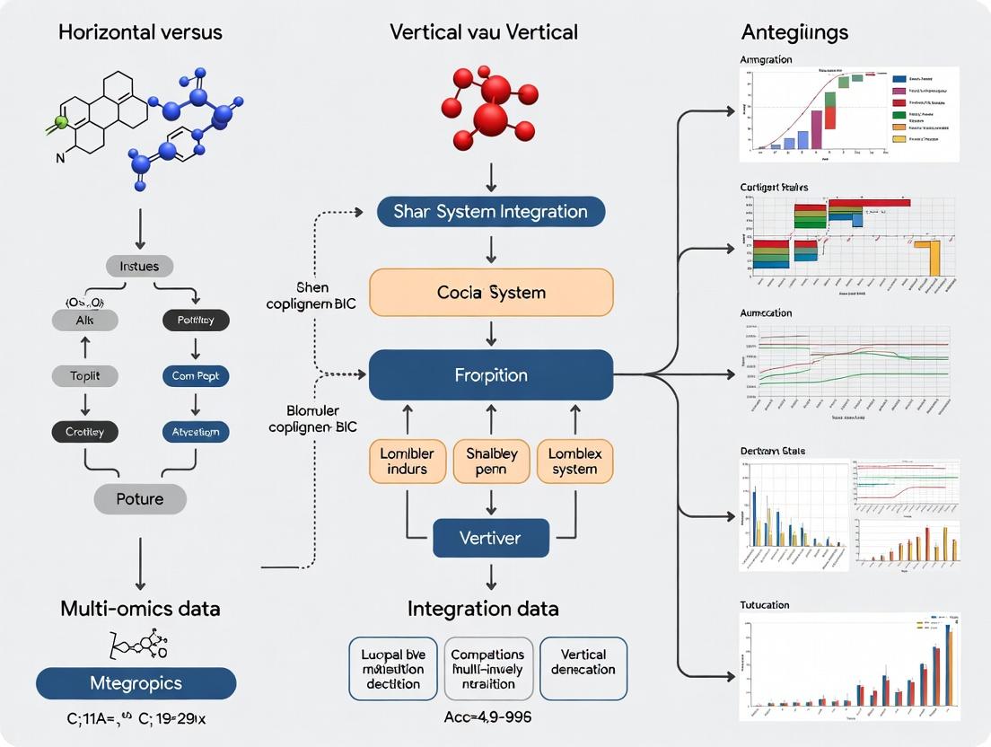
This article provides a comprehensive guide for researchers and drug development professionals navigating the two primary paradigms of multi-omics data integration.
Horizontal vs Vertical Multi-Omics Integration: Choosing the Right Strategy for Precision Medicine Research
Abstract
This article provides a comprehensive guide for researchers and drug development professionals navigating the two primary paradigms of multi-omics data integration. We explore the foundational concepts of horizontal (sample-matched) and vertical (feature-matched) integration, detail state-of-the-art methodologies and their applications in biomarker discovery and disease subtyping, address common computational and biological challenges, and offer a comparative validation framework. The goal is to empower scientists to select and optimize the appropriate integration strategy for robust, translatable biological insights in biomedical research.
Demystifying Multi-Omics Integration: A Primer on Horizontal and Vertical Strategies
Application Notes: Paradigm Definitions in Multi-Omics
Multi-omics integration is a cornerstone of systems biology, aiming to construct a comprehensive view of biological systems. Two principal paradigms govern the approach: Horizontal and Vertical Integration.
Horizontal Integration (HI): Also called "data-level" or "meta-omics" integration, HI involves the simultaneous analysis of multiple different omics datasets (e.g., genomics, transcriptomics, proteomics, metabolomics) acquired from the same set of biological samples. The goal is to identify correlated patterns and interactions across different molecular layers within a defined cohort, building network-level understanding.
Vertical Integration (VI): Also termed "feature-level" or "multi-scale" integration, VI focuses on tracing a biological signal or relationship (e.g., a genetic variant's effect) across multiple molecular levels for the same biological entity (e.g., a single gene or pathway). It connects causal chains from one molecular layer to the next (e.g., SNP → Gene Expression → Protein Abundance → Metabolite Level).
Quantitative Comparison of Integration Paradigms
Table 1: Core Characteristics of Horizontal vs. Vertical Integration
| Aspect | Horizontal Integration | Vertical Integration |
|---|---|---|
| Primary Goal | Discover coordinated patterns & networks across omics layers. | Establish causal, mechanistic flows across omics layers. |
| Sample Relationship | Multiple omics measured in the same cohort of samples. | Relationships traced for specific features across linked assays. |
| Temporal Dimension | Often cross-sectional (single time point). | Can incorporate longitudinal or perturbation time-series data. |
| Typical Methods | Multivariate statistics, similarity-based fusion, graph networks. | Bayesian networks, structural equation modelling, mechanistic models. |
| Key Challenge | Technical noise/batch effects alignment, heterogeneous data scales. | Requiring a priori biological knowledge or linkage models. |
| Primary Output | Molecular subtypes, predictive biomarkers, inter-omics networks. | Mechanistic hypotheses, driver identification, pathway causality. |
Table 2: Common Computational Tools & Their Applications (2024)
| Tool/Package | Primary Paradigm | Key Function | Language |
|---|---|---|---|
| MOFA+ | Horizontal | Factor analysis for multi-view data. | R/Python |
| mixOmics | Horizontal | Multivariate exploration & integration. | R |
| DIABLO | Horizontal | Multi-omics data integration for classification. | R |
| MONGREL | Vertical | Multi-omics hierarchical regression for causal inference. | R/Stan |
| Multi-Omic | Both | Bayesian network learning across omics. | Python |
| Graphical Model | |||
| CausalPath | Vertical | Infer causal signaling from phosphoproteomics & other data. | Web/Java |
Experimental Protocols
Protocol for a Horizontally Integrated Multi-Omics Cohort Study
Objective: To identify molecular subtypes of a disease (e.g., breast cancer) by integrating genomic, transcriptomic, and metabolomic data from the same patient tumor samples.
Workflow Summary:
- Sample Collection & Preparation: Collect tumor tissue biopsies from N=200 patients under standardized SOPs. Aliquot tissue for DNA, RNA, and metabolite extraction.
- Multi-Omic Data Generation:
- Genomics (DNA): Perform Whole Exome Sequencing (WES) to identify somatic mutations and copy number variations. Use a platform like Illumina NovaSeq. Process with GATK best practices.
- Transcriptomics (RNA): Perform RNA-Seq (poly-A selected) on the same samples. Use Illumina platform. Align to reference genome (STAR) and quantify gene-level counts (featureCounts).
- Metabolomics: Perform untargeted Liquid Chromatography-Mass Spectrometry (LC-MS) on tissue lysates. Use both positive and negative ionization modes.
- Data Preprocessing & Normalization:
- Genomics: Create a binary mutation matrix (1/0 for presence/absence of non-silent mutations in driver genes) and a segmented copy number matrix.
- Transcriptomics: TMM normalization, log2-CPM transformation, and batch correction (e.g., using ComBat).
- Metabolomics: Peak alignment, missing value imputation (minimum value), log-transformation, and Pareto scaling.
- Horizontal Integration Analysis: Use the MOFA+ framework.
- Input: Genomic matrix (mutations), Transcriptomic matrix (log-CPM), Metabolomic matrix (scaled intensities).
- Train the MOFA model to decompose variation into a set of common latent factors.
- Cluster patients based on their factor values to define molecular subtypes.
- Interpret factors by identifying heavily weighted features (e.g., Factor 1 driven by TP53 mutations, immune gene expression, and lactate levels).
Title: Workflow for Horizontal Multi-Omics Integration
Protocol for a Vertical Integration Study on a Genetic Perturbation
Objective: To mechanistically trace the effects of a specific gene knockout (e.g., MYC) across the transcriptome, proteome, and phosphoproteome in a cell line model.
Workflow Summary:
- Perturbation & Experimental Design: Generate isogenic MYC knockout (KO) and wild-type (WT) control cell lines using CRISPR-Cas9. Culture biological replicates (n=6) for each condition.
- Multi-Layer Profiling from Same Culture:
- Transcriptome: Harvest cells for total RNA extraction. Perform RNA-Seq (poly-A). Library prep with kits like Illumina TruSeq Stranded mRNA.
- Proteome & Phosphoproteome: From the same cell pellet, lyse cells. Digest lysates with trypsin. Perform Tandem Mass Tag (TMT) labeling for multiplexing.
- Global Proteome: Fractionate one aliquot of labeled peptides by high-pH reverse-phase HPLC and analyze by LC-MS/MS.
- Phosphoproteome: Enrich phosphorylated peptides from another aliquot using TiO2 or Fe-IMAC beads, then fractionate and analyze by LC-MS/MS.
- Data Processing:
- RNA-Seq: Differential expression analysis (e.g., DESeq2). Output: Log2 fold changes (KO vs WT) for genes.
- Proteomics: MS data processed with MaxQuant/SearchGUI. Differential abundance tested (e.g., Limma). Output: Log2 fold changes for proteins and phospho-sites.
- Vertical Integration Analysis: Construct a Bayesian Network or use CausalPath.
- Map features: MYC gene → MYC transcript → MYC protein.
- Input differential data into CausalPath with a prior knowledge network (e.g., SIGNOR, KEGG). The tool statistically tests for consistent downstream effects.
- Output: A validated cascade showing MYC KO leading to reduced E2F transcript targets, subsequently altering cell cycle protein abundance, and finally changing phosphorylation of key CDK substrates.
Title: Vertical Integration Tracing a Perturbation
The Scientist's Toolkit: Research Reagent Solutions
Table 3: Essential Reagents & Kits for Multi-Omics Integration Studies
| Item | Function & Application |
|---|---|
| AllPrep DNA/RNA/Protein Mini Kit (Qiagen) | Simultaneous co-isolation of genomic DNA, total RNA, and protein from a single tissue or cell sample. Critical for minimizing sample variance in HI studies. |
| Tandem Mass Tag (TMT) 16/18-plex (Thermo Fisher) | Isobaric labeling reagents for multiplexed quantitative proteomics. Allows combined analysis of up to 18 samples in one MS run, enabling robust VI across conditions with high precision. |
| TruSeq Stranded mRNA Library Prep Kit (Illumina) | Standardized library preparation for RNA-Seq. Ensures high-quality transcriptomic data, a foundational layer for both HI and VI. |
| KAPA HyperPrep Kit (Roche) | Flexible library prep for WES/WGS. Provides uniform coverage for genomic variant detection, a key input for integration. |
| TiO2 Mag Sepharose (Cytiva) or Fe-IMAC Beads | Magnetic beads for highly efficient phosphopeptide enrichment. Enables deep phosphoproteome coverage for vertical signaling studies. |
| Seahorse XFp / XFe96 Analyzer (Agilent) | Measures cellular metabolic fluxes (OCR, ECAR) in live cells. Functional metabolomic data for validating/grounding integrated molecular findings. |
| Single-Cell Multiome ATAC + Gene Exp. (10x Genomics) | Emerging technology allowing simultaneous assay of chromatin accessibility (ATAC) and gene expression in single nuclei. Represents the next frontier in horizontal integration. |
The integrative analysis of multi-omics data is a cornerstone of modern systems biology, pivotal for unraveling complex biological mechanisms in disease and therapeutics. The prevailing strategies are categorized as horizontal (sample-matched) and vertical (feature-matched) integration. Horizontal integration correlates multiple omics layers (e.g., transcriptomics, proteomics, metabolomics) across a common set of biological samples. Vertical integration connects different molecular layers along the central dogma (e.g., genomic variant to gene expression to protein abundance) for shared biological features or genes across potentially different sample cohorts. This application note details the experimental design, protocols, and analytical considerations for generating and utilizing these two distinct data structures.
Comparative Framework: Definitions and Applications
Table 1: Core Characteristics of Sample-Matched vs. Feature-Matched Designs
| Characteristic | Sample-Matched (Horizontal) Integration | Feature-Matched (Vertical) Integration |
|---|---|---|
| Primary Aim | Understand coordinated multi-layer changes across a cohort (e.g., patient stratification). | Establish causal or regulatory chains from genome to phenome for specific genes/pathways. |
| Sample Requirement | Identical samples subjected to multiple omics assays. | Samples can differ but must share relevant features (e.g., specific genetic variants). |
| Typical Data Structure | Multi-assay matrix: Samples (rows) x Multi-omics features (columns). | Linked datasets via feature anchors (e.g., gene ID, genomic coordinate). |
| Key Analytical Challenge | Batch effect correction across assay platforms, data scaling. | Harmonizing annotations, resolving context-specific (e.g., tissue) discordance. |
| Primary Application | Biomarker discovery, molecular subtyping, phenotypic prediction. | Mechanistic disease modeling, understanding GWAS hits, identifying drug targets. |
| Common Tools | MOFA+, DIABLO, mixOmics, Integrative NMF. | Multi-omics QTL mapping, PRIORitizE, NetWAS, linear mixed models. |
Experimental Protocols
Protocol 3.1: Generating a Sample-Matched Multi-Omics Dataset from Tumor Tissue
Objective: To extract DNA, RNA, and protein from the same tumor tissue sample for genomic, transcriptomic, and proteomic profiling.
Materials:
- Fresh-frozen or optimally preserved tissue (e.g., RNAlater).
- AllPrep DNA/RNA/Protein Mini Kit (Qiagen).
- RNeasy MinElute Cleanup Kit (Qiagen).
- BCA Protein Assay Kit.
- DNase I.
- Platform-specific library prep kits (e.g., Illumina for WES/RNA-seq).
Procedure:
Tissue Homogenization:
- Weigh 10-30 mg of tissue. Place in a tube with lysis buffer and homogenize using a rotor-stator homogenizer. Keep lysate cool.
Simultaneous Extraction (AllPrep):
- Follow manufacturer's protocol. Lysate is loaded onto an AllPrep DNA spin column. Flow-through (contains RNA and protein) is saved.
- DNA Column: Wash and elute genomic DNA. Proceed to Whole Exome Sequencing (WES) library prep.
- RNA from Flow-Through: Add ethanol to flow-through, apply to RNeasy column. Perform on-column DNase I digestion. Wash and elute RNA. Assess integrity (RIN > 7). Proceed to RNA-seq library prep.
- Protein from Flow-Through: Precipitate protein from the RNA extraction flow-through using acetone. Resuspend pellet. Quantify via BCA assay. Proceed to proteomic preparation (e.g., tryptic digestion for LC-MS/MS).
Quality Control & Sequencing/Mass Spec:
- QC DNA (Fragment Analyzer), RNA (Bioanalyzer), and protein yield.
- Perform WES (150bp paired-end), RNA-seq (e.g., 100M reads), and LC-MS/MS (e.g., TMT-labeled, data-dependent acquisition) using platform-standard protocols.
Protocol 3.2: Establishing a Feature-Matched Linkage from GWAS to Proteomics
Objective: To validate and characterize the functional protein-level consequences of a genetic variant identified in a Genome-Wide Association Study (GWAS).
Materials:
- GWAS summary statistics for a trait of interest.
- Genotyping data (e.g., SNP array) from a cohort with plasma proteomic data (e.g., SomaScan or Olink).
- PLINK software.
- R/Bioconductor with
coloc,MendelianRandomizationpackages.
Procedure:
Variant Selection and Cohort Identification:
- Identify lead SNP from GWAS (p < 5e-8). Define its linkage disequilibrium (LD) block using reference panels (e.g., 1000 Genomes).
- Identify an independent cohort where subjects have been genotyped (for the same SNP/region) and have quantified plasma protein levels (feature match = genomic region & gene).
Proteomic Quantitative Trait Locus (pQTL) Mapping:
- For each protein quantified in the proteomic platform, perform linear regression of protein abundance (normalized, log-transformed) on the SNP genotype (additive model), adjusting for covariates (age, sex, principal components).
- pQTL is significant if p < (0.05 / number of tested proteins in the platform).
Colocalization Analysis:
- Use
colocin R to assess if the GWAS signal and the pQTL signal share a common causal variant. A high posterior probability (PP.H4 > 0.8) suggests colocalization.
- Use
Mendelian Randomization (MR):
- If a significant cis-pQTL is found and colocalizes, use the SNP as an instrumental variable in MR to test if the protein has a causal effect on the GWAS trait. Use TwoSampleMR or MR-Base.
Visualizing Integration Strategies and Workflows
Sample-Matched (Horizontal) Integration Workflow
Feature-Matched (Vertical) Integration Logic
The Scientist's Toolkit: Key Research Reagents & Platforms
Table 2: Essential Solutions for Multi-Omics Sample Processing
| Item | Function | Example Product/Brand |
|---|---|---|
| All-in-One Nucleic Acid/Protein Kits | Co-extraction of DNA, RNA, and protein from a single tissue lysate, preserving sample integrity. | Qiagen AllPrep, Norgen's All-in-One Purification Kit. |
| Single-Cell Multi-Omic Kits | Enable simultaneous profiling of transcriptome and epigenome from the same single cell. | 10x Genomics Multiome ATAC + Gene Expression, Parse Biosciences Single Cell Multiome. |
| High-Multiplex Immunoassays | Quantify 1000s of proteins from minute sample volumes for large cohort proteomics. | SomaScan (Somalogic), Olink Explore, Proximity Extension Assay. |
| Isobaric Mass Tag Kits | Multiplex samples for quantitative proteomics, increasing throughput and reducing batch effects. | TMT (Thermo Fisher), iTRAQ (AB Sciex). |
| Spatial Multi-omics Platforms | Map transcriptomic and proteomic data within tissue architecture from the same section. | 10x Visium, Nanostring GeoMx DSP, Akoya CODEX. |
| Cell-Free DNA/RNA Collection Tubes | Stabilize blood samples for downstream plasma-based genomic and transcriptomic assays. | Streck cfDNA BCT, PAXgene Blood ccfDNA Tube. |
Within the broader thesis on horizontal versus vertical multi-omics data integration, the choice of approach is a fundamental strategic decision. This document provides application notes and experimental protocols to guide researchers in selecting and implementing the appropriate methodology.
- Horizontal (Patient-Centric) Integration: Integrates multiple omics layers (e.g., genomics, transcriptomics, proteomics) across a cohort of patients or samples. The primary axis of integration is the biological subject, aiming to build a comprehensive, cross-omic profile for each individual to stratify populations, identify biomarkers, or understand inter-individual variability.
- Vertical (Pathway-Centric) Integration: Integrates multiple omics layers within a specific biological pathway, process, or system. The primary axis of integration is the biological mechanism, aiming to reconstruct detailed, causal flow of information (e.g., from genetic variant to mRNA to protein to metabolite) for a defined pathway.
Decision Framework: When to Use Each Approach
The following table summarizes the key objectives, applications, and data requirements that dictate the choice of approach.
Table 1: Decision Framework for Horizontal vs. Vertical Integration
| Aspect | Horizontal (Patient-Centric) Approach | Vertical (Pathway-Centric) Approach |
|---|---|---|
| Primary Objective | Identify patient subtypes, predictive/prognostic biomarkers, or comprehensive molecular signatures correlated with phenotype. | Elucidate mechanistic drivers, causal relationships, and regulatory dynamics within a specific biological system. |
| Core Question | "What are the multi-omic differences between patient groups A and B?" | "How does a genetic perturbation in Pathway X alter the transcriptome, proteome, and metabolome downstream?" |
| Ideal Use Case | Cohort studies (e.g., TCGA, clinical trials), population health, precision oncology, complex disease stratification. | Functional validation studies, pathway pharmacology, toxicology, understanding drug mechanism of action (MoA). |
| Typical Study Design | Many subjects/samples (n > 100), fewer omics layers (2-3), matched samples per subject. | Fewer experimental units (n < 20), deeper omics layers (3+), often with controlled perturbations (e.g., knock-out, inhibition). |
| Data Structure | Wide: Samples as rows, multi-omic features (e.g., mutations, genes, proteins) as columns. | Deep: Features linked to a pathway as rows, multi-omic measurements across conditions/time as columns. |
| Key Analytical Methods | Multi-omic clustering, supervised classification, multivariate regression, network-based stratification. | Pathway enrichment, multi-omic Bayesian networks, time-series integration, kinetic modeling. |
| Main Output | Patient clusters, multi-omic signatures, biomarker panels for diagnosis/stratification. | Annotated pathway maps with multi-omic measurements, predictive models of pathway flux. |
Application Notes & Experimental Protocols
Protocol 3.1: Implementing a Horizontal (Patient-Centric) Study
Objective: To identify multi-omic subtypes of a disease (e.g., breast cancer) from a cohort of patient tumors.
Workflow Summary: Sample Collection → Multi-omic Data Generation → Data Alignment & Preprocessing → Horizontal Integration & Clustering → Subtype Characterization & Validation.
Title: Horizontal Integration Workflow for Patient Stratification
Detailed Protocol Steps:
- Cohort & Sample Selection: Select a well-annotated cohort (e.g., n=200 patients) with matched tumor tissue. Ensure appropriate IRB approval and informed consent.
- Multi-Omic Data Generation:
- Genomics (WES): Extract tumor and matched germline DNA. Perform exome capture and sequencing on an Illumina platform (150bp paired-end, >100x coverage). Call variants (SNVs, Indels) using GATK best practices.
- Transcriptomics (RNA-Seq): Extract total RNA, assess RIN >7. Prepare stranded mRNA-seq libraries. Sequence to a depth of ~50 million reads per sample. Align to reference genome (STAR) and quantify gene expression (featureCounts).
- Proteomics (LC-MS/MS): Perform tissue lysis, protein digestion (trypsin), and peptide cleanup. Use TMTpro 16-plex labeling for multiplexing. Fractionate by high-pH reverse-phase HPLC. Analyze fractions by LC-MS/MS on an Orbitrap Eclipse. Identify and quantify proteins using SequestHT in Proteome Discoverer.
- Data Preprocessing: For each omics layer, perform quality control, batch correction (ComBat), normalization (e.g., VSN for proteomics, TMM for RNA-Seq), and feature reduction (e.g., remove low-variance genes).
- Horizontal Integration: Use the patient ID as the primary key. Align data into a list of matrices where each matrix
[i]is an omics dataset withmpatients (rows) andn_ifeatures (columns). All matrices share the same row order (patients). - Clustering Analysis: Apply the Similarity Network Fusion (SNF) algorithm.
- Calculate patient similarity matrices
W^(i)for each omic using Euclidean distance and a patient similarity kernel. - Fuse all
W^(i)into a single integrated patient networkW. - Apply spectral clustering on
Wto obtain patient cluster assignments (k=3-5).
- Calculate patient similarity matrices
- Subtype Characterization: Perform differential analysis (DESeq2 for RNA-Seq, limma for proteomics) between clusters. Conduct pathway enrichment (GSEA, MsigDB) on differential features. Correlate clusters with clinical outcomes (survival analysis).
The Scientist's Toolkit: Key Reagents for Protocol 3.1
| Item | Function | Example (Vendor) |
|---|---|---|
| Allprep DNA/RNA/miRNA Kit | Simultaneous purification of genomic DNA and total RNA from a single tissue sample, ensuring matched multi-omic material. | Qiagen #80204 |
| TMTpro 16plex Label Reagent Set | Isobaric chemical tags for multiplexing up to 16 samples in a single LC-MS/MS run, reducing quantitative variability. | Thermo Fisher Scientific #A44520 |
| TruSeq DNA Exome & Stranded mRNA Prep Kits | Standardized library preparation kits for WES and RNA-Seq, ensuring reproducibility across large cohorts. | Illumina #20020616 / #20020595 |
| Sera-Mag Magnetic Beads | For PCR cleanup and library size selection; critical for efficient NGS library preparation. | Cytiva #29343052 |
| Trypsin, Sequencing Grade | High-purity protease for consistent and complete protein digestion prior to MS analysis. | Promega #V5111 |
Protocol 3.2: Implementing a Vertical (Pathway-Centric) Study
Objective: To delineate the multi-omic impact of inhibiting the MAPK/ERK signaling pathway in a cancer cell line model.
Workflow Summary: Pathway Selection & Perturbation → Multi-Omic Time-Course → Vertical Data Alignment → Causal Network Inference → Mechanistic Model.
Title: Vertical Integration Workflow for Pathway Analysis
Detailed Protocol Steps:
- Pathway Definition & Perturbation: Select a well-annotated pathway (e.g., KEGG MAPK signaling). Treat a sensitive cell line (e.g., A375 melanoma) with a specific MEK inhibitor (e.g., Trametinib, 100 nM). Include a DMSO vehicle control.
- Multi-Omic Time-Course Sampling: Harvest cells at multiple time points post-treatment (e.g., 0, 15min, 1h, 6h, 24h) in biological triplicate for each omic.
- Multi-Omic Data Generation:
- Phospho-Proteomics: Lyse cells, digest proteins, enrich phosphopeptides using Fe-NTA or TiO2 magnetic beads. Analyze by LC-MS/MS (Orbitrap). Quantify phosphosite levels (MaxQuant).
- Transcriptomics: Extract RNA and prepare sequencing libraries as in Protocol 3.1.
- Metabolomics: Perform methanol extraction of polar metabolites. Analyze by Hydrophilic Interaction Liquid Chromatography (HILIC) coupled to a high-resolution mass spectrometer (e.g., Q Exactive HF). Process with XCMS.
- Vertical Data Alignment: Map all measured entities (phosphosites, transcripts, metabolites) to the KEGG MAPK pathway map (hsa04010). Create a unified data table where rows are pathway components (e.g., "MAPK1", "ELK1") and columns are multi-omic measurements across time points and conditions.
- Causal Network Inference: Construct a Dynamic Bayesian Network (DBN) using the time-series data.
- Discretize the continuous multi-omic data.
- Use the bnlearn R package (
structure learningwithdtabcalgorithm) to infer probabilistic relationships between entities across time lags, constrained by prior pathway knowledge. - This infers directional edges (e.g., "Phospho-ERK at t-1 → FOS mRNA at t").
- Model Building & Validation: Integrate DBN output with literature knowledge to refine a mechanistic model. Validate predictions (e.g., of a key downstream transcription factor) using orthogonal methods like ChIP-qPCR or a CRISPRi knockdown experiment.
The Scientist's Toolkit: Key Reagents for Protocol 3.2
| Item | Function | Example (Vendor) |
|---|---|---|
| Phosphoprotein Enrichment Kits (Fe-NTA/TiO2) | Selective enrichment of phosphopeptides from complex digests, essential for phosphoproteomics. | Thermo Fisher Scientific #88807 / GL Sciences #5010-21309 |
| Trametinib (MEK Inhibitor) | High-potency, selective tool compound for perturbing the MAPK/ERK pathway. | Selleckchem #S2673 |
| HILIC Chromatography Columns | Stationary phase for separating polar metabolites in LC-MS based metabolomics. | Waters #186004742 |
| KAPA mRNA HyperPrep Kit | Efficient, rapid library prep from low RNA inputs, suitable for time-course experiments. | Roche #08098140702 |
| MetaXpress Software | For high-content image analysis if pathway validation includes immunofluorescence assays. | Molecular Devices |
Application Notes: Horizontal vs. Vertical Integration in Disease Research
In the thesis comparing horizontal (multi-assay across a cohort) and vertical (deep, multi-layered on a single sample) multi-omics integration, the choice of strategy is fundamentally dictated by the biological question. This note details their application across three therapeutic areas.
Table 1: Integration Strategy Selection Based on Research Question
| Disease Area | Exemplary Research Question | Optimal Integration Strategy | Primary Rationale & Data Types |
|---|---|---|---|
| Oncology | Identifying robust molecular subtypes and prognostic biomarkers across a heterogeneous patient population. | Horizontal Integration | Enables discovery of consensus patterns (e.g., immune-hot vs. -cold tumors) by clustering across many patients. Data: Bulk RNA-seq, DNA methylation, somatic mutations from TCGA/ICGC cohorts. |
| Oncology | Unraveling the complete mechanism of action of a targeted therapy in a specific in vitro model. | Vertical Integration | Connects the drug's primary target to downstream functional effects within the same biological system. Data: Proteomics (target engagement), phospho-proteomics (signaling), RNA-seq (transcriptional response). |
| Neurology | Discovering peripheral biomarkers (e.g., in blood) for central nervous system pathology in Alzheimer's disease. | Horizontal Integration | Correlates diverse molecular features from an accessible tissue (blood) with clinical imaging/outcomes across a cohort. Data: Plasma proteomics, metabolomics, miRNA-seq from longitudinal studies like ADNI. |
| Neurology | Modeling the cell-type-specific dysregulation in a post-mortem brain sample from a Parkinson's disease patient. | Vertical Integration | Builds a causal, layer-by-layer understanding within a single, critically relevant tissue sample. Data: snRNA-seq (cell type), paired snATAC-seq (chromatin accessibility), and spatial transcriptomics from adjacent section. |
| Complex Diseases (e.g., RA, IBD) | Stratifying patients into endotypes for targeted clinical trial recruitment. | Horizontal Integration | Identifies clusters of patients sharing multi-omics profiles, predicting drug response. Data: Serum metabolomics, synovial tissue RNA-seq, immunophenotyping from trial baseline data. |
| Complex Diseases | Deconstructing the tumor-immune-stroma interactome in a single rheumatoid arthritis synovial biopsy. | Vertical Integration | Maps the local cellular crosstalk and signaling networks driving inflammation in a specific tissue microenvironment. Data: CITE-seq (transcriptome + surface proteins), secretome analysis from the same biopsy culture. |
Detailed Experimental Protocols
Protocol 1: Horizontal Integration for Oncology Subtyping Objective: To identify integrative molecular subtypes in breast cancer using public cohort data.
- Data Acquisition: Download matched tumor data (RNA-seq, DNA methylation 450k array, somatic copy number alterations) for ~1000 samples from The Cancer Genome Atlas (TCGA) BRCA cohort via the Genomic Data Commons (GDC) API.
- Preprocessing & Dimensionality Reduction:
- RNA-seq: Fragments Per Kilobase Million (FPKM) normalization, log2 transformation. Perform principal component analysis (PCA), retain top 50 PCs.
- Methylation: M-value calculation from beta values, ComBat batch correction. Retain top 50 PCs.
- CNAs: Segment log2 ratios, summarize to gene-level GISTIC scores. Retain top 50 PCs.
- Multi-Omics Clustering: Use the
MoClustermethod (from themovicsR package) on the concatenated PCA matrices (150 features total). Apply non-negative matrix factorization (NMF) to define clusters (k=2-10). Select optimal k via consensus clustering metrics (cophenetic correlation, silhouette width). - Subtype Characterization: For each cluster, perform differential analysis per platform. Annotate subtypes using known pathways (e.g., PI3K/AKT, immune response), PAM50 classification, and survival analysis (Kaplan-Meier, log-rank test).
Protocol 2: Vertical Integration for Drug Mechanism of Action Objective: To delineate the signaling cascade induced by a KRAS G12C inhibitor in a lung adenocarcinoma cell line.
- Cell Treatment & Lysis: Culture NCI-H358 cells. Treat with 1 µM MRTX849 (adagrasib) or DMSO for 1, 6, and 24 hours (n=4). Wash with PBS and lyse cells in a denaturing buffer.
- Multi-Layer Protein Extraction:
- Phospho-Proteomics: Enrich phosphopeptides from 1mg of total protein lysate using Fe-NTA magnetic beads. Desalt and dry.
- Global Proteomics: Use the flow-through from phospho-enrichment, followed by clean-up.
- LC-MS/MS Analysis: Reconstitute peptides and analyze on a timsTOF Pro with a NanoElute UHPLC. Use PASEF method. Libraries for DIA-NN are generated from parallel deep DDA runs.
- Vertical Data Integration:
- Kinase-Substrate Mapping: Input significantly changing phosphosites (p<0.01, |log2FC|>1) into the Kinase-Substrate Enrichment Analysis (KSEA) tool. Identify activated/inhibited upstream kinases (e.g., ERK1/2, SHP2).
- Pathway Linking: Integrate KSEA results with significant changes from global proteomics (e.g., upregulation of DUSP6, downregulation of Cyclin D1) using causal network tools (CausalPath). Validate top predictions (e.g., p-ERK/ERK ratio) by western blot.
Mandatory Visualizations
Diagram 1: Horizontal vs Vertical Integration Workflow
Diagram 2: Vertical MoA Analysis for KRASi
The Scientist's Toolkit: Research Reagent Solutions
Table 2: Essential Reagents for Featured Multi-Omics Protocols
| Item | Function in Protocol | Example Product/Catalog |
|---|---|---|
| Phosphopeptide Enrichment Beads | Selective isolation of phosphorylated peptides from complex digests for phosphoproteomics. | Fe-NTA Magnetic Agarose Beads (Thermo Fisher, 78601) |
| CITE-seq Antibody Conjugation Kit | Enables labeling of antibodies with oligonucleotide barcodes for simultaneous protein and RNA detection at single-cell level. | TotalSeq-C Antibody Labeling Kit (BioLegend, 688102) |
| Single-Nucleus ATAC-seq Kit | Provides reagents for nuclei isolation, transposition, and library prep for chromatin accessibility profiling. | Chromium Next GEM Single Cell ATAC Kit (10x Genomics, 1000175) |
| DIA-MS Spectral Library Kit | Contains standardized HeLa digests for generating comprehensive spectral libraries for Data-Independent Acquisition proteomics. | Pierce HeLa Protein Digest Standard (Thermo Fisher, PCO001) |
| Multi-Omics Integration Software | Platform for performing horizontal (NMF, iCluster) and vertical (causal inference) integration analyses. | Movics R Package; CausalPath Web Tool |
| Cohort Data Portal Access | Source for matched, clinically annotated multi-omics data from large patient cohorts (e.g., TCGA, ADNI). | GDC Data Portal; ADNI LONI Image & Data Archive |
Within the ongoing research discourse comparing horizontal (across samples) and vertical (across molecular layers within a single sample) multi-omics integration, the foundational step is the rigorous definition and preparation of input data. The choice of integration approach is fundamentally constrained by the nature, scale, and quality of the omics data types available. This application note delineates the essential prerequisites for each paradigm, providing protocols for initial data assessment and curation to ensure robust downstream integration and biological inference.
The following tables summarize the core data requirements for horizontal and vertical multi-omics integration strategies.
Table 1: Data Type Suitability and Scale
| Omics Data Type | Typical Assay | Horizontal Integration (Across Samples) | Vertical Integration (Across Layers) |
|---|---|---|---|
| Genomics | Whole Genome Sequencing (WGS), SNP arrays | Essential. Requires consistent variant calling across a large cohort (n > 100s). | Foundation Layer. Serves as static reference for regulatory or functional variation. |
| Transcriptomics | RNA-Seq, Microarrays | Core Data. Expression matrices (genes x samples) for correlation/prediction. | Core Layer. Dynamic layer linking genotype to phenotype. Requires matched sample. |
| Epigenomics | ChIP-Seq, ATAC-Seq, Methylation arrays | Cohort-wide. Histone mark, accessibility, or methylation profiles across samples. | Regulatory Layer. Explains transcriptomic variation. Must be from same biological system. |
| Proteomics | Mass Spectrometry (LC-MS/MS), RPPA | Highly Valuable. Protein abundance or post-translational modification data. | Functional Effector Layer. Critical for mechanistic models. Matching is critical. |
| Metabolomics | LC/MS, GC/MS, NMR | Phenotypic Anchor. End-point small molecule profiles across cohorts. | Phenotypic Output Layer. Captures final biochemical activity. Technical variability is high. |
Table 2: Minimum Quality and Replication Requirements
| Prerequisite | Horizontal Integration | Vertical Integration |
|---|---|---|
| Sample Size | Large cohorts (100s-1000s) for statistical power. | Can be deep-dive on smaller N (e.g., 10-50), but requires perfect matching. |
| Sample Matching | Can be meta-analysis of disparate studies with batch correction. | Absolute Mandate. All omics layers must derive from the same biological specimen (or aliquots). |
| Data Completeness | Tolerates missing data per layer if sample N is large. | Missing data in any layer for a sample can severely compromise the integrated model. |
| Technical Replication | Important for assessing assay robustness within cohort. | Crucial for verifying measurement accuracy within the same sample. |
| Minimum Sequencing Depth/ Coverage | RNA-Seq: >20M reads/sample; WGS: >30X; Proteomics: Depth to ID 5000+ proteins. | Often requires greater depth per sample to detect low-abundance, layer-crossing signals. |
| Key QC Metric | Batch effect assessment (PCA, surrogate variable analysis). | Pairwise correlation of measurements from the same sample across platforms (e.g., RNA-protein). |
Experimental Protocols for Foundational Data Generation
Protocol 1: Generating a Vertically Integrated Multi-omics Sample from Tissue
Objective: To obtain genomic, transcriptomic, proteomic, and metabolomic data from a single tissue specimen.
Materials:
- Fresh-frozen tissue sample (≥50 mg)
- AllPrep DNA/RNA/Protein Mini Kit (Qiagen)
- Methanol, acetonitrile, water (LC-MS grade)
- RIPA lysis buffer with protease/phosphatase inhibitors
- DNase I, RNase inhibitors
Procedure:
- Cryopulverization: Under liquid N2, pulverize frozen tissue using a mortar and pestle or cryomill. Keep powder frozen.
- Aliquoting: Rapidly weigh and aliquot powder for (a) DNA/RNA, (b) protein, (c) metabolomics.
- Nucleic Acid & Protein Co-extraction: Process aliquot (a) per AllPrep protocol. Elute DNA and RNA in separate tubes. Quantify via Qubit and Bioanalyzer/TapeStation.
- Protein Extraction: Homogenize aliquot (b) in RIPA buffer. Centrifuge at 14,000g, 20min, 4°C. Collect supernatant. Quantify via BCA assay. Aliquot for LC-MS/MS.
- Metabolite Extraction: To aliquot (c), add 500µL of 80% methanol (-80°C). Vortex, sonicate, incubate at -80°C for 1hr. Centrifuge at 21,000g, 15min, 4°C. Collect supernatant for LC-MS.
- Sequencing/Profiling: Process DNA for WGS, RNA for RNA-Seq (stranded, poly-A selected). Process protein extracts for tryptic digestion and LC-MS/MS. Analyze metabolites on HRAM LC-MS.
Protocol 2: Cohort-Level (Horizontal) Multi-omics Data Curation and QC
Objective: To aggregate and quality-control omics data from multiple public or in-house studies for horizontal integration.
Procedure:
- Metadata Harmonization: Map all sample metadata to a common ontology (e.g., NCBI Biosample attributes).
- Batch Detection: For each omics data type (e.g., gene expression matrix), perform PCA. Color-code by study of origin, sequencing batch, or processing date.
- Batch Correction (if needed): Apply a method like ComBat (parametric empirical Bayes) to adjust for non-biological technical variation, preserving biological signal.
- Missing Data Imputation: For proteomics or metabolomics data with missing values, use methods like k-nearest neighbors (KNN) or MissForest, only within assays, not across layers.
- Platform/Gene ID Alignment: Map all molecular features (genes, proteins, metabolites) to common identifiers (e.g., gene symbol, UniProt ID, InChIKey). Use resources like HGNC, UniProt, HMDB.
Visualizing Data Integration Workflows
Title: Horizontal integration workflow across a cohort.
Title: Vertical integration workflow for a single sample.
The Scientist's Toolkit: Key Research Reagent Solutions
| Item | Function in Multi-omics Research |
|---|---|
| AllPrep DNA/RNA/Protein Mini Kit (Qiagen) | Enables simultaneous isolation of genomic DNA, total RNA, and native protein from a single sample aliquot, crucial for vertical integration. |
| MTBE/Methanol/Water Solvent System | A robust metabolite extraction protocol for untargeted metabolomics, offering broad coverage of polar and non-polar metabolites. |
| TMTpro 16plex (Thermo Fisher) | Isobaric labeling reagents for high-throughput proteomics, allowing multiplexing of up to 16 samples in one LC-MS run, reducing batch effects in horizontal studies. |
| DNase I (RNase-free) | Essential for removing genomic DNA contamination during RNA extraction, ensuring pure RNA for transcriptomics. |
| Phase Lock Gel Tubes | Improve recovery and purity during phenol-chloroform extractions, commonly used in proteomics and metabolomics workflows. |
| ERCC RNA Spike-In Mix (Thermo Fisher) | Synthetic RNA controls added before RNA-Seq library preparation to monitor technical variability and enable normalization across horizontal study batches. |
| Pierce Quantitative Colorimetric Peptide Assay | Accurate quantification of peptide yield prior to LC-MS/MS, ensuring equal loading and improving quantitative reproducibility. |
| Sera-Mag Magnetic Beads (Cytiva) | Used for SPRI-based clean-up and size selection in NGS library prep, ensuring consistent yield and fragment size across genomics/transcriptomics samples. |
Tools & Techniques: Implementing Horizontal and Vertical Multi-Omics Integration in Practice
This protocol details a horizontal (sample-wise) multi-omics integration workflow, framed within a comparative thesis investigating horizontal versus vertical (feature-wise) data fusion strategies. Horizontal integration correlates the same set of samples across multiple omics layers (genomics, transcriptomics, proteomics), seeking a unified sample representation. This contrasts with vertical integration, which models biological relationships across different molecular levels for a given feature set. The presented workflow progresses from classical statistical learning (Multi-Kernel Learning) to modern deep learning architectures (Deep Neural Networks) for robust, non-linear sample fusion, enabling advanced biomarker discovery and patient stratification in translational research and drug development.
Core Methodological Frameworks
Multi-Kernel Learning (MKL) for Late Integration
MKL combines multiple kernel matrices, each representing similarity between samples for one omics data type, into an optimal composite kernel for downstream prediction (e.g., disease subtyping).
Key Equation: K_combined = ∑_{m=1}^{M} β_m K_m, where K_m is the kernel matrix for omics modality m, β_m is its learned weight (β_m ≥ 0, ∑β_m = 1), and M is the total number of omics types.
Table 1: Common Kernel Functions for Omics Data
| Kernel Type | Function | Best For | Key Parameter | ||||
|---|---|---|---|---|---|---|---|
| Linear | K(x_i, x_j) = x_i^T x_j |
Dense, normalized data (e.g., gene expression) | None | ||||
| Radial Basis Function (RBF) | `K(xi, xj) = exp(-γ | xi - xj | ^2)` | Capturing complex, non-linear similarities | γ (bandwidth) |
||
| Polynomial | K(x_i, x_j) = (x_i^T x_j + c)^d |
Modeling feature interactions | Degree (d), coefficient (c) |
Deep Neural Network (DNN) for Early Integration
DNNs enable early fusion by concatenating raw or reduced feature vectors from each omics type at the input layer, allowing high-level representations to be learned through non-linear transformations in hidden layers.
Table 2: Comparison of MKL vs. DNN Fusion Approaches
| Aspect | Multi-Kernel Learning (MKL) | Deep Neural Network (DNN) |
|---|---|---|
| Integration Stage | Late (kernel-level) | Early (input-level) or Intermediate |
| Interpretability | High (kernel weights β_m) |
Lower (black-box) |
| Data Requirements | Lower (works well with smaller N) | High (requires large N to avoid overfitting) |
| Handles Non-linearity | Yes (via kernel choice) | Excellently (via activation functions) |
| Feature Interaction | Limited to kernel definition | Complex, learned interactions across omics |
Experimental Protocols
Protocol 3.1: Standardized Multi-Omics Data Preprocessing for Horizontal Fusion
Goal: Prepare coherent sample-matched datasets from diverse omics sources.
Input: Raw data matrices (samples x features) for Genomics (e.g., SNPs), Transcriptomics (RNA-seq counts), Proteomics (Abundance).
Reagents/Software: R/Python, sva/ComBat (R), scikit-learn (Python).
Steps:
- Sample Alignment: Ensure a common set of
Nsamples exists across allMomics datasets. Log and document any exclusions. - Per-Omics Normalization:
- Genomics (SNPs): Standardize to mean=0, variance=1 per SNP.
- Transcriptomics: TMM normalization (edgeR) followed by log2(CPM+1) transformation.
- Proteomics: Quantile normalization and log2 transformation.
- Batch Effect Correction: Apply
ComBat(or similar) within each omics dataset to adjust for technical batches, using sample-wise omics data as the input matrix. - Feature Filtering: Retain top
kfeatures per omics layer based on variance or association with phenotype to reduce dimensionality (k=5000typical). - Output:
Mcleaned, sample-aligned, and scaled matricesX_m∈R^{N x k_m}.
Protocol 3.2: Implementating SimpleMKL for Classification
Goal: Fuse omics datasets to predict a binary clinical outcome.
Input: Preprocessed matrices X_1...X_M from Protocol 3.1, binary phenotype vector y ∈ {0,1}^N.
Reagents/Software: SimpleMKL toolbox, SHOGUN toolbox, or scikit-learn with custom MKL.
Steps:
- Kernel Computation: For each omics matrix
X_m, compute a kernel matrixK_mof sizeN x N. For continuous data, an RBF kernel is recommended. Use cross-validation to tune itsγparameter. - Model Training: Implement the SimpleMKL algorithm:
a. Initialize all kernel weights
β_m = 1/M. b. Solve the standard SVM dual problem using the current combined kernelK_combined. c. Compute gradient of the SVM objective w.r.tβ_m. d. Updateβ_mvia reduced gradient descent, projecting onto the simplex (β_m ≥ 0, ∑β_m = 1). e. Iterate steps b-d until convergence of the objective. - Validation: Perform nested cross-validation. The outer loop splits data into train/test; the inner loop on the training set optimizes SVM
Cparameter, RBFγ, and finalβ_m. - Output: Optimal kernel weights
β_m, final classifier, and cross-validated performance metrics (AUC, Accuracy).
Protocol 3.3: Deep Neural Network for Early Integration & Classification
Goal: Use a feedforward DNN to integrate omics data at the input layer.
Input: Preprocessed matrices X_1...X_M from Protocol 3.1, phenotype vector y.
Reagents/Software: PyTorch or TensorFlow/Keras, scikit-learn, Hyperopt or Optuna for tuning.
Steps:
- Input Concatenation: For each sample
i, concatenate feature vectors from allMomics layers to create a unified input vector:z_i = concat(x_i^1, x_i^2, ..., x_i^M). - Network Architecture Design:
a. Input Layer: Size =
∑ k_m(total features from all omics). b. Hidden Layers: 2-4 fully connected (dense) layers with decreasing neurons (e.g., 1024 → 512 → 256). Use ReLU activation and Batch Normalization. c. Output Layer: Single neuron with sigmoid activation for binary classification. d. Regularization: Incorporate Dropout (rate=0.5) after each hidden layer and L2 weight regularization. - Model Training: Use Adam optimizer, Binary Cross-Entropy loss. Implement a
10%validation split for early stopping (patience=20 epochs). Train for a maximum of 200 epochs. - Hyperparameter Tuning: Use Bayesian optimization (Hyperopt) to search learning rate, dropout rate, L2 coefficient, and layer sizes.
- Output: Trained DNN model, test set performance metrics, and sample-level learned representations from the penultimate layer.
Visualizations
Title: Multi-Kernel Learning (MKL) Fusion Workflow
Title: Deep Neural Network for Early Omics Fusion
The Scientist's Toolkit
Table 3: Essential Research Reagent Solutions for Multi-Omics Fusion Studies
| Item/Category | Example Product/Software | Primary Function in Workflow |
|---|---|---|
| Batch Effect Correction | sva/ComBat (R), Harmony (R/Py) |
Removes non-biological technical variation within each omics dataset, critical for valid horizontal integration. |
| Kernel Computation Library | scikit-learn (Python), kernlab (R) |
Provides optimized functions to compute diverse kernel matrices (Linear, RBF, Polynomial) from feature matrices. |
| MKL Solver | SimpleMKL (MATLAB), SHOGUN (C++/Py) |
Implements optimization algorithm to learn optimal kernel weights (β_m) for combining omics-specific kernels. |
| Deep Learning Framework | PyTorch, TensorFlow with Keras |
Enables flexible design, training, and evaluation of DNN architectures for early integration of omics data. |
| Hyperparameter Optimization | Optuna, Hyperopt, Weights & Biases |
Automates the search for optimal model parameters (e.g., learning rate, network depth, dropout) for MKL/DNN. |
| Unified Data Structure | MultiAssayExperiment (R), MuData (Python) |
Provides a standardized container for sample-aligned multi-omics data, ensuring consistency across analysis steps. |
| Omics-Specific Normalization | edgeR/DESeq2 (RNA-seq), limma (Proteomics) |
Performs appropriate, statistically sound normalization for raw count or abundance data before integration. |
1. Introduction & Thesis Context Within the broader thesis contrasting horizontal (across cohorts) and vertical (across omics layers within the same sample) integration, this protocol details a robust vertical integration workflow. It enables the causal linking of multi-omic features from disparate molecular layers (e.g., genome, epigenome, transcriptome, proteome) derived from the same biological specimen, moving beyond correlation to infer regulatory mechanisms driving phenotype.
2. Overall Workflow Protocol
- Input: Matched multi-omics datasets (e.g., Whole Genome Sequencing (WGS)/Whole Exome Sequencing (WES), DNA methylation, RNA-Seq, Proteomics) from the same set of samples (N > 50 recommended).
- Stage 1: Unsupervised Vertical Integration & Dimensionality Reduction.
- Aim: Identify latent structures and sample clusters driven by coherent cross-omic patterns.
- Protocol: Multi-Omics Factor Analysis (MOFA+).
- Data Preparation: Format each omics dataset as a samples-by-features matrix. Perform omics-specific normalization (e.g., VST for RNA-Seq, beta-mixture quantile normalization for methylation). Handle missing data via MOFA+'s internal probabilistic framework.
- Model Training: Set the number of Factors (K) using automatic relevance determination or cross-validation. Train the model to decompose data into Factors (latent variables) and corresponding weights per omics view.
- Output Interpretation: Correlate Factors with sample covariates (e.g., disease status) to annotate. Analyze top-weighted features for each Factor in each omics layer to generate biological hypotheses.
- Stage 2: Supervised Vertical Linking for Candidate Driver Identification.
- Aim: Statistically link specific "driver" features from one layer (e.g., genetic variant, methylated CpG) to "target" features in a downstream layer (e.g., gene expression, protein abundance).
- Protocol: Sparse Multi-Block Partial Least Squares (sMBPLS) Regression.
- Block Definition: Define blocks: X1 (genetic variants in cis-regions), X2 (methylation in promoter/enhancers), Y (gene expression/protein levels of the target gene).
- Model Fitting: Use k-fold cross-validation to tune sparsity parameters (λ) for each block to select non-redundant, predictive features. Fit sMBPLS to extract latent components that maximally covary between combined X blocks and Y.
- Significance Testing: Perform permutation testing (≥1000 permutations) on the extracted component's covariance to calculate a p-value. Apply false discovery rate (FDR) correction across all tested gene loci.
- Stage 3: Mechanistic Validation & Network Construction.
- Aim: Integrate prior biological knowledge to construct testable pathway models from statistically linked features.
- Protocol: Knowledge-Primed Causal Network Mapping.
- Seed Network Generation: Use Stage 2 results (e.g., a significant SNP-CpG-Gene triplet) as seeds in a knowledge graph (e.g., STRING, KEGG, Reactome) via APIs.
- Contextual Pruning: Prune the extended network using tissue-specific interaction data (e.g., from GTEx) and chromatin interaction data (e.g., Hi-C) to retain spatially plausible edges.
- Hypothesis Output: The final sub-network proposes a mechanistic chain (e.g., SNP→TF binding site alteration→methylation change→expression change→protein activity change).
3. Data Tables
Table 1: Comparison of Vertical Integration Methods Applied in Workflow
| Method | Type | Primary Objective | Key Output | Software/Package |
|---|---|---|---|---|
| MOFA+ | Unsupervised | Dimensionality reduction; identify latent factors | Factors explaining variance across omics; sample clustering | R/Python MOFA2 |
| sMBPLS | Supervised | Predictive linking of blocks of features | Sparse model of cross-omic predictors for an outcome; p-values | R sgPLS |
| mixOmics | Both | Diverse DIABLO framework for classification | Integrated signature for sample discrimination | R mixOmics |
Table 2: Example Results from a sMBPLS Analysis Linking Genotype to Expression
| Target Gene (Y) | Top SNP Predictor (X1) | Beta (X1) | Top Methylation Predictor (X2) | Beta (X2) | Model p-value (FDR-corrected) | Explained Variance (R²Y) |
|---|---|---|---|---|---|---|
| EGFR | rs17337023 | -0.87 | cg02801887 | 0.42 | 2.1e-05 | 0.31 |
| TP53 | rs1042522 | 0.91 | cg11073992 | -0.38 | 4.7e-04 | 0.26 |
| VEGFA | rs699947 | 0.45 | cg16785077 | 0.51 | 1.3e-03 | 0.22 |
4. Visualization Diagrams
Diagram 1: Vertical vs Horizontal Integration Context
Diagram 2: Multi-Stage Vertical Integration Workflow
Diagram 3: sMBPLS Model for Cross-Omic Feature Linking
5. The Scientist's Toolkit: Research Reagent Solutions
| Item / Reagent | Function in Vertical Integration Workflow |
|---|---|
| PAXgene Tissue System | Stabilizes RNA, DNA, and proteins simultaneously from a single tissue biopsy, ensuring matched multi-omic input. |
| Single-Cell Multiome ATAC + Gene Expression Kit | Enables vertical integration at single-cell resolution by capturing chromatin accessibility and transcriptome from the same cell. |
| TMTpro 16plex Isobaric Label Reagents | Allows multiplexed quantitative proteomics of up to 16 samples, crucial for profiling matched sample cohorts cost-effectively. |
| CETSA & PTMscan Kits | Provide functional readouts (protein thermal stability, post-translational modifications) to validate proteomic predictions from upstream omics. |
| CRISPR Screening Libraries (e.g., Kinome) | Enable functional validation of predicted driver genes or regulatory elements identified in the integration workflow. |
| MOFA2 R/Bioconductor Package | Core tool for unsupervised factor analysis across heterogeneous omics data types. |
| Cytoscape with STRING/Reactome Apps | Platform for visualizing and enriching knowledge-primed causal networks from linked feature lists. |
Multi-omics integration strategies are fundamentally categorized as horizontal (integration across different omics layers from the same samples) or vertical (integration across different levels of biological information, from molecular to phenotypic, often for the same entity). This review critically assesses four prominent frameworks within this dichotomy, guiding researchers in tool selection for their specific integration paradigm.
Framework Comparison & Application Notes
Quantitative Comparison Table
| Framework | Primary Integration Type | Core Algorithm/Method | Key Output | Scalability (Samples/Features) | Language/Platform | Best For |
|---|---|---|---|---|---|---|
| MOFA+ | Horizontal | Statistical Bayesian Factor Analysis | Latent factors, feature weights | ~1,000s samples, 10,000s features | R/Python | Unsupervised discovery of shared & unique variation across omics. |
| mixOmics | Horizontal & Vertical | Projection-based (PCA, PLS, DIABLO) | Component plots, variable selection | ~100s samples, 1,000s features | R | Supervised & unsupervised integration with strong visualization. |
| netDx | Vertical | Patient similarity networks, machine learning | Diagnostic models, feature importance | ~1,000s samples, 10,000s+ features | R/BioConductor | Building interpretable predictive models from multi-modal data. |
| iCluster | Horizontal | Joint latent variable model (penalized regression) | Integrated clusters, subtype discovery | ~100s-1,000 samples, 10,000s features | R | Integrative clustering for discrete subgroup identification. |
Detailed Application Notes
MOFA+: A Bayesian framework for horizontal integration. It decomposes multi-omics data into a set of latent factors that capture the common and dataset-specific sources of variation. It is exceptionally robust to missing data and noise, making it ideal for large-scale cohort studies like TCGA. It does not directly incorporate phenotypic outcomes (vertical integration).
mixOmics: Provides a versatile suite for both horizontal (e.g., DIABLO for multi-omics classification) and vertical (e.g., PLS for linking omics to clinical traits) integration. Its strength lies in powerful visualizations (e.g., circos plots, relevance networks) to interpret complex associations.
netDx: A vertically-oriented framework that builds patient-specific similarity networks for each data type (e.g., mRNA, methylation, clinical) and integrates them to predict clinical outcomes. It generates highly interpretable models, showing which data types and features drive predictions.
iCluster: A horizontal integration tool specifically designed for integrative clustering. It uses a joint latent variable model with lasso-type penalties to identify coherent multi-omics subtypes, crucial for cancer classification and biomarker discovery.
Experimental Protocols
Protocol 1: Multi-omics Subtype Discovery using iCluster (Horizontal Integration)
Objective: Identify integrated molecular subtypes from mRNA expression, DNA methylation, and copy number variation data.
- Data Preprocessing: Independently normalize each omics dataset. Features are typically centered and scaled. Filter low-variance features.
- Data Formatting: Create a list object in R where each element is a sample-by-feature matrix for one omics type. Ensure identical sample ordering.
- Parameter Tuning: Use the
tune.iCluster()function to perform cross-validation and select the optimal lambda (penalty) parameters and number of latent components (K). - Model Fitting: Run the
iCluster()function with the optimal K and lambda values. - Cluster Assignment: Extract the cluster assignments for each sample from the fitted model.
- Validation: Assess cluster stability via bootstrapping. Perform survival analysis (Kaplan-Meier) or correlate with known clinical phenotypes to validate biological relevance.
Protocol 2: Building a Predictive Diagnostic Model with netDx (Vertical Integration)
Objective: Integrate gene expression, histopathology images, and clinical data to predict patient survival groups.
- Define Patient Similarity: For each data type, design a custom similarity metric. Examples:
- Gene Expression: 1 - Pearson correlation distance.
- Clinical Data: Normalized Euclidean distance.
- Build Similarity Networks: For each data type, create a patient similarity network (graph) where nodes are patients and edge weights are defined by the similarity metric.
- Feature Selection: Use a supervised approach (e.g., iterative feature pruning) to select features within each data type that best correlate with the outcome label.
- Integrated Model Training: Use the integrated network (combined from selected features across data types) within a machine learning classifier (e.g, support vector machine on graphs) to predict the outcome.
- Model Interpretation: Use pathway analysis on selected gene networks and examine weights from clinical data networks to interpret the model's decision logic.
Visualizations
Title: iCluster Workflow for Horizontal Integration
Title: Horizontal vs. Vertical Multi-omics Integration
The Scientist's Toolkit: Key Research Reagent Solutions
| Item | Function/Application in Multi-omics Integration |
|---|---|
| R/BioConductor | Primary computational environment for statistical analysis and execution of MOFA+, mixOmics, netDx, and iCluster. |
| Single-cell RNA-seq Kit (e.g., 10x Genomics) | Generates transcriptomic data for one omics layer, often integrated with surface protein (CITE-seq) or ATAC-seq data horizontally. |
| DNA Methylation Array (e.g., Illumina EPIC) | Provides genome-wide methylation profiles for integration with gene expression data to study regulatory mechanisms. |
| Proteomics Reagents (e.g., TMT Isobaric Labels) | Enable multiplexed quantitative proteomics, creating a protein abundance layer for integration with mRNA data. |
| High-Quality DNA/RNA Extraction Kits | Foundational step to ensure high-integrity, multi-omic data from the same biological sample (critical for horizontal integration). |
| Clinical Data Management System (CDMS) | Source of curated phenotypic and outcome data essential for vertical integration models (e.g., in netDx). |
Within the broader thesis comparing horizontal (multi-omics per sample) versus vertical (single-omics across large cohorts) data integration strategies for biomarker discovery, this protocol focuses on a hybrid approach. This method leverages vertical cohort-derived multi-omics features to build and validate horizontal, patient-specific composite signatures. The goal is to move beyond single-molecule biomarkers to robust, systems-level signatures that enhance diagnostic accuracy and prognostic prediction.
Core Experimental Protocol: A Hybrid Integration Workflow
Protocol 2.1: Multi-Omic Data Acquisition and Pre-processing
- Objective: To generate consistent, analysis-ready datasets from publicly available cohorts (vertical data).
- Steps:
- Cohort Selection: Identify relevant disease cohorts from repositories (TCGA, GEO, EGA). Prioritize studies with matched mRNA-seq, DNA methylation (e.g., Illumina EPIC array), and proteomic (e.g., RPPA or mass spectrometry) data.
- Data Download: Use genomic data portals (e.g., UCSC Xena, cBioPortal) or
GEOquery/SRAtoolkitin R/Bioconductor. - Uniform Pre-processing:
- RNA-seq: Align to reference genome (STAR), quantify transcripts (featureCounts), normalize (TPM, DESeq2's median of ratios).
- Methylation: Process IDAT files (
minfipackage), perform functional normalization, filter probes (detection p-value, SNPs, cross-reactive). Define beta-values. - Proteomics: Normalize to internal controls/median polish, log2-transform.
- Sample Matching: Retain only samples with data across all omics layers. Annotate with clinical variables (diagnosis, stage, survival).
Protocol 2.2: Vertical Integration for Feature Selection
- Objective: To identify candidate features from each omic layer associated with the clinical outcome.
- Steps:
- Univariate Screening: For each omic layer separately, perform statistical testing (e.g., Cox regression for survival, limma for diagnosis) against the clinical endpoint.
- Multi-Omic Network Integration: Construct a knowledge-guided multi-omics network.
- Nodes: Include significant features from Step 1.
- Edges: Define relationships (e.g., gene-protein identity, cis-gene methylation-expression correlation, protein-protein interaction from STRING DB).
- Module Detection: Use community detection algorithms (e.g., Louvain, Walktrap) on the integrated network to identify tightly connected modules spanning omics types.
- Representative Feature Selection: From each significant module, select the top-ranked feature per omic layer (based on initial p-value and network centrality) as a candidate for the composite signature.
Protocol 2.3: Composite Signature Construction & Validation
- Objective: To build a single, sample-level (horizontal) prognostic index from the selected multi-omic features.
- Steps:
- Signature Training: In a designated training cohort (e.g., 70% of main cohort), fit a multivariate model (Coxnet/Lasso-Cox for survival, logistic regression for diagnosis) using the candidate features from Protocol 2.2.
- Compute Prognostic/Diagnostic Index: For any new patient (horizontal data), apply the model:
PI = Σ (Feature_Value_i * Model_Coefficient_i). This PI is the composite signature score. - Threshold Determination: In the training set, use maximally selected rank statistics (survival) or Youden's index (diagnosis) to define optimal PI cut-off for risk/status stratification.
- Validation: Test the locked signature in the held-out test cohort (30%) and independent, publicly available validation cohorts. Assess performance via time-dependent ROC (prognosis) or standard AUC (diagnosis).
Data Tables
Table 1: Performance Comparison of Signature Types in a Simulated Validation Cohort
| Signature Type | # Features | Diagnosis AUC (95% CI) | Prognostic C-index (95% CI) | Data Integration Strategy |
|---|---|---|---|---|
| Transcript-only | 12 | 0.82 (0.78-0.86) | 0.65 (0.60-0.70) | Vertical (single-omic) |
| Methylation-only | 10 | 0.79 (0.75-0.83) | 0.68 (0.63-0.73) | Vertical (single-omic) |
| Composite Multi-omic | 8 | 0.91 (0.88-0.94) | 0.76 (0.72-0.80) | Hybrid (Vertical -> Horizontal) |
Table 2: Example Composite Signature for Breast Cancer Prognosis
| Feature | Omic Layer | Model Coefficient | Biological Interpretation |
|---|---|---|---|
| ESR1 | Gene Expression | -0.52 | Luminal differentiation marker |
| AKT1 | Protein (Phospho) | +0.31 | Activated PI3K pathway signal |
| BRCA1 CpG Island | Methylation (Beta) | +0.48 | Epigenetic silencing |
| miR-21-5p | microRNA Expression | +0.23 | Oncogenic miRNA, therapy resistance |
Visualizations
Title: Hybrid Multi-Omic Integration Workflow for Biomarker Discovery
Title: Example Composite Signature Biological Pathway
The Scientist's Toolkit: Key Research Reagent Solutions
| Item/Category | Example Product/Technology | Function in Protocol |
|---|---|---|
| Nucleic Acid Extraction | Qiagen AllPrep Kit | Simultaneous purification of DNA, RNA, and protein from a single tissue sample, preserving horizontal sample integrity. |
| Methylation Profiling | Illumina Infinium MethylationEPIC v2.0 BeadChip | Genome-wide CpG site methylation quantification at single-nucleotide resolution for vertical cohort analysis. |
| Proteomic Assay | Olink Target 96/384 Panels | High-specificity, multiplex immunoassay for relative protein quantification in serum/plasma, suitable for large cohorts. |
| Multi-Omic Data Portal | UCSC Xena Browser | Platform for downloading and visually exploring pre-processed vertical cohort data (TCGA, GTEx, etc.). |
| Network Analysis | Cytoscape with STRING App | Visualization and analysis of feature interaction networks for integrated module detection. |
| Statistical Modeling | R glmnet package |
Implementation of Lasso and Elastic-Net regression for building parsimonious composite signature models. |
Modern drug development is fundamentally a data integration challenge. The thesis contrasting horizontal (across-sample) and vertical (within-sample) multi-omics integration provides a critical framework. Horizontal integration, analyzing one omics layer (e.g., genomics) across many patients, excels in patient stratification and identifying population-level targets. Vertical integration, profiling multiple omics layers (genomics, transcriptomics, proteomics) within the same sample/patient, is paramount for elucidating complete mechanistic pathways and understanding the functional consequences of genetic alterations. Effective drug development requires a strategic synthesis of both approaches: horizontal to define cohorts and validate targets across populations, and vertical to deconvolute causal biology within a defined system.
Application Notes & Protocols
Target Identification: Integrating GWAS with Functional Genomics
Application Note: Target identification leverages horizontal integration of large-scale genomic datasets (e.g., GWAS summary statistics across hundreds of thousands of individuals) with vertical integration of functional omics from model systems to prioritize causal genes and druggable pathways.
Protocol 1.1: Computational Prioritization of Causal Genes from GWAS Loci
- Objective: To move from a GWAS-associated genomic locus to a high-confidence, druggable target gene.
- Materials: GWAS summary statistics, reference epigenomic annotations (e.g., ENCODE, ROADMAP), expression/protein Quantitative Trait Locus (eQTL/pQTL) data, druggable genome database (e.g., DGIdb).
- Methodology:
- Locus Definition: For lead GWAS variant(s), define a credible interval (e.g., ±500 kb) or use statistical fine-mapping to identify a set of potentially causal variants.
- Functional Annotation Overlay: Annotate variants using epigenomic data (chromatin accessibility, histone marks) from relevant cell types/tissues to highlight regulatory regions.
- Colocalization Analysis: Perform statistical colocalization (e.g., using
colocR package) between GWAS signals and eQTL/pQTL datasets to identify genes whose expression is likely influenced by the same causal variant. - Pathway & Network Enrichment: Input prioritized genes into pathway (KEGG, Reactome) and protein-protein interaction network analyses to identify enriched, druggable modules.
- Druggability Assessment: Cross-reference final gene list with databases of known drug targets, bioactive compounds, and protein structures to assess tractability.
Table 1: Key Data Sources for Genomic Target Identification
| Data Type | Example Source | Primary Use in Target ID |
|---|---|---|
| GWAS Summary Stats | UK Biobank, GWAS Catalog | Identify disease-associated genomic loci (Horizontal) |
| Epigenomic Maps | ENCODE, ROADMAP Epigenomics | Annotate regulatory potential of variants (Vertical) |
| eQTL/pQTL Data | GTEx, PancanQTL, UKB-PPP | Link variants to gene/protein expression (Vertical) |
| Druggable Genome | DGIdb, ChEMBL, Target Central | Assess pharmacological tractability |
| CRISPR Screens | DepMap, Project Score | Identify essential genes in disease models (Vertical) |
Title: Target ID workflow combining horizontal GWAS and vertical functional omics.
Mechanism of Action (MoA) Elucidation: Vertical Multi-Omics Profiling
Application Note: Deconvoluting MoA requires deep vertical integration, measuring the molecular cascade from genetic perturbation or drug treatment through transcriptome, proteome, and phosphoproteome in relevant cellular or tissue samples.
Protocol 2.1: Multi-Omics Profiling for Drug MoA Deconvolution
- Objective: To comprehensively characterize the molecular effects of a drug candidate in a primary cell line model.
- Materials: Target cell line, drug compound and vehicle control, multi-omics profiling platforms (RNA-seq, LC-MS/MS for proteomics/phosphoproteomics).
- Methodology:
- Experimental Design: Treat cells with three concentrations of drug (IC10, IC50, IC90) and vehicle control in biological triplicate. Harvest cells at multiple time points (e.g., 2h, 8h, 24h).
- Sample Processing:
- RNA: Extract total RNA, perform poly-A selection, and prepare stranded RNA-seq libraries.
- Protein/Phosphoprotein: Lyse cells in urea-based buffer with phosphatase/protease inhibitors. Digest proteins with trypsin. Enrich phosphopeptides using TiO2 or Fe-IMAC columns.
- Data Acquisition: Sequence RNA libraries (minimum 30M reads/sample). Analyze peptides via LC-MS/MS on a high-resolution mass spectrometer.
- Vertical Data Integration:
- Perform differential analysis for each omics layer individually (DESeq2 for RNA,
limmafor proteomics). - Apply multi-omics integration tools (e.g.,
MOFA+,Integrative NMF) to identify latent factors representing coordinated changes across molecular layers. - Perform joint pathway analysis (e.g., using
multiGSEA) on integrated factor loadings.
- Perform differential analysis for each omics layer individually (DESeq2 for RNA,
- Mechanistic Inference: Overlay significantly changing phosphoproteins on kinase-substrate networks (e.g., PhosphoSitePlus) to infer kinase activity. Integrate with transcriptomic changes to map upstream regulators and downstream effectors.
Table 2: Multi-Omics MoA Study Quantitative Results (Example)
| Molecular Layer | Total Features Measured | Significantly Altered Features (vs. Control, 24h) | Top Enriched Pathway (FDR < 0.05) |
|---|---|---|---|
| Transcriptomics (RNA-seq) | ~20,000 genes | 1,542 up, 1,187 down | mTORC1 signaling (p=3.2e-09) |
| Proteomics (LC-MS/MS) | ~8,000 proteins | 210 up, 310 down | Autophagy (p=1.7e-05) |
| Phosphoproteomics | ~25,000 phosphosites | 890 up, 1,450 down | AGC kinase substrates (p=5.4e-12) |
Title: Vertical multi-omics workflow for drug mechanism of action.
Patient Stratification: Horizontal Integration for Biomarker Discovery
Application Note: Stratifying patients likely to respond to a therapy relies on horizontal integration of clinical data with molecular profiling (often a single dominant omics layer) across a large, heterogeneous patient cohort to identify predictive biomarkers.
Protocol 3.1: Development of a Transcriptomic-Based Predictive Biomarker Signature
- Objective: To identify and validate an RNA expression signature that predicts response to a targeted therapy from pre-treatment tumor biopsies.
- Materials: Archived FFPE tumor biopsies from a completed Phase II/III trial with documented clinical response (Responders vs. Non-Responders). RNA-seq or Nanostring nCounter platform.
- Methodology:
- Cohort Definition: Select matched responder (R) and non-responder (NR) samples (e.g., n=50 each) from the trial population. Ensure balanced clinical covariates (age, sex, prior therapy).
- Profiling: Extract RNA and perform whole-transcriptome RNA-seq or profile using a targeted oncology-focused gene expression panel.
- Signature Discovery (Training Set):
- Using 2/3 of samples, perform differential expression analysis (R vs. NR).
- Apply regularized regression (LASSO or Elastic Net) to identify a minimal gene set predictive of response, using cross-validation to prevent overfitting.
- Generate a continuous signature score (e.g., linear combination of normalized gene expression).
- Signature Validation (Test Set):
- Apply the locked model to the held-out 1/3 of samples.
- Assess performance: Calculate Area Under the ROC Curve (AUC), sensitivity, and specificity. Determine an optimal score cutoff.
- Clinical Assay Development: Translate the discovered signature into a clinically deployable assay (e.g., RT-qPCR panel or diagnostic Nanostring cartridge).
Table 3: Performance Metrics of a Hypothetical Predictive Biomarker Signature
| Metric | Training Set (n=67) | Independent Test Set (n=33) | Acceptable Threshold |
|---|---|---|---|
| AUC (95% CI) | 0.89 (0.82-0.95) | 0.85 (0.72-0.96) | >0.75 |
| Sensitivity | 88% | 83% | >80% |
| Specificity | 82% | 79% | >75% |
| Signature Size | 12 genes | 12 genes (locked) | Minimized |
Title: Horizontal integration workflow for predictive biomarker development.
The Scientist's Toolkit: Research Reagent Solutions
Table 4: Essential Reagents & Kits for Multi-Omics in Drug Development
| Item | Function & Application | Example Vendor/Product |
|---|---|---|
| Poly(A) RNA Selection Beads | Isolate mRNA from total RNA for RNA-seq library prep, reducing ribosomal RNA background. | NEBNext Poly(A) mRNA Magnetic Isolation Module |
| Phosphopeptide Enrichment Kits | Selective enrichment of phosphorylated peptides from complex digests for phosphoproteomics. | Thermo Fisher Titanium Dioxide (TiO2) Spin Tips |
| Isobaric Mass Tag Kits (TMT/IBT) | Enable multiplexed quantitative proteomics, allowing parallel analysis of 6-18 samples in one MS run. | Thermo Fisher TMTpro 16plex |
| Single-Cell RNA-seq Kit | Profile gene expression in individual cells for patient stratification in heterogeneous tissues (e.g., tumors). | 10x Genomics Chromium Next GEM Single Cell 3' |
| CRISPR Screening Library | Genome-wide or targeted gRNA libraries for functional genomics and target identification/validation. | Horizon Discovery DECIPHER pooled library |
| Multiplex Immunoassay Panels | Simultaneously quantify dozens of proteins (cytokines, chemokines, phospho-proteins) in serum/tissue lysates for MoA/PD studies. | Meso Scale Discovery (MSD) U-PLEX Assays |
| Cell Viability/Proliferation Assay | High-throughput measurement of drug response (IC50) in cell lines or primary cells. | Promega CellTiter-Glo Luminescent Assay |
Overcoming Challenges: Practical Solutions for Multi-Omics Data Integration Pitfalls
Tackling Technical Noise and Batch Effects in Horizontal Cohort Studies
Horizontal multi-omics integration involves the analysis of multiple molecular layers (e.g., genomics, transcriptomics, proteomics) across a single, often large, cohort of individuals. This approach is central to systems biology in population-scale studies, such as those in epidemiology or clinical trial biomarker discovery. In contrast, vertical integration focuses on deep multi-omics from a single subject or small sample set. The primary challenge in horizontal studies is the confounding of true biological signals with non-biological technical variation introduced by batch effects, platform differences, reagent lots, and personnel shifts. This Application Note provides detailed protocols for identifying, diagnosing, and mitigating these artifacts to ensure robust biological inference.
Table 1: Quantitative Impact of Common Technical Confounders in Horizontal Omics Studies
| Technical Confounder | Typical Measurement (e.g., Transcriptomics) | Estimated % Variance Explained (Range) | Primary Diagnostic Method |
|---|---|---|---|
| Processing Batch | Samples processed in different weeks | 10-40% | PCA, colored by batch |
| Sequencing Lane/Library Prep Batch | Different Illumina lanes or prep kits | 5-25% | Correlation matrix, batch-wise PCA |
| Sample Isolation Date | Time between sample collection & processing | 5-30% | Linear model (Date ~ PC) |
| Operator/Technician | Different personnel performing assay | 3-15% | PERMANOVA on sample distances |
| Reagent Lot | Different lots of extraction kits, arrays | 8-35% | Differential analysis by lot ID |
| RNA Integrity Number (RIN) | RNA quality metric | 15-50% | Correlation with first principal component |
| Instrument Drift | Mass spectrometer or array scanner calibration changes over time | 5-20% | Time-series analysis of QC samples |
Experimental Protocols for Noise Diagnosis & Correction
Protocol 3.1: Pre-Experimental Design for Batch Minimization
Objective: To structure a cohort study from inception to minimize technical confounding.
- Randomization: Assign biological samples of different groups (e.g., case/control) randomly across all processing batches, sequencing lanes, and technicians.
- Balancing: Ensure each batch contains a proportional mix of all biological conditions. Use randomized block design.
- QC Sample Integration:
- Prepare a large aliquot of a homogeneous "reference" sample (e.g., pooled from many subjects, commercial control).
- Spike this identical QC sample into every processing batch (recommended: 3-5 replicates per batch).
- These QC samples are processed identically to experimental samples and serve as a benchmark for technical variance.
Protocol 3.2: Post-Hoc Batch Effect Diagnosis using PCA and PVCA
Objective: To quantify the proportion of total data variance attributable to technical factors.
Materials:
- Normalized, but not batch-corrected, omics data matrix (features x samples).
- Sample metadata table with batch and biological covariates.
Procedure:
- Perform Principal Component Analysis (PCA):
- Center and scale the data.
- Compute the top N principal components (PCs, typically N=20).
- Generate a PCA scores plot (PC1 vs. PC2, etc.), coloring samples by suspected batch variable (e.g., processing date). Clustering by color indicates a strong batch effect.
- Perform Principal Variance Components Analysis (PVCA):
- Using the top N PCs and their variance explained, fit a linear mixed model for each PC:
PC ~ Fixed_Factor_1 + ... + Fixed_Factor_k + (1|Batch_Random_Factor). - Aggregate the variance components contributed by each factor across all PCs, weighted by the variance explained of each PC.
- The output is the percentage of total variance attributable to each biological and technical factor.
- Using the top N PCs and their variance explained, fit a linear mixed model for each PC:
Protocol 3.3: Batch Effect Correction using ComBat (Empirical Bayes)
Objective: To remove batch-specific mean and variance shifts while preserving biological signal.
Materials:
svaR package (orcombatin Python).- Log-transformed, normalized data matrix.
- Batch variable (categorical).
- Optional: Model matrix of biological covariates to protect.
Procedure:
- Data Preparation: Load your data matrix
dat(genes/features in rows, samples in columns). Definebatchas a vector of batch IDs. Definemodas a model matrix of biological covariates (e.g.,model.matrix(~disease_status, data=metadata)). - Run ComBat:
- Validation:
- Re-run PCA on
corrected_data. - Generate PCA plot colored by batch. Successful correction shows no batch clustering.
- Generate PCA plot colored by biological condition. Biological separation should be maintained or enhanced.
- Re-run PCA on
Mandatory Visualizations
Diagram 1: Horizontal vs. Vertical Integration in Cohort Studies
Diagram 2: Batch Effect Diagnosis & Correction Workflow
The Scientist's Toolkit: Research Reagent Solutions
Table 2: Essential Reagents & Materials for Batch Effect Mitigation
| Item | Function in Protocol | Example Product/Kit | Key Consideration |
|---|---|---|---|
| Universal Reference RNA | Serves as a homogeneous QC sample spiked into every batch to track technical variance. | Human Universal Reference Total RNA (Agilent), External RNA Controls Consortium (ERCC) spike-ins. | Must be abundant, stable, and representative of your sample type. |
| Process Control Spike-Ins | Synthetic RNAs/proteins added to each sample at known concentration to monitor extraction efficiency and dynamic range. | SIRV Spike-In RNA Variants (Lexogen), UPS2 Proteomics Standard (Sigma). | Should be non-human/non-model organism to distinguish from endogenous signal. |
| Multi-Batch DNA/RNA Extraction Kit | Using a single, high-yield kit lot for an entire study minimizes reagent-induced variance. | AllPrep DNA/RNA/miRNA Universal Kit (Qiagen), MagMAX Total Nucleic Acid Isolation Kit (Thermo). | Purchase all required kits from a single manufacturing lot. |
| Library Preparation Master Mix | A single, large-volume master mix for all library preps reduces pipetting error and reagent variability. | KAPA HyperPrep Kit (Roche), NEBNext Ultra II DNA Library Prep Kit (NEB). | Aliquot master mix to avoid freeze-thaw cycles. |
| Barcoded Index Adapters (Unique Dual Indexing) | Allows pooling of samples from multiple batches before sequencing, eliminating lane effects. | IDT for Illumina UDI sets, Twist Dual Indexed Adapters. | UDI strategy is critical to prevent index hopping from creating artificial batch effects. |
| Mass Spectrometry Internal Standard | For proteomics/metabolomics, a labeled standard added to all samples enables quantitative normalization. | Stable Isotope Labeled Amino Acids in Cell Culture (SILAC), heavy-labeled peptide standards. | Ideally, add standards early in the protocol (e.g., during lysis). |
Addressing Missing Data and Incomplete Multi-Omic Profiles
Horizontal integration refers to the combination of the same type of omics data (e.g., genomics) across different samples or cohorts. Vertical integration, in contrast, combines different omics layers (e.g., genomics, transcriptomics, proteomics) from the same biological sample. Both paradigms are critically hampered by missing data, which arises from technical variability, cost constraints, sample limitations, and analytical dropouts. Effective strategies for handling missingness are prerequisite for robust integrative analysis and for accurate biological inference in both horizontal and vertical research frameworks.
Types and Mechanisms of Missing Data in Multi-Omics
Missing data mechanisms are classified as:
- Missing Completely at Random (MCAR): Missingness is independent of both observed and unobserved data.
- Missing at Random (MAR): Missingness depends on observed data but not on unobserved data.
- Missing Not at Random (MNAR): Missingness depends on the unobserved data itself (e.g., low-abundance proteins not detected).
The prevalence and mechanism vary by omics layer and technology.
Table 1: Common Sources of Missing Data by Omics Layer
| Omics Layer | Primary Technology | Common Causes of Missingness | Typical Mechanism |
|---|---|---|---|
| Genomics (WES/WGS) | Next-Generation Sequencing | Low coverage regions, mapping errors, variant calling thresholds. | Often MCAR/MAR |
| Transcriptomics | RNA-Seq, Microarrays | Lowly expressed genes, dropout in single-cell RNA-seq. | Frequently MNAR |
| Proteomics | Mass Spectrometry | Low-abundance peptides, ionization efficiency, dynamic range limits. | Predominantly MNAR |
| Metabolomics | LC/GC-MS, NMR | Low concentration, inefficient extraction, compound ID challenges. | Predominantly MNAR |
| Epigenomics | ChIP-Seq, Bisulfite Seq | Antibody efficiency (ChIP), incomplete bisulfite conversion. | MAR/MNAR |
Application Notes & Protocols for Data Imputation
Protocol: Systematic Assessment of Missing Data Patterns
Objective: To characterize the extent and potential mechanism of missingness prior to imputation.
- Calculate Missingness Matrix: For each omics dataset (features x samples), generate a binary matrix (1=missing, 0=observed).
- Quantify Per-Feature/Per-Sample Missingness: Compute the percentage of missing values for each molecular feature (e.g., gene) and each sample.
- Criteria: Often, features with >50% missingness and samples with >80% missingness are considered for removal.
- Visualize Pattern: Use a heatmap of the missingness matrix to identify systematic patterns (e.g., whole blocks missing suggests batch effects).
- Test for MCAR: Apply statistical tests like Little's MCAR test or use visualization (e.g., distribution of observed vs. missing values for a subset of features).
Protocol: Imputation for Vertical Integration (Multi-Omic Profiles of the Same Sample)
Objective: To infer missing values in one omic layer using information from other, jointly measured omic layers from the same sample. Methodology: Multi-Omic Factor Analysis (MOFA+) Based Imputation.
- Input: Matrices for multiple omics types (e.g., mRNA, methylation, protein) from n common samples.
- Model Training: Run MOFA+ on the observed data only to decompose variation into a set of common latent factors.
- Imputation: For a missing entry in omic layer k for sample i, use the model's estimate based on the sample's factor weights and the omic-specific loadings.
- Validation (Critical Step):
- Artificially mask 10-20% of observed values ("ground truth").
- Perform imputation and compare imputed values to ground truth using metrics: Root Mean Square Error (RMSE) for continuous data, classification error for binary data.
- Compare biological consistency (e.g., pathway enrichment) of analyses pre- and post-imputation.
Table 2: Selected Imputation Methods for Multi-Omic Data
| Method Category | Example Algorithms | Best Suited For | Key Considerations |
|---|---|---|---|
| Matrix Factorization | MissForest, Matrix Completion (SVT) | Horizontal integration, single-omics with complex patterns. | Preserves data structure, can be computationally heavy. |
| K-Nearest Neighbors | KNN-impute (sample/feature-based) | Both horizontal & vertical, when similar profiles exist. | Choice of 'k' and distance metric is critical. |
| Multi-Omic Leverage | MOFA+, MINT, DrImpute | Vertical integration, leveraging inter-omic correlations. | Requires aligned multi-omic samples. |
| Deep Learning | Autoencoders, GAIN | Large-scale datasets with non-linear relationships. | Requires significant data, risk of overfitting. |
| Bayesian Methods | Bayesian PCA, LPD | All types, provides uncertainty estimates. | Computationally intensive, complex implementation. |
Protocol: Imputation for Horizontal Integration (Same Omics Across Cohorts)
Objective: To handle batch-specific missingness when aggregating datasets from different studies. Methodology: Reference-Based Imputation Using a Master Dataset.
- Define a Reference: Designate a high-quality, deeply profiled dataset as the "master" set.
- Feature Alignment: Align features (e.g., genes, SNPs) across the master and the incomplete "target" dataset.
- Correlation-Based Imputation:
- For each sample in the target set, identify the k most correlated samples in the master set based on the shared observed features.
- Impute missing values in the target sample as a weighted average of values from the k neighbor master samples.
- Batch Correction Post-Imputation: Apply ComBat or Harmony to remove remaining technical variation after imputation.
Visualization of Workflows
Diagram 1: Missing Data Imputation Workflow (82 chars)
Diagram 2: Vertical vs. Horizontal Imputation Logic (78 chars)
The Scientist's Toolkit: Research Reagent Solutions
Table 3: Essential Tools for Managing Missing Multi-Omic Data
| Tool/Reagent Category | Specific Example(s) | Function in Context |
|---|---|---|
| Statistical Software/Packages | R: mice, missForest, impute, MOFA2Python: scikit-learn, fancyimpute, autoimpute |
Provides algorithmic implementations for MCAR/MAR imputation, matrix completion, and deep learning-based methods. |
| Multi-Omic Integration Suites | MOFA+, MINT, mixOmics, LinkedOmics |
Specifically designed to model shared variation across omics layers, enabling informed imputation for vertical integration. |
| Quality Control Kits | Bioanalyzer Kits (Agilent), Qubit dsDNA/RNA HS Assay (Thermo Fisher) | Accurate quantification and quality assessment of input material reduces technical missingness at source. |
| Proteomics Sample Preparation | TMT/Isobaric Tags (Thermo Fisher), Data-Independent Acquisition (DIA) Kits | Multiplexing and advanced MS methods increase proteome coverage, reducing missing values. |
| Spike-In Controls | ERCC RNA Spike-Ins (Thermo Fisher), Proteomics Spike-Ins (e.g., Biognosys' PQ500) | Distinguish technical zeros (dropouts) from biological zeros, informing MNAR modeling. |
| Benchling / Labvantage LIMS | Digital Lab Notebooks and LIMS | Tracks sample provenance and protocol steps to identify sources of batch-driven missingness (MAR). |
1. Introduction within Multi-Omics Integration The integration of high-dimensional multi-omics data (genomics, transcriptomics, proteomics, metabolomics) is central to systems biology and precision medicine. A fundamental challenge is the "dimensionality curse," where the number of features (p) vastly exceeds the number of samples (n). This p >> n scenario leads to model overfitting, reduced generalizability, and inflated computational costs. Within the thesis framework contrasting horizontal (across samples, single-omics) versus vertical (across omics layers, multi-omics per sample) integration, feature selection and regularization are critical for deriving robust, biologically interpretable models. Horizontal integration often faces sheer feature volume, while vertical integration must manage both high dimensionality and complex cross-omics relationships.
2. Quantitative Comparison of Feature Selection & Regularization Methods
Table 1: Comparison of Key Strategies for High-Dimensional Multi-Omics Data
| Strategy Category | Specific Method | Primary Use Case | Key Strength | Key Limitation | Typical Software/Package |
|---|---|---|---|---|---|
| Filter Methods | Variance Threshold | Pre-processing | Fast, model-agnostic | Removes only low-variance features | Scikit-learn (Python) |
| Correlation-based | Pre-processing | Simple, interpretable | Ignores feature interactions | Scikit-learn, statsmodels | |
| ANOVA F-test | Univariate selection | Good for categorical outcomes | Univariate, ignores multivariate effects | Scikit-learn, Stats | |
| Wrapper Methods | Recursive Feature Elimination (RFE) | Model-specific selection | Considers model performance | Computationally expensive, risk of overfit | Scikit-learn, caret (R) |
| Sequential Feature Selection | Targeted feature number | Flexible direction (forward/backward) | Greedy algorithm, may miss optima | Scikit-learn, mlr3 (R) | |
| Embedded Methods | LASSO (L1) Regression | Linear models | Simultaneous selection & regularization, sparse solutions | Limited to linear relationships | Glmnet (R), Scikit-learn |
| Elastic Net (L1+L2) | Linear models | Balances selection (L1) and group stability (L2) | Two hyperparameters to tune | Glmnet, Scikit-learn | |
| Random Forest Feature Importance | Tree-based models | Handles non-linearity, provides importance scores | Bias towards high-cardinality features | RandomForest (R), Scikit-learn | |
| Regularization | Ridge (L2) Regression | Linear models | Handles multicollinearity, stabilizes coefficients | Does not perform feature selection | Glmnet, Scikit-learn |
| Dropout (Neural Nets) | Deep learning | Prevents co-adaptation in neurons | Requires large samples, computationally heavy | TensorFlow, PyTorch |
Table 2: Performance Metrics on Simulated Multi-Omics Data (n=100, p=10,000 per omics layer)
| Method | Avg. Model Accuracy (CV) | Avg. Features Selected | Runtime (s) | Interpretability Score (1-5) |
|---|---|---|---|---|
| Univariate (ANOVA) | 0.72 | 500 | < 1 | 4 |
| LASSO | 0.88 | 45 | 15 | 5 |
| Elastic Net (α=0.5) | 0.89 | 68 | 18 | 4 |
| Random Forest | 0.91 | (Importance) | 120 | 3 |
| RFE (SVM) | 0.86 | 75 | 300 | 3 |
3. Experimental Protocols
Protocol 1: Embedded Feature Selection for Vertical Integration Using Sparse Multi-Block PLS Objective: Identify discriminative features across multiple omics layers (e.g., mRNA, miRNA, protein) that correlate with a clinical outcome.
- Data Pre-processing: For each omics block, perform log-transformation, quantile normalization, and autoscaling (mean-centered, unit variance).
- Model Formulation: Apply sMBPLS (sparse Multi-Block Partial Least Squares) using the
mixOmicsR package. This introduces L1 penalty on each block's loadings vectors. - Hyperparameter Tuning: Use 10-fold cross-validation to optimize:
- Number of latent components (ncomp, range 1-10).
- Sparsity penalty (keepX, range 10-100 features per block) per component.
- Feature Extraction: Run the tuned model on the full training set. Extract selected features with non-zero loadings for each component and block.
- Validation: Assess model performance on a held-out test set using ROC-AUC and precision-recall metrics. Perform pathway enrichment (e.g., with g:Profiler) on selected features for biological validation.
Protocol 2: Stability Selection with LASSO for Horizontal Integration Objective: Obtain a robust, consensus set of features from a single high-throughput omics dataset (e.g., RNA-seq).
- Subsampling: Generate 100 random subsamples of the data, each containing 80% of the samples.
- LASSO Application: On each subsample, run LASSO regression (
glmnet) across a predefined, wide regularization lambda path. - Selection Probability: For each feature, calculate its selection probability as the proportion of subsamples where its coefficient is non-zero.
- Thresholding: Define a stable feature set as those with a selection probability above a threshold (e.g., π_thr = 0.8). This threshold controls the per-family error rate.
- Final Model: Train a final Ridge or standard linear model using only the stable feature set to obtain unbiased coefficient estimates.
4. Visualization: Workflows and Pathway
Feature Selection and Regularization Workflow for Multi-Omics Data
Logical Relationship: From Dimensionality Curse to Robust Models
5. The Scientist's Toolkit: Research Reagent Solutions
Table 3: Essential Reagents and Tools for Multi-Omics Feature Selection Experiments
| Item | Function/Application | Example Product/Code |
|---|---|---|
| High-Throughput Sequencing Reagents | Generate foundational genomics/transcriptomics data for feature space. | Illumina NovaSeq 6000 S4 Reagent Kit |
| Proteomics Multiplexing Kits | Enable simultaneous protein quantification across many samples (reduces n concerns). | TMTpro 18-plex Mass Tag Label Reagent |
| Nucleic Acid/Protein Normalization Beads | Critical pre-processing step to reduce technical variance before analysis. | SPRIselect Beads (Beckman Coulter) |
| Single-Cell Multi-Omics Kit | Allows vertical integration from a single cell, generating matched multi-omics data. | 10x Genomics Multiome ATAC + Gene Expression |
| Statistical Software Suite | Core platform for implementing feature selection and regularization algorithms. | R (with glmnet, mixOmics, caret packages) |
| High-Performance Computing (HPC) License | Essential for computationally intensive wrapper methods and large-scale cross-validation. | SLURM workload manager on cluster |
| Pathway Analysis Database Subscription | Validates biological relevance of selected feature sets post-analysis. | Ingenuity Pathway Analysis (QIAGEN) or Metascape |
In the domain of multi-omics data integration, the dichotomy between horizontal (across samples) and vertical (across omics layers per sample) integration strategies presents a significant analytical challenge. While powerful machine learning models can predict clinical outcomes from these integrated datasets, they often operate as black boxes. This document provides application notes and protocols to move from these opaque predictions to interpretable, biologically validated insights, which is crucial for translational research and drug development.
Key Concepts and Data Presentation
Table 1: Comparison of Multi-Omics Integration Strategies
| Feature | Horizontal Integration | Vertical Integration |
|---|---|---|
| Primary Dimension | Across many samples/patients | Across multiple omics layers per sample |
| Typical Goal | Identify patient subgroups, population-level biomarkers | Understand mechanistic drivers within an individual |
| Interpretability Challenge | Black-box clustering or classification; biological meaning of clusters is unclear | Causal relationships between omics layers are model-dependent |
| Key Validation Approach | Survival analysis, correlation with known clinical phenotypes | Perturbation experiments (e.g., CRISPR), pathway enrichment |
| Common Model Types | Unsupervised clustering (k-means, NMF), supervised classifiers | Multi-modal deep learning (autoencoders), Bayesian networks |
Table 2: Quantitative Metrics for Model Interpretability & Validation
| Metric Category | Specific Metric | Target Value/Interpretation | Relevant Integration Type |
|---|---|---|---|
| Model Simplicity | Number of features used | <50 for high interpretability (sparse models) | Both |
| Stability | Jaccard Index (feature stability) | >0.7 across bootstrap resamples | Both |
| Biological Concordance | Overlap with known pathways (e.g., KEGG) | p-value < 0.01 (adjusted) after multiple testing correction | Vertical |
| Clinical Utility | Hazard Ratio (Cox PH model) | HR > 2.0 or < 0.5, with p-value < 0.05 | Horizontal |
| Predictive Performance | AUC-ROC (classification) | >0.8, but not at the expense of interpretability | Both |
Experimental Protocols
Protocol 3.1: Explainable AI (XAI) for Feature Attribution in a Horizontally Integrated Model
Objective: To identify which integrated genomic and proteomic features drive a black-box classifier's prediction of drug response. Materials: Pre-processed multi-omics dataset (RNA-seq, RPPA), trained ensemble model (e.g., Random Forest), SHAP (SHapley Additive exPlanations) Python library. Procedure:
- Model Training: Train a classifier (e.g., Random Forest) on the horizontally integrated dataset (samples x [RNA features + protein features]) to predict binary drug response.
- SHAP Value Calculation:
a. Instantiate a
shap.TreeExplainerusing the trained model. b. Calculate SHAP values for all samples in the test set usingexplainer.shap_values(X_test). - Global Interpretation:
a. Generate a summary plot:
shap.summary_plot(shap_values, X_test). This ranks features by their mean absolute SHAP value across all samples. b. Aggregate SHAP values per omics layer to assess the relative contribution of genomics vs. proteomics. - Local Interpretation:
a. Select a specific patient sample of interest (e.g., a responder misclassified as non-responder).
b. Generate a force plot:
shap.force_plot(explainer.expected_value, shap_values[sample_index,:], X_test.iloc[sample_index,:])to visualize how each feature pushed the model's prediction from the base value. - Biological Hypothesis Generation: Take the top 20 features by mean absolute SHAP and perform pathway over-representation analysis (see Protocol 3.3).
Protocol 3.2: In Silico Causal Network Inference from Vertically Integrated Data
Objective: To infer a directed network representing putative regulatory interactions between genes (transcriptome) and metabolites (metabolome).
Materials: Paired transcriptomics and metabolomics data from the same set of samples, R/Bioconductor packages CausalIntegrator or ParallelPC, prior knowledge database (e.g., Recon3D metabolic model).
Procedure:
- Data Preparation: Ensure data matrices (genes x samples, metabolites x samples) are aligned by sample ID. Apply appropriate normalizations (e.g., VST for RNA, Pareto scaling for metabolites).
- Constraint-Based Causal Discovery:
a. Use the PC (Peter-Clark) algorithm (as implemented in
pcalgpackage) with fused data. b. Set genes as potential "parents" and metabolites as potential "children" based on biological plausibility. c. Use a significance level (alpha) of 0.01 for conditional independence tests. - Prior Knowledge Integration: a. Download known gene-metabolite interactions from Recon3D or similar resource. b. Use these as required or forbidden edges to constrain the causal search.
- Network Evaluation: a. Perform bootstrap resampling (n=100) to assess edge stability. b. Retain only edges present in >70% of bootstrap networks.
- Output: A directed acyclic graph (DAG) file in
.dotor.graphmlformat, listing stable causal edges (e.g., "Gene A -> Metabolite B").
Protocol 3.3: Wet-Lab Validation via CRISPRi and Functional Assays
Objective: To biologically validate a top predictive gene identified from an interpretable multi-omics model. Materials: Relevant cell line, lentiviral CRISPR interference (CRISPRi) system (dCas9-KRAB), sgRNA constructs, qPCR reagents, cell viability assay (e.g., CellTiter-Glo). Procedure:
- sgRNA Design: Design 3 sgRNAs targeting the promoter region of the candidate gene. Include a non-targeting control (NTC) sgRNA.
- Lentiviral Production & Transduction: a. Co-transfect HEK293T cells with packaging plasmids (psPAX2, pMD2.G) and the sgRNA lentivector. b. Harvest virus supernatant at 48 and 72 hours. c. Transduce target cells with viral supernatant plus polybrene (8 µg/mL). d. Select with puromycin (2 µg/mL) for 72 hours.
- Knockdown Validation: a. Extract total RNA 96 hours post-transduction using a silica-membrane kit. b. Synthesize cDNA and perform qPCR with TaqMan probes for the target gene. Normalize to GAPDH. Aim for >70% knockdown.
- Phenotypic Assay: a. Seed validated cells in 96-well plates. b. Treat with the drug of interest (or DMSO) across a 8-point dose range. c. After 72h, measure cell viability using CellTiter-Glo reagent according to manufacturer's instructions.
- Analysis: Calculate IC50 values. A significant shift in IC50 (e.g., >2-fold) in knockdown cells versus NTC cells validates the gene's role in modulating drug response.
Mandatory Visualizations
Title: From Black-Box Predictions to Mechanistic Understanding
Title: Horizontal vs. Vertical Integration for Interpretable Insights
The Scientist's Toolkit: Research Reagent Solutions
Table 3: Essential Reagents and Tools for Validation Experiments
| Item Name | Supplier (Example) | Function in Validation |
|---|---|---|
| dCas9-KRAB Lentiviral System | Addgene | Enables stable, transcriptome-wide CRISPR interference for gene knockdown validation. |
| SHAP (SHapley Additive exPlanations) Library | GitHub (shap) | Python library to explain output of any machine learning model, attributing predictions to input features. |
| CellTiter-Glo 3D | Promega | Luminescent cell viability assay for 3D cultures or organoids post-perturbation. |
| Isobaric Tags (TMTpro 18-plex) | Thermo Fisher | Allows multiplexed quantitative proteomics of up to 18 samples to validate protein-level predictions. |
| CausalNetwork Toolbox | Bioconductor | R package suite for constraint-based and Bayesian causal discovery from observational data. |
| Synaptic Vesicle Glycoprotein 2A (SV2A) Tracers | AAA Pharma | PET imaging tracers for in vivo validation of target engagement in neurological drug development. |
| Organoid Starter Kit | STEMCELL Technologies | Enables generation of patient-derived organoids for functional validation in a near-physiological context. |
| NanoString GeoMx DSP | NanoString | Enables spatially resolved multi-omics (RNA/protein) from tissue sections to validate spatial hypotheses. |
Application Notes
In the context of horizontal (across different sample types) versus vertical (across different omics layers per sample) multi-omics integration research, managing computational resources is paramount. The scale of data from technologies like single-cell RNA-seq, spatial transcriptomics, and mass spectrometry-based proteomics presents unique challenges. Horizontal integration of datasets from multiple studies compounds data volume and batch effect complexities, while vertical integration demands co-processing of heterogeneous data types with varying noise structures and dimensionalities. Efficient resource allocation directly impacts the feasibility and statistical power of these integrative analyses.
Key Computational Challenges & Resource Benchmarks
The table below summarizes quantitative benchmarks for processing large multi-omics datasets, highlighting resource demands for different integration scenarios.
Table 1: Computational Resource Benchmarks for Multi-omics Pipelines
| Analysis Type / Tool | Dataset Scale | Approx. Memory (GB) | Approx. CPU Cores | Approx. Wall-Time | Primary Challenge |
|---|---|---|---|---|---|
| Horizontal scRNA-seq Integration (e.g., Seurat, Harmony) | 1M cells, 10 studies | 128-256 | 32-64 | 4-12 hours | Batch correction, kNN graph construction |
| Vertical CITE-seq Integration (RNA + Protein) | 100k cells, 200 surface proteins | 64-128 | 16-32 | 1-2 hours | Modality weighting, imputation |
| Vertical Multi-omics (WNN) | 50k cells (RNA + ATAC) | 128+ | 24 | 3-6 hours | Sparse data alignment, joint embedding |
| Bulk RNA-seq + Proteomics Vertical Integration (e.g., MOFA+) | 500 samples, 20k genes & 300 proteins | 32 | 8 | 30-60 mins | Dimensionality disparity, missing data |
| Spatial Transcriptomics + Proteomics | 1 slide (5000 spots, 50 plex protein) | 64 | 16 | 2-4 hours | Spatial registration, resolution matching |
Experimental Protocols
Protocol 1: Scalable Horizontal Integration of scRNA-seq Datasets Using a Cloud-Based Workflow
Objective: To integrate single-cell transcriptomic data from multiple independent studies (horizontal integration) while optimizing for computational cost and scalability.
Data Acquisition & Curation:
- Download raw count matrices (in MTX/H5AD format) from public repositories (e.g., GEO, ArrayExpress) for
Nstudies. - Use a Snakemake or Nextflow workflow to automate the download and validation of metadata.
- Download raw count matrices (in MTX/H5AD format) from public repositories (e.g., GEO, ArrayExpress) for
Preprocessing & Quality Control (Parallelized):
- For each study independently, using a containerized tool (e.g.,
scannyin Docker):- Filter cells:
min_genes = 200,max_genes = 5000, mitochondrial percent < 20%. - Filter genes: Require expression in at least 10 cells.
- Normalize data per cell using total count normalization to 10,000 reads, followed by log1p transformation.
- Filter cells:
- Execute this step in parallel on a cloud compute cluster (e.g., Google Cloud Batch, AWS Batch), allocating one task per study.
- For each study independently, using a containerized tool (e.g.,
Feature Selection & Integration:
- Concatenate all filtered matrices, retaining only the union of high-variance genes (top 5000) identified per dataset.
- Perform integration using a memory-efficient algorithm. Two primary strategies are recommended:
- Strategy A (Harmony): Run PCA on the concatenated matrix (50 components). Apply Harmony (
max.iter.harmony = 20) to the PCA embedding to remove study-specific effects. This step is performed on a high-memory node. - Strategy B (SCVI): Train a scVI model on the raw concatenated counts, using the study identity as a batch key (
n_layers=2,n_latent=30,gene_likelihood='zinb'). Training is performed on a GPU-equipped node (e.g., NVIDIA T4) for 400 epochs.
- Strategy A (Harmony): Run PCA on the concatenated matrix (50 components). Apply Harmony (
Downstream Analysis & Visualization:
- Construct a shared nearest-neighbor graph from the integrated embedding (Harmony PCA or scVI latent space).
- Perform Leiden clustering and UMAP visualization on a standard compute node.
- Export results (clusters, embeddings, markers) to Parquet/HD5 formats for efficient storage.
Protocol 2: Vertical Integration of Transcriptomics and Proteomics Using a Multi-Task Learning Framework
Objective: To jointly analyze paired bulk transcriptome and proteome profiles from the same biological samples (vertical integration) with a focus on pipeline reproducibility and resource efficiency.
Data Preparation & Normalization:
- RNA-seq Data: Process raw FASTQ files through a Salmon quasi-mapping pipeline to obtain transcript-level TPMs. Summarize to gene-level using
tximport. Apply variance stabilizing transformation (VST) using DESeq2. - Proteomics Data: Load protein intensity matrices from mass spectrometry output (e.g., MaxQuant
proteinGroups.txt). Filter for contaminants and reverse decoys. Impute missing values using ak-nearest neighbor method (k=10). Apply quantile normalization. - Alignment: Match samples by unique identifier, creating a paired data object where rows are samples and columns are features (genes + proteins).
- RNA-seq Data: Process raw FASTQ files through a Salmon quasi-mapping pipeline to obtain transcript-level TPMs. Summarize to gene-level using
Vertical Integration with MOFA2:
- Create a
MultiAssayExperimentobject in R containing the two matched omics views. - Train the MOFA2 model:
object <- create_mofa(data). Set training options to leverage sparse data structures (use_basilisk=TRUE). Run the model withn_factors = 15. - Monitor convergence of the evidence lower bound (ELBO). Scale training to multiple cores (
cores = 8) to reduce runtime.
- Create a
Interpretation & Resource Tracking:
- Extract factor values and inspect variance explained per view and per factor.
- Perform automated pipeline profiling using the
runsvdrR package or Linuxtimeand/usr/bin/time -vcommands to log peak memory usage and CPU time for each step.
Visualizations
Diagram 1: H vs V Multi-omics Integration Workflow
Diagram 2: Scalable Pipeline Cloud Architecture
The Scientist's Toolkit: Research Reagent Solutions
Table 2: Essential Computational Tools & Resources for Multi-omics Integration
| Tool/Resource Name | Category | Primary Function in Pipeline | Key Consideration for Scalability |
|---|---|---|---|
| Nextflow / Snakemake | Workflow Orchestration | Defines portable, reproducible pipelines. Enables seamless execution on HPC, cloud, or local. | Native support for cloud APIs and containerized execution. |
| Docker / Singularity | Containerization | Packages software, dependencies, and environment into a single unit for consistent execution. | Eliminates "works on my machine" issues; essential for cluster deployment. |
| Scanpy (Python) | Single-Cell Analysis | Provides scalable, AnnData-based functions for preprocessing, integration, and analysis of large cell numbers. | Efficient sparse matrix operations; integrates with Dask for out-of-core computation. |
| MOFA2 (R/Python) | Multi-Omics Integration | Bayesian framework for vertical integration of multiple omics views. Identifies latent factors. | Handles missing data naturally; benefits from multi-core CPU parallelization. |
| scVI (Python) | Deep Learning / Integration | Probabilistic generative model for scRNA-seq data. Excels at horizontal integration and denoising. | Requires GPU for training on large datasets (>100k cells); significant speedup. |
| Harmony (R/Python) | Batch Correction | Fast, linear method for integrating datasets across technical batches (horizontal integration). | Low memory footprint compared to some neural net methods; CPU-efficient. |
| Parquet / H5AD Format | Data Storage | Columnar (Parquet) or hierarchical (H5AD) file formats for efficient storage of large matrices. | Enables rapid reading of subsets of data; critical for cloud-native pipelines. |
| Google Cloud Life Sciences / AWS Batch | Cloud Compute Services | Managed services for executing batch workloads across thousands of vCPUs or GPUs. | Auto-scaling eliminates need to manage physical clusters; pay-per-use. |
Benchmarking Success: How to Validate and Compare Integration Strategies for Robust Results
Application Notes for Multi-Omics Integration Research
Within horizontal (multi-layer data from the same cohort) versus vertical (deep profiling of few samples) multi-omics integration strategies, robust validation is paramount to distinguish technical artifacts from true biological signals and to ensure translational relevance. These frameworks address distinct aspects of model reliability and biological causality.
Cross-Validation: Assessing Model Generalizability
Purpose: To evaluate the predictive performance and stability of a computational model derived from multi-omics integration, preventing overfitting. This is critical for horizontal integration studies where sample number is a key limitation.
Key Quantitative Insights: Table 1: Common Cross-Validation Schemes in Multi-Omics Research
| Scheme | Typical Use Case | Key Advantage | Reported Performance Metric (Example Range) | Consideration for Multi-Omics |
|---|---|---|---|---|
| k-Fold (k=5/10) | Model tuning & comparison | Efficient use of limited data | AUC: 0.65-0.95, Accuracy: 70-95% | Can be biased if batch effects are present within folds. |
| Leave-One-Out (LOOCV) | Very small cohorts (n<30) | Low bias estimate | Stable but high variance estimates | Computationally intensive for large n; sensitive to outliers. |
| Repeated k-Fold | Stabilizing performance estimate | Reduces variability of estimate | AUC Std. Dev. can decrease by 0.02-0.05 | Better for assessing model robustness. |
| Stratified k-Fold | Imbalanced class outcomes | Preserves class distribution in folds | Improves minority class recall by 5-15% | Must be applied per omics layer if imbalances differ. |
| Grouped CV | Paired samples or family data | Prevents data leakage | Prevents inflated accuracy by 10-30% | Essential for vertical integration with repeated measures. |
Protocol 1.1: Nested Cross-Validation for Integrated Model Development Objective: To perform unbiased model selection and performance evaluation when tuning hyperparameters (e.g., fusion weights, regularization strength) in a multi-omics pipeline.
- Define Outer Loop: Split the full dataset (e.g., N=100 samples) into k outer folds (e.g., k=5). Reserve one fold as the test set, use the remaining k-1 folds as the development set.
- Define Inner Loop: On the development set, perform a second, independent k-fold split (e.g., k=4). This is the validation/tuning set.
- Model Training & Tuning: For each combination of hyperparameters: a. Train the multi-omics integration model (e.g., MOFA+, iCluster, or a custom neural network) on the training set of the inner loop. b. Apply the trained model to the held-out validation set and compute the chosen metric (e.g., AUC, RMSE). c. Repeat for all inner loop splits and average the performance for that hyperparameter set.
- Select Best Hyperparameters: Choose the set yielding the best average validation performance.
- Final Assessment: Retrain a model on the entire development set using the optimal hyperparameters. Evaluate it on the held-out outer test set. Record this score.
- Iterate & Summarize: Repeat steps 2-5 for each outer fold. The final model performance is the average of the outer test set scores. The final model for deployment is retrained on all data using hyperparameters selected via a final full inner CV.
Validation with Independent Cohorts
Purpose: To establish the portability and generalizability of multi-omics signatures across different populations, platforms, and protocols. This is the gold standard for verifying horizontal integration findings.
Key Quantitative Insights: Table 2: Considerations for Independent Cohort Validation
| Aspect | Common Challenge | Mitigation Strategy | Impact on Validation Outcome |
|---|---|---|---|
| Batch & Technical Variation | Different sequencing platforms/centers | Combat normalization, batch correction (e.g., ComBat, limma). | Uncorrected batch effects can reduce correlation of signatures by >50%. |
| Demographic/Clinical Heterogeneity | Differing age, ethnicity, disease subtype | Stratified analysis or covariate adjustment. | Signature may validate only in specific subpopulations. |
| Sample Processing | Varying tissue preservation (FFPE vs frozen), extraction kits | Use platform-agnostic features (e.g., pathway scores). | Technical bias can lead to false negative validation. |
| Effect Size Attenuation | "Winner's Curse" from discovery overfitting | Expect moderate attenuation (e.g., 20-40% reduction in hazard ratio). | Critical for setting realistic thresholds for successful validation. |
Protocol 2.1: Meta-Analysis for Cross-Cohort Validation of a Prognostic Signature Objective: To validate a 50-gene prognostic signature derived from horizontal TCGA integration in two independent cohorts (GEO: GSE12345, EGA: EGAS00001067890).
- Data Harmonization: a. Download normalized expression matrices and clinical survival data for the independent cohorts. b. Map gene identifiers to a common nomenclature (e.g., Ensembl ID). c. For each cohort, standardize the expression of each of the 50 genes to a mean of 0 and SD of 1 across all samples.
- Signature Score Calculation:
a. For each sample
i, calculate the signature scoreS_ias a weighted sum:S_i = Σ (w_j * expr_ij)wherew_jis the Cox coefficient from the discovery analysis for genej, andexpr_ijis the standardized expression. b. Dichotomize samples within each cohort into "High-Risk" and "Low-Risk" groups based on the cohort-specific median ofS_i. - Statistical Validation: a. Perform Kaplan-Meier survival analysis for each cohort separately. Log-rank test p-value < 0.05 is considered successful validation for that cohort. b. Perform a multivariate Cox proportional hazards regression within each independent cohort, adjusting for key clinical variables (e.g., age, stage). A significant (p < 0.05) independent hazard ratio (HR > 1) for the signature score confirms additive prognostic value.
- Meta-Analysis:
a. If validated individually, pool the Cox regression results (coefficient and standard error) for the continuous signature score from each cohort using a fixed-effects inverse-variance model (e.g.,
metaforpackage in R). b. A summary HR with 95% CI not crossing 1 and a p-value < 0.05 constitutes strong cross-cohort evidence.
Validation with Functional Assays
Purpose: To establish causal or mechanistic links predicted by vertical multi-omics integration (e.g., linking a somatic mutation to a phosphoproteomic change and a phenotypic outcome).
Protocol 3.1: CRISPR-Cas9 Gene Editing with Subsequent Multi-Omics Profiling
Objective: To functionally validate a candidate driver gene X identified from vertical integration of WGS, RNA-seq, and ATAC-seq on a patient-derived organoid (PDO).
- Design and Synthesis of gRNAs: Design two independent gRNAs targeting early exons of gene
Xand a non-targeting control (NTC) gRNA. Clone into a lentiviral CRISPR-Cas9 (or Cas9-sgRNA) vector with a puromycin resistance marker. - Lentiviral Production & Transduction:
a. Produce lentivirus in HEK293T cells using standard packaging plasmids.
b. Transduce target PDO cells (dissociated into single cells) with virus for gene
XgRNAs and NTC at a low MOI (<1) in the presence of polybrene (8 µg/mL). c. At 48 hours post-transduction, select with puromycin (dose determined by kill curve) for 72 hours. - Validation of Knockout: a. Genomic: Extract genomic DNA from a cell aliquot. Perform T7 Endonuclease I assay or Sanger sequencing of the target region to confirm indel formation. b. Protein: Harvest cell lysates. Perform western blotting with an antibody against protein X to confirm loss of expression (use β-actin as loading control).
- Phenotypic Assay:
a. Seed equal numbers of NTC and
X-KO cells in 3D Matrigel. b. Monitor organoid growth and morphology over 7-14 days. Quantify size (area/diameter) using brightfield microscopy and image analysis software (e.g., ImageJ). c. Perform a cell viability assay (e.g., CellTiter-Glo 3D) at endpoint. - Vertical Multi-Omics Follow-up (Tier 1 Validation):
a. Profile the NTC and
X-KO organoids using the original vertical stack (e.g., RNA-seq, ATAC-seq, and maybe targeted phospho-proteomics). b. Analysis: Confirm that theX-KO model recapitulates the molecular relationships observed in the original patient sample (e.g., similar downstream transcriptional program, chromatin accessibility changes). c. Integrate new data with the original model to refine the proposed mechanism.
The Scientist's Toolkit: Research Reagent Solutions
Table 3: Essential Reagents for Multi-Omics Validation Experiments
| Reagent / Material | Supplier Examples | Function in Validation Workflow |
|---|---|---|
| CRISPR-Cas9 Lentiviral System | Addgene, Santa Cruz Biotechnology, Synthego | Enables stable gene knockout/activation for functional validation in cell lines or primary models. |
| Patient-Derived Organoid (PDO) Culture Kit | STEMCELL Technologies, Thermo Fisher, Corning | Provides defined matrices and media for cultivating physiologically relevant ex vivo models for functional assays. |
| CellTiter-Glo 3D Cell Viability Assay | Promega | Quantifies metabolically active cells in 3D culture formats, crucial for measuring phenotypic consequences of perturbations. |
| Multiplex Immunoblotting System (e.g., Jess) | ProteinSimple | Allows quantitative protein/phospho-protein detection from minute lysate volumes, enabling validation of proteomic predictions. |
| TruSeq Stranded Total RNA Library Prep Kit | Illumina | Standardized, high-quality library preparation for RNA-seq follow-up on engineered models. |
| Nextera DNA Flex Library Prep Kit | Illumina | Efficient library preparation for ATAC-seq or whole-genome sequencing from limited cell numbers. |
| ComBat or limma R/Bioconductor Packages | Open Source | Statistical tools for batch effect correction when harmonizing data from independent cohorts. |
| Survival R Package | Open Source | Core statistical toolkit for performing Kaplan-Meier and Cox proportional hazards analyses in cohort validation. |
Diagrams
In the context of horizontal (multi-assay on the same samples) versus vertical (multi-layer on the same biological unit) multi-omics integration research, a critical framework for evaluation is required. This application note details the experimental and computational protocols for assessing integration methods based on three core comparative metrics: predictive performance for a phenotype of interest, stability across technical or biological replicates, and biological coherence of the derived features or clusters.
Core Comparative Metrics Framework
Table 1: Definitions and Measurement Scales for Core Metrics
| Metric | Definition | Measurement Scale | Ideal Outcome |
|---|---|---|---|
| Predictive Performance | Ability of the integrated model to accurately predict a predefined clinical or phenotypic outcome (e.g., disease status, survival). | AUC-ROC (Classification), C-index (Survival), RMSE (Regression) | High Accuracy (AUC > 0.85) |
| Stability | Robustness of the integration output (e.g., selected features, patient clusters) to perturbations in the input data (e.g., batch effects, subsampling). | Jaccard Index (Features), Adjusted Rand Index (Clusters), Normalized Dispersion Score. | High Consistency (Index > 0.8) |
| Biological Coherence | Relevance of the integrated results to established biological knowledge (e.g., pathway enrichment, known gene-disease links). | Enrichment FDR (-log10), Functional Coherence Score, Number of Validated Findings. | High Enrichment (-log10(FDR) > 3) |
Experimental Protocols
Protocol 1: Benchmarking Predictive Performance
Objective: To evaluate the prognostic power of a vertically (genome, transcriptome, proteome from same tumor) vs. horizontally (transcriptome across cohort) integrated model for predicting patient survival.
- Data Partition: Split cohort (e.g., TCGA) into training (70%), validation (15%), and hold-out test (15%) sets, stratified by outcome.
- Integration & Modeling: Apply integration methods (e.g., MOFA+, Data Fusion, DIABLO) on training data. Train a Cox proportional-hazards or survival-SVM model on the derived latent factors.
- Validation Tuning: Use the validation set for hyperparameter optimization (e.g., number of factors, regularization).
- Testing: Apply the finalized model to the hold-out test set. Calculate the concordance index (C-index) and generate Kaplan-Meier curves for risk groups.
- Comparison: Benchmark against models built on single-omics data and simple early concatenation.
Protocol 2: Quantifying Stability via Subsampling
Objective: To assess the reproducibility of features selected through different integration paradigms.
- Perturbation: Perform 100 iterations of bootstrap subsampling (e.g., 80% of samples) on the full dataset.
- Feature Selection: Run the chosen integration and feature selection algorithm on each subsample.
- Aggregation: Record the set of selected biomarkers (e.g., genes, proteins, metabolites) from each iteration.
- Calculation: Compute the pairwise Jaccard Index between all iteration pairs. Report the mean and standard deviation.
- Output: A stable method will yield a high mean Jaccard Index (>0.7).
Protocol 3: Assessing Biological Coherence
Objective: To determine if a horizontally integrated patient subtype has coherent pathway activity.
- Cluster Definition: Identify patient clusters from the integrated latent space (e.g., via k-means).
- Differential Analysis: For each cluster, perform differential expression analysis against all others for each omics layer.
- Pathway Enrichment: Input ranked gene lists (by p-value) into a pre-ranked GSEA analysis using the MSigDB Hallmark pathways.
- Integration of Enrichment: Combine pathway enrichment results across omics layers using Fisher's combined probability test or similar.
- Validation: Check enriched pathways against known disease biology from curated databases (e.g., DisGeNET).
Visualizations
Diagram Title: Multi-omics Integration Evaluation Framework
Diagram Title: Three Core Experimental Protocols
The Scientist's Toolkit
Table 2: Essential Research Reagent Solutions & Computational Tools
| Item | Function & Application | Example Product/Software |
|---|---|---|
| Multi-omics Integration Software | Implements algorithms for horizontal/vertical data fusion, dimensionality reduction, and joint analysis. | MOFA+, mixOmics (DIABLO), STATIS, MultiNMF |
| Stability Analysis Package | Provides functions for subsampling, result aggregation, and calculation of stability indices (Jaccard, ARI). | fpc R package, scikit-bootstrap in Python, custom scripts. |
| Pathway Knowledgebase | Curated database of gene sets, pathways, and disease associations for biological coherence testing. | MSigDB, KEGG, Reactome, DisGeNET |
| Enrichment Analysis Tool | Performs statistical over-representation or gene set enrichment analysis (GSEA). | clusterProfiler (R), GSEA software, Enrichr. |
| Benchmarking Dataset | Public, well-annotated multi-omics cohort with clinical outcomes for controlled comparison. | TCGA (cancer), CPTAC (proteogenomic), ROSMAP (neuro). |
| Containerization Platform | Ensures reproducibility of computational workflows across different computing environments. | Docker, Singularity, Code Ocean capsule. |
This application note is framed within a broader thesis on horizontal versus vertical multi-omics data integration. Horizontal integration (also called multi-assay or late integration) analyzes multiple omics layers from different sets of samples, often to increase statistical power or identify cross-cohort patterns. Vertical integration (early or single-sample integration) focuses on analyzing multiple omics layers measured on the same biological samples to construct a unified molecular profile. The choice of approach has profound implications for biological insight, computational methodology, and translational application in drug development.
Key Definitions and Conceptual Workflow
Diagram 1: Horizontal vs Vertical Integration Workflow (98 chars)
Table 1: Comparative Analysis of Horizontal vs. Vertical Approaches Applied to TCGA and UK Biobank
| Aspect | Horizontal Integration | Vertical Integration |
|---|---|---|
| Primary Data Structure | Multi-omics data from different sample sets (e.g., TCGA RNA-seq + UKB GWAS). | Multiple omics layers from the same sample set (e.g., TCGA patient with RNA, DNAme, CNV). |
| Typical Goal | Increase statistical power, validate findings across cohorts, discover population-level associations. | Understand coordinated molecular changes per sample, define multi-omics subtypes, causal inference. |
| Key TCGA Application | Pan-cancer analysis identifying common transcriptional programs across 33 cancer types. | Identification of integrated molecular subtypes within a single cancer (e.g., BRCA, GBM). |
| Key UK Biobank Application | Meta-analysis of GWAS with external functional genomics (e.g., ENCODE, GTEx) for variant interpretation. | Integrating genetics, plasma proteomics, and imaging data on the same individuals for phenotypic prediction. |
| Common Algorithms | Meta-analysis (e.g., random effects), Cross-dataset normalization (ComBat), Multivariate regression. | Multi-view clustering (iNMF, MOFA+), Kernel fusion, Bayesian networks, Deep learning (autoencoders). |
| Statistical Challenge | Batch effects, population stratification, heterogeneous data formats and protocols. | High dimensionality, missing data, modality-specific noise, computational complexity. |
| Drug Development Utility | Target prioritization and validation across independent cohorts; biomarker generalizability. | Patient stratification for clinical trials; understanding resistance mechanisms via multi-omics pathways. |
| Example Finding (TCGA) | A pan-cancer immune signature predictive of survival across 10 solid tumors (horizontal meta-analysis). | The four integrated subtypes of Glioblastoma (Proneural, Neural, Classical, Mesenchymal). |
| Example Finding (UK Biobank) | Polygenic risk scores (PRS) for heart disease refined by external metabolomics data. | Integrated polygenic-phosphoproteomic score for insulin resistance prediction in individuals. |
Table 2: Performance Metrics from Recent Benchmarking Studies (2023-2024)
| Study (Dataset) | Integration Approach | Primary Task | Key Metric | Horizontal Result | Vertical Result |
|---|---|---|---|---|---|
| Rappoport et al. (TCGA Pan-Cancer) | Horizontal: Meta-analysis of cancer types. Vertical: Single-cancer multi-omics. | Subtype Discovery & Survival Prediction | Adjusted Rand Index (ARI) / C-index | ARI: 0.18 (pan-cancer clusters) | ARI: 0.42 (cancer-specific clusters) |
| Zitnik et al. (TCGA + GTEx) | Horizontal: Tissue-aware integration. Vertical: Patient-level fusion. | Gene Function Prediction | AUC-PR (Area Under Precision-Recall Curve) | AUC-PR: 0.71 | AUC-PR: 0.89 |
| Pomello et al. (UK Biobank + TOPMed) | Horizontal: Cross-cohort GWAS meta-analysis. Vertical: Genotype + Proteome in same individuals. | Novel Locus Discovery | Number of novel trait-associated loci | 15 novel loci for plasma proteins | 8 novel cis-acting pQTLs with mechanistic insight |
| Singh et al. (TCGA BRCA) | Horizontal: Compare BRCA to other cancers. Vertical: Full multi-omics on BRCA. | Drug Response Prediction | Root Mean Square Error (RMSE) | RMSE: 1.45 (less accurate) | RMSE: 0.92 (more accurate) |
Detailed Experimental Protocols
Protocol 4.1: Horizontal Integration for Cross-Dataset Biomarker Validation
Objective: To discover and validate a pan-cancer transcriptional signature using RNA-seq data from multiple TCGA cohorts and an independent dataset from UK Biobank's cancer outcomes.
Materials: See "Scientist's Toolkit" (Section 6). Input Data: TCGA RNA-seq count matrices (e.g., for 5 cancer types), UK Biobank linked-e-health records and/or genomic data.
Procedure:
- Data Acquisition & Curation:
- Download RNA-seq (FPKM/UQ) and clinical data for selected TCGA cancers via the Genomic Data Commons (GDC) API or UCSC Xena browser.
- Obtain relevant cancer phenotype and genomic data from UK Biobank (Application 44584).
- Preprocessing & Normalization:
- For each TCGA cohort separately: log2-transform FPKM/UQ values, filter lowly expressed genes.
- Apply cross-platform normalization (e.g., ComBat-seq or limma's removeBatchEffect) to merge TCGA cohorts into a single horizontal matrix (Genes x (SamplesCohort1 + SamplesCohort2 + ...)).
- For UK Biobank data, perform analogous preprocessing and align gene identifiers to TCGA.
- Discovery Analysis (on TCGA Meta-Cohort):
- Perform differential expression analysis across cancer types or versus normal tissue using a linear model (e.g.,
limma-voom). - Select top N genes (e.g., 100) as a "pan-cancer signature."
- Perform differential expression analysis across cancer types or versus normal tissue using a linear model (e.g.,
- Horizontal Validation:
- Map the signature genes to the orthogonal UK Biobank dataset.
- Calculate a signature score (e.g., single-sample GSEA or mean z-score) for each UK Biobank sample.
- Test the association between the signature score and cancer incidence/outcome using Cox proportional hazards or logistic regression, adjusting for covariates (age, sex, population structure).
- Output: A validated multi-cohort gene signature with association statistics from independent data.
Protocol 4.2: Vertical Integration for Multi-Omics Patient Subtyping
Objective: To identify molecular subtypes within a single cancer (e.g., Colon Adenocarcinoma [COAD]) by integrating DNA methylation, RNA-seq, and miRNA-seq from the same TCGA patients.
Materials: See "Scientist's Toolkit" (Section 6). Input Data: Matched TCGA-COAD data: Illumina HM450K methylation beta-values, RNA-seq counts, miRNA-seq counts for the same set of ~300 patients.
Procedure:
- Data Acquisition & Matching:
- Download matched multi-omics data for TCGA-COAD from the GDC. Filter to patients with data for all three modalities.
- Modality-Specific Preprocessing:
- Methylation: Filter probes (remove cross-reactive, SNP-associated). Perform functional normalization (
minfipackage). Get M-values for analysis. - RNA-seq: TMM normalization, convert to log2-CPM. Select top 5000 most variable genes.
- miRNA-seq: RPM normalization, log2-transform. Select top 500 most variable miRNAs.
- Methylation: Filter probes (remove cross-reactive, SNP-associated). Perform functional normalization (
- Vertical Integration via Multi-Omic Factorization:
- Use MOFA+ (Multi-Omics Factor Analysis) or Integrative NMF (iNMF).
- Format data as a list of three matrices (samples x features) with aligned sample IDs.
- Train the model to infer a set of latent factors (e.g., k=10-15) that capture shared and specific variation across omics.
- Cluster patients in the latent factor space using k-means or hierarchical clustering.
- Subtype Characterization:
- Define final clusters as "integrated subtypes."
- Analyze factor loadings to interpret biological drivers (e.g., Factor 1: Hypermethylated/immune silent; Factor 2: miRNA-regulated proliferation).
- Perform survival analysis (Kaplan-Meier) and differential pathway analysis (GSEA) per subtype.
- Output: Defined multi-omics subtypes, their clinical correlates, and key driving molecular features.
Signaling Pathway Visualization
Diagram 2: Multi-omics Mapping onto PI3K-AKT-mTOR Pathway (99 chars)
The Scientist's Toolkit: Research Reagent Solutions
Table 3: Essential Materials and Tools for Multi-Omics Integration Studies
| Item / Solution | Category | Function / Purpose | Example Vendor/Software |
|---|---|---|---|
| R/Bioconductor | Software Environment | Primary platform for statistical analysis, visualization, and implementation of integration algorithms. | R Foundation, Bioconductor Project |
| Python (SciPy/PyPI) | Software Environment | Alternative platform with extensive machine learning (scikit-learn, PyTorch) and bioinformatics libraries. | Python Software Foundation |
| MOFA+ | Analysis Toolbox | Bayesian framework for vertical integration of multi-omics data. Discovers latent factors. | GitHub: "bioFAM/MOFA2" |
| LinkedOmics | Data Resource | Web portal for analyzing multi-omics data within TCGA samples (vertical focus). | linkedomics.org |
| UCSC Xena Browser | Data Resource | Platform for visual exploration and analysis of horizontal (pan-cancer) TCGA and other public data. | xena.ucsc.edu |
| UK Biobank Research Analysis Platform (RAP) | Data Resource | Cloud-based environment for secure, large-scale analysis of UK Biobank's integrated phenotypic and genomic data. | UK Biobank |
| ComBat / sva | Analysis Toolbox | Empirical Bayes method for adjusting batch effects in horizontal integration studies. | Bioconductor: sva package |
| Census of Immune Cells (CIBERSORTx) | Analysis Toolbox | Deconvolutes horizontal transcriptomic data to infer cell-type abundances, enabling immune-focused integration. | Stanford / cibersortx.stanford.edu |
| Multi-omics Factor Analysis (MOMA) Cloud | Analysis Toolbox | Cloud-based service for running vertical integration pipelines without local compute. | (Various academic offerings) |
| Illumina EPIC Array | Wet-lab Reagent | Genome-wide DNA methylation profiling platform, generating data for vertical integration. | Illumina |
| Olink Explore | Wet-lab Reagent | High-throughput proteomics platform for measuring ~3000 proteins in plasma/serum, used in UK Biobank. | Olink Proteomics |
| 10x Genomics Multiome | Wet-lab Reagent | Single-cell assay combining ATAC-seq and GEX sequencing, enabling vertical integration at single-cell resolution. | 10x Genomics |
Within the landscape of multi-omics data integration research, two principal paradigms exist. Horizontal integration refers to the combination of the same type of omics data (e.g., transcriptomics) across multiple samples or conditions. Vertical integration involves the combination of multiple types of omics data (e.g., genomics, proteomics, metabolomics) from the same biological sample or cohort. The central thesis of contemporary research posits that while each approach has distinct strengths, hybrid models that strategically combine horizontal and vertical elements offer superior power for biomarker discovery, pathway elucidation, and therapeutic target identification. This document provides application notes and protocols for implementing such hybrid models.
Foundational Concepts and Data Typology
Horizontal Elements: Intra-omics comparisons (e.g., mRNA expression across 100 patients). Enables identification of population-level variations and subtypes. Vertical Elements: Inter-omics relationships from co-measured samples (e.g., linking somatic mutations to protein abundance in a tumor). Uncovers mechanistic insights and causal relationships.
Table 1: Comparative Analysis of Integration Paradigms
| Aspect | Vertical Integration | Horizontal Integration | Hybrid Model |
|---|---|---|---|
| Primary Data Relationship | Multiple omics layers per subject/sample. | Single omics layer across multiple subjects/conditions. | Multi-layer data across a cohort (N subjects x M omics layers). |
| Key Strength | Mechanistic, causal inference within a system. | Population heterogeneity, robust biomarker discovery. | Contextualized biomarkers; stratification with mechanistic insight. |
| Typical Challenge | Cohort size limited by cost of multi-omics profiling. | Findings may be correlative, lacking mechanistic basis. | Computational complexity, data harmonization, missing data. |
| Example Method | Multi-omics factor analysis (MOFA), Pathway enrichment. | Differential expression, clustering, Cox regression. | Supervised vertical integration within horizontally-defined groups. |
Hybrid Model Architectures: Application Notes
Architecture A: Horizontally-Stratified Vertical Integration
Description: The cohort is first stratified into subgroups using horizontal analysis of a key omics layer (e.g., transcriptomic subtypes). Vertical integration is then performed within each subgroup to identify subtype-specific multi-omics drivers. Use Case: Identifying distinct resistance mechanisms in different molecular subtypes of breast cancer.
Architecture B: Vertically-Informed Horizontal Meta-Analysis
Description: Vertical integration on a discovery cohort identifies key multi-omics signatures (e.g., a cis-QTL-gene-protein triad). This signature is then validated horizontally across multiple independent cohorts or studies. Use Case: Validating a pharmacogenomic biomarker across multiple clinical trial arms.
Architecture C: Joint Dimensionality Reduction & Factorization
Description: Models like MOFA+ are applied to a cohort with multiple omics measured per subject. This is intrinsically hybrid: it learns latent factors that explain variation vertically (across omics) and horizontally (across samples) simultaneously. Use Case: Deconvolving sources of variation in a complex disease cohort (genetic, environmental, technical).
Experimental Protocols for a Standard Hybrid Analysis
Protocol 1: Implementing Architecture A for Cancer Subtyping and Driver Identification
Objective: To identify subtype-specific master regulators by combining transcriptomic clustering with integrated genomic and proteomic analysis.
Step 1: Cohort Assembly & Preprocessing.
- Assemble a cohort of N tumor samples with matched whole-exome sequencing (WES), RNA-Seq, and quantitative proteomics (e.g., TMT-LC/MS) data.
- Preprocess each data layer independently:
- WES: Somatic variant calling (MuTect2), copy number alteration analysis (GISTIC2.0).
- RNA-Seq: Alignment (STAR), quantification (featureCounts), TPM normalization, batch correction (ComBat).
- Proteomics: Protein abundance matrix generation, normalization, log2 transformation, imputation (MissForest for <20% missingness).
Step 2: Horizontal Stratification (Transcriptomic).
- Perform unsupervised clustering on the top 5000 most variable genes from RNA-Seq using ConsensusClusterPlus (R package).
- Determine optimal cluster number (k) via consensus cumulative distribution function (CDF) and delta area plot.
- Validate clusters using silhouette width and correlation with known clinical/pathological variables.
Step 3: Vertical Integration within Subtypes.
- For each transcriptomic subtype
S:- Subset all three omics data matrices to samples belonging to
S. - Integrative Network Analysis:
- Construct a subtype-specific co-expression network from RNA-Seq data using WGCNA (Weighted Gene Co-expression Network Analysis). Identify gene modules.
- Overlay genomic alterations: For each module, test for enrichment of samples with specific mutations (Fisher's exact test) or copy number events (linear model).
- Anchor to proteomics: Correlate module eigengene(s) with the abundance of corresponding proteins. Identify modules where the mRNA-protein correlation is high, suggesting direct regulatory impact.
- Multi-Omics Pathway Enrichment:
- Input: 1) Somatic mutations (per gene, per sample), 2) Differential expression (subtype vs. others), 3) Differential protein abundance.
- Use tools like PARADIGM (Pathway Recognition Algorithm using Data Integration on Genomic Models) or custom enrichment across KEGG/Reactome. Prioritize pathways showing concerted alteration at DNA, RNA, and protein levels.
- Subset all three omics data matrices to samples belonging to
Step 4: Hybrid Validation.
- In-silico validation: Apply the multi-omics signatures from Step 3 to independent public datasets (e.g., TCGA, CPTAC) using Single Sample Predictor methods.
- Experimental validation: Design functional experiments (e.g., CRISPRi knockdown of identified master regulator in subtype-matched cell lines) and assay with transcriptomics and proteomics to confirm downstream effects.
Diagram 1: Hybrid Analysis Workflow for Architecture A
The Scientist's Toolkit: Research Reagent Solutions
Table 2: Essential Reagents & Tools for Hybrid Multi-Omics Studies
| Item | Function & Application |
|---|---|
| TMTpro 16plex Kit (Thermo) | Tandem Mass Tag reagents for multiplexed quantitative proteomics of up to 16 samples simultaneously, enabling cohort-scale vertical integration with proteomics. |
| Chromium Next GEM Single Cell Multiome ATAC + Gene Exp (10x Genomics) | Enables simultaneous profiling of chromatin accessibility (ATAC-seq) and gene expression (RNA-seq) from the same single nucleus, a powerful vertical integration at the single-cell level. |
| Twist Bioscience Pan-Cancer Panel | Targeted NGS panel for harmonized horizontal analysis of somatic variants across large, diverse cancer cohorts. |
| Bio-Plex Pro Human Cytokine 27-plex Assay (Bio-Rad) | Multiplex immunoassay for quantifying secreted proteins (e.g., cytokines), providing a bridge between cellular omics and phenotypic/horizontal clinical data. |
| MOFA+ (R/Python Package) | Bayesian statistical tool for unsupervised integration of multiple omics data types across large sample sets (core hybrid model implementation). |
| Cell Painting Kit (Broad Institute) | High-content imaging assay generating morphological profiles; can be treated as a phenotypic "omics" layer for horizontal screening and vertical integration with molecular data. |
Data Presentation from a Recent Hybrid Study
A recent (2023) study applied a hybrid model to The Cancer Genome Atlas (TCGA) Pan-Cancer Atlas data, integrating copy number, mRNA, and miRNA expression across 33 cancer types (horizontal) to identify pan-cancer and cancer-specific regulatory networks (vertical).
Table 3: Summary of Key Quantitative Findings from a Pan-Cancer Hybrid Study
| Network Type | Number of Identified Master Regulators | Median mRNA-miRNA Correlation (ρ) | Percent Validated in CPTAC Proteomics Data | Associated with Poor Survival (p<0.01) |
|---|---|---|---|---|
| Pan-Cancer Core | 47 | -0.68 | 89% | 74% |
| Tissue-Specific | 112 | -0.71 to -0.92 (range) | 76% | 81% |
| Cancer-Subtype Specific | 58 | -0.65 to -0.89 (range) | 82% | 93% |
Protocol 2: MOFA+ Analysis for Hybrid Dimensionality Reduction (Architecture C)
Objective: To decompose the variation in a multi-omics cohort into shared and data-type-specific latent factors.
Step 1: Data Input Preparation.
- For each omics view
m(e.g.,m1=methylation,m2=RNA-seq,m3=proteomics), create a samples-by-features matrix. - Perform necessary preprocessing: filtering of low-variance features, centering, and scaling.
- Handle missing values: MOFA+ can handle missingness, but extensive missingness should be imputed beforehand.
Step 2: Model Training.
Step 3: Factor Interpretation.
- Correlate factors with sample metadata (e.g., disease status, batch) to annotate them.
- Plot factor values per group (
plot_factor). - Examine loadings for each factor and view to identify driving features (
plot_weights,plot_top_weights). - Perform pathway enrichment on high-loading features for interpretable factors.
Step 4: Downstream Hybrid Utilization.
- Use factors as covariates in horizontal analyses (e.g., differential expression) to account for multi-omics driven heterogeneity.
- Cluster samples based on factor values to define integrative subtypes.
Diagram 2: MOFA+ Model Schematic for Hybrid Integration
Hybrid models represent the next evolutionary step in multi-omics integration, moving beyond the horizontal vs. vertical dichotomy. By systematically combining the breadth of horizontal studies with the depth of vertical integration, researchers can achieve enhanced statistical power, more robust biomarker discovery, and mechanistically contextualized findings. The protocols and frameworks outlined here provide a actionable foundation for implementing such models in translational research and drug development pipelines.
Within the framework of horizontal versus vertical multi-omics integration research, selecting an appropriate strategy is paramount. Horizontal integration analyzes multiple omics layers (e.g., genomics, transcriptomics, proteomics) across a cohort of biological samples. Vertical integration, or multi-modal single-cell analysis, measures multiple omics modalities from the same cell or sample. This guide provides a structured decision matrix to navigate this critical choice.
Integration Strategy Decision Matrix
The following matrix synthesizes current research to guide strategy selection based on project goals, sample type, and resource considerations.
Table 1: Decision Matrix for Multi-Omics Integration Strategy
| Decision Factor | Horizontal Integration | Vertical Integration | Key Considerations |
|---|---|---|---|
| Primary Biological Question | Cohort-level patterns, biomarker discovery across populations, systems-level interactions. | Causal mechanisms within a single cell, direct genotype-to-phenotype mapping, cellular heterogeneity. | Define whether population variance or single-cell deterministic links are the target. |
| Sample Type & Availability | Bulk tissue or large cell populations from distinct samples. Can utilize existing cohort data. | Requires specialized protocols for single-cells or nuclei with multi-omics capture. Sample often limiting. | Vertical methods (e.g., CITE-seq, ATAC-seq + RNA-seq) require fresh or specially preserved samples. |
| Data Structure | Matched group-level profiles (e.g., 100 patients with both WGS and RNA-seq). | Paired measurements from the same single cell (e.g., chromatin accessibility and transcriptome). | Horizontal data is typically larger in sample size (N) but may have missing paired data points. |
| Computational Complexity | High-dimensional integration across cohorts; challenges in batch effect correction and dimensionality. | Technical noise from sparse, low-count data; integration of inherently different data types (e.g., peaks vs. counts). | Both require advanced statistical methods, but the nature of the noise and algorithms differ significantly. |
| Typical Costs | Can be high but distributed; often leverages existing large-scale omics projects. | Very high per sample due to specialized assays and sequencing depth requirements. | Cost-benefit analysis should factor in the unique biological insight from paired measurements. |
| Optimal Use Case Example | Identifying a plasma proteomic signature correlated with a genomic variant and a metabolic profile across a patient cohort. | Determining which open chromatin regions are directly linked to gene expression changes in individual tumor cells. |
Experimental Protocols for Key Integration Approaches
Protocol 3.1: Horizontal Integration Workflow for Cohort-Level Analysis
Objective: To integrate genomic, transcriptomic, and proteomic data from a matched patient cohort to identify cross-omics biomarkers.
Materials & Reagents:
- Cohort samples (tissue, blood).
- DNA/RNA/Protein extraction kits (e.g., AllPrep, TRIzol).
- Next-generation sequencing platforms.
- Proteomics platform (e.g., LC-MS/MS).
- Computational infrastructure (High-performance cluster).
Procedure:
- Sample Processing: For each subject, perform parallel DNA (for WES/WGS), RNA (for RNA-seq), and protein (for MS) extractions from aliquots of the same biological specimen.
- Data Generation:
- Sequence DNA and RNA using standard NGS protocols.
- Process proteins using tryptic digestion and liquid chromatography-mass spectrometry (LC-MS/MS).
- Individual Omics Processing:
- Genomics: Align sequences, call variants, annotate.
- Transcriptomics: Align RNA-seq reads, quantify gene expression (TPM/FPKM).
- Proteomics: Identify peptides, quantify protein abundance.
- Data Harmonization: Normalize each dataset separately. Annotate all features to a common identifier space (e.g., Gene Symbol).
- Integration Analysis: Apply integration method (see Table 2).
- Early Integration: Concatenate normalized matrices (genes + proteins + SNP scores) and perform PCA or use deep learning autoencoders.
- Intermediate Integration: Use Multi-Omics Factor Analysis (MOFA+) or Similarity Network Fusion (SNF) to extract latent factors from all modalities.
- Late Integration: Perform separate analyses (e.g., GWAS, differential expression) and integrate results via pathway over-representation or correlation networks.
Diagram: Horizontal Integration Workflow
Protocol 3.2: Vertical Integration Workflow for Single-Cell Multi-Omics
Objective: To obtain paired transcriptome and chromatin accessibility profiles from the same single nucleus.
Materials & Reagents:
- Fresh or frozen tissue sample.
- Nuclei isolation buffer (e.g., Nuclei EZ Lysis Buffer).
- Single-cell multi-omics kit (e.g., 10x Genomics Multiome ATAC + Gene Expression).
- Dual-indexed sequencing primers.
- Bioanalyzer/TapeStation.
Procedure:
- Nuclei Isolation: Homogenize tissue and isolate intact nuclei using a lysis buffer and differential centrifugation. Filter through a flow cytometry-compatible strainer.
- Multi-Omics Tagmentation & Partitioning:
- Use the Tn5 transposase loaded with sequencing adapters (from kit) to tagment accessible chromatin in nuclei.
- Co-encapsulate single nuclei, gel beads with barcoded oligonucleotides, and RT/amplification reagents in droplets.
- Perform reverse transcription to generate barcoded cDNA from mRNA.
- Perform PCR to amplify barcoded DNA fragments from tagmented chromatin.
- Library Construction: Separate cDNA (for GEX) and ATAC amplicons. Construct sequencing libraries following kit protocol with appropriate index PCR.
- Sequencing: Pool libraries and sequence on a high-throughput platform (e.g., Illumina NovaSeq). Recommended: 50,000 reads/nucleus for ATAC, 20,000 reads/nucleus for GEX.
- Computational Processing & Integration:
- Demultiplexing: Use cellranger-arc (10x) or similar to assign reads to individual nuclei using shared barcodes.
- Individual Analysis: Generate gene expression count matrix and peak-by-cell matrix.
- Joint Analysis: Use the natural pairing via shared barcode. Perform integrative embedding (e.g., Seurat's Weighted Nearest Neighbors, scVI) that uses both modalities to cluster cells. Link peaks to genes using correlation or regression models (e.g., Cicero, Signac).
Diagram: Vertical Integration Workflow
The Scientist's Toolkit: Key Research Reagents & Solutions
Table 2: Essential Materials for Multi-Omics Integration Studies
| Item | Function | Example Product/Kit |
|---|---|---|
| Multi-Omics DNA/RNA/Protein Co-Extraction Kit | Enables simultaneous isolation of multiple molecular species from a single, often limiting, sample specimen. Minimizes sample-to-sample technical variation for horizontal studies. | Qiagen AllPrep, Promega Maxwell RSC Trio. |
| Single-Cell Multi-Omics Library Prep Kit | Provides all reagents for vertically profiling 2+ modalities (e.g., ATAC + GEX, CITE-seq) from single cells with shared cell barcodes. Critical for generating naturally paired data. | 10x Genomics Chromium Multiome, BD Rhapsody Multiomic. |
| Multiplexed Antibody-Conjugated Oligos | For CITE-seq/REAP-seq. Allows vertical integration of surface protein abundance with transcriptome by using antibody-bound DNA barcodes. | BioLegend TotalSeq, BD AbSeq. |
| Cross-Linking Reagents | For assays like ChIP-seq or PLIC-seq. Preserves protein-DNA interactions, enabling vertical integration of transcription factor binding with chromatin state. | Formaldehyde, DSG. |
| Indexed Sequencing Primers & Beads | For multiplexing samples in horizontal cohort studies. Unique dual indices allow pooling of many libraries, reducing batch effects and cost. | Illumina IDT for Illumina, CleanPlex. |
| Spatial Transcriptomics Slide | For novel horizontal-vertical hybrid integration. Captures omics data (transcriptome) with 2D spatial context, allowing integration with histopathology images. | 10x Visium, Nanostring GeoMx. |
| Benchmark Datasets | Gold-standard, publicly available multi-omics datasets (horizontal or vertical) for method validation and comparison. | TCGA (horizontal), 10x PBMC Multiome (vertical). |
Quantitative Comparison of Integration Methods
Table 3: Performance Metrics of Common Multi-Omics Integration Algorithms
| Algorithm | Type | Key Strength | Reported Accuracy/Score* | Computational Demand |
|---|---|---|---|---|
| MOFA+ | Horizontal / Vertical (Intermediate) | Extracts interpretable latent factors from multiple omics. Handles missing data. | High (F1 ~0.85 on benchmark tasks). | Moderate. |
| Weighted Nearest Neighbors (WNN) | Vertical (Late) | Uses information from each modality to refine cell-cell distances in single-cell data. | ARI > 0.7 on complex tissue datasets. | Low to Moderate. |
| Similarity Network Fusion (SNF) | Horizontal (Intermediate) | Fuses sample similarity networks from each omic. Robust to noise and scale. | Cluster accuracy ~90% vs. single-omic. | High (large N). |
| Seurat v5 Integration | Horizontal (Late) | Anchors and aligns datasets for batch correction and joint analysis of scRNA-seq. | Consistently high batch correction (kBET > 0.8). | Moderate. |
| Multi-omics Autoencoder | Horizontal / Vertical (Early) | Deep learning for non-linear dimensionality reduction and integration. | Reconstruction loss < 0.1 on normalized data. | Very High (GPU required). |
| Cobolt | Vertical (Generative) | Probabilistic generative model for paired single-cell multi-omics. Imputes missing modalities. | High imputation correlation (r > 0.6). | Moderate. |
*Scores are illustrative from recent literature (2023-2024) and are dataset-dependent.
Conclusion
Horizontal and vertical multi-omics integration are complementary, not competing, strategies, each illuminating different facets of biological complexity. The choice hinges on the specific research question: horizontal integration excels at patient classification and prediction by finding consensus patterns across omics, while vertical integration is superior for understanding mechanistic interactions and regulatory networks. Future directions point towards dynamic, context-aware hybrid models, integration of single-cell and spatial omics, and a stronger emphasis on causal inference to move from correlation to actionable biological mechanisms. Ultimately, a thoughtful, question-driven selection of integration paradigm, coupled with rigorous validation, is paramount for unlocking the transformative potential of multi-omics in precision medicine and therapeutic development.