Navigating the Maze: A Comprehensive Guide to Multi-Omics Data Integration Challenges and Solutions in 2024
This article provides a comprehensive overview of the central challenges in multi-omics data integration for researchers, scientists, and drug development professionals.
Navigating the Maze: A Comprehensive Guide to Multi-Omics Data Integration Challenges and Solutions in 2024
Abstract
This article provides a comprehensive overview of the central challenges in multi-omics data integration for researchers, scientists, and drug development professionals. We explore the foundational complexities of diverse, high-dimensional data types and their biological context. We then examine current methodological approaches, from early to late integration and AI-driven techniques, and their applications in disease subtyping and biomarker discovery. The guide also addresses critical troubleshooting steps for data harmonization, noise reduction, and computational bottlenecks. Finally, we cover validation frameworks and comparative analyses of tools to ensure biological robustness and reproducibility. This roadmap equips professionals to effectively leverage integrated multi-omics for transformative biomedical insights.
Understanding the Multi-Omics Landscape: Core Challenges and Data Complexity
The advent of high-throughput technologies has ushered in the era of multi-omics, a holistic approach to biological investigation that integrates multiple layers of molecular information. This guide defines the core omics layers and their associated technologies, framed by the central thesis that the primary challenge in modern systems biology is not data generation, but the meaningful integration of heterogeneous, multi-scale, and noisy omics datasets to derive actionable biological insights. Successful integration is critical for researchers and drug development professionals aiming to understand complex disease mechanisms and identify robust biomarkers.
Core Omics Layers: Definitions and Technologies
Each omics layer captures a distinct dimension of biological state and function, each with its own data characteristics and noise profiles that complicate integration.
Table 1: The Core Omics Layers
| Omics Layer | Analysed Molecule | Key Technologies | Provides Insight Into | Primary Challenges for Integration |
|---|---|---|---|---|
| Genomics | DNA | Whole-Genome Sequencing (WGS), Whole-Exome Sequencing (WES), SNP arrays | Genetic blueprint, variants, predispositions | Static data; requires functional interpretation via other layers. |
| Epigenomics | Chromatin modifications, DNA methylation | ChIP-seq, ATAC-seq, Bisulfite sequencing | Gene regulation, heritable phenotypic changes without DNA sequence alteration. | Tissue/cell-type specific; dynamic; complex correlation with expression. |
| Transcriptomics | RNA (coding & non-coding) | RNA-seq (bulk & single-cell), Microarrays | Gene expression levels, alternative splicing, regulatory RNAs. | mRNA levels poorly correlate with protein abundance (r≈0.4-0.7). |
| Proteomics | Proteins & Peptides | Mass Spectrometry (LC-MS/MS), Affinity-based arrays (e.g., Olink), RPPA | Protein abundance, post-translational modifications (PTMs), protein-protein interactions. | Dynamic range (>10^6); lack of amplification; PTM complexity. |
| Metabolomics | Small molecule metabolites (<1,500 Da) | LC-MS, GC-MS, NMR | Metabolic activity, endpoints of cellular processes, closest to phenotype. | High chemical diversity; rapid turnover; database coverage is incomplete. |
Key Methodologies and Experimental Protocols
Detailed workflows are essential for understanding the source of technical variance in each dataset.
Protocol 2.1: Bulk RNA-Sequencing (Transcriptomics)
- Objective: Profile the transcriptome (mRNA) of a tissue or cell population.
- Steps:
- Total RNA Extraction: Use guanidinium thiocyanate-phenol-chloroform (e.g., TRIzol) to isolate RNA, followed by DNase I treatment.
- Poly-A Selection: Isolate mRNA using oligo(dT) magnetic beads.
- Library Preparation: Fragment RNA, synthesize cDNA, add adapters, and amplify via PCR.
- Sequencing: Perform paired-end sequencing on an Illumina platform (e.g., NovaSeq).
- Bioinformatics: Align reads to a reference genome (STAR, HISAT2), quantify gene counts (featureCounts), and perform differential expression analysis (DESeq2, edgeR).
Protocol 2.2: Label-Free Quantitative Proteomics (LC-MS/MS)
- Objective: Identify and quantify proteins in a complex lysate.
- Steps:
- Protein Extraction & Digestion: Lyse cells in RIPA buffer, reduce disulfide bonds (DTT), alkylate (iodoacetamide), and digest with trypsin (overnight, 37°C).
- Desalting: Clean up peptides using C18 solid-phase extraction tips.
- LC-MS/MS Analysis: Inject peptides onto a C18 nanoLC column coupled online to a high-resolution tandem mass spectrometer (e.g., Thermo Orbitrap).
- Data Acquisition: Use Data-Dependent Acquisition (DDA): full MS scan followed by fragmentation (MS2) of the most intense ions.
- Data Processing: Identify proteins by searching MS2 spectra against a protein database (MaxQuant, Proteome Discoverer). Quantify based on precursor ion intensity.
Protocol 2.3: Untargeted Metabolomics (LC-MS)
- Objective: Detect a broad range of metabolites in a biofluid (e.g., plasma).
- Steps:
- Sample Preparation: Deproteinize plasma with cold methanol (1:4 ratio), vortex, incubate at -20°C, and centrifuge to pellet proteins.
- Chromatography: Separate metabolites using reversed-phase (C18) and hydrophilic interaction liquid chromatography (HILIC) columns in separate runs.
- Mass Spectrometry: Analyze using a high-resolution Q-TOF or Orbitrap mass spectrometer in both positive and negative electrospray ionization modes.
- Feature Extraction: Use software (XCMS, MZmine) to detect chromatographic peaks, align samples, and create a feature-intensity table.
- Identification: Annotate features by matching exact mass and MS/MS spectra to libraries (e.g., HMDB, METLIN).
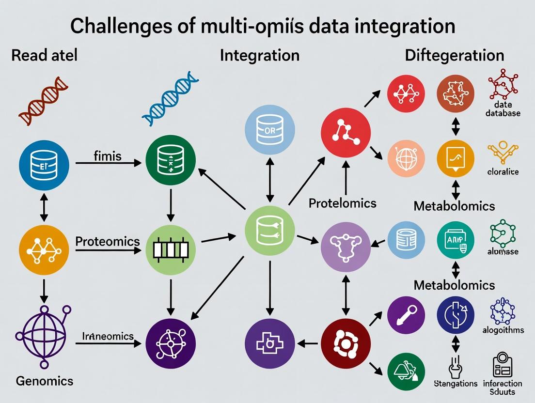
Visualization of Multi-Omics Workflow and Integration Challenge
Title: Multi-Omics Data Generation and Integration Pipeline
Title: Biological Information Flow and Integration Hurdles
The Scientist's Toolkit: Key Research Reagent Solutions
Table 2: Essential Reagents and Kits for Multi-Omics Research
| Reagent/Kits | Supplier Examples | Function in Multi-Omics Workflow |
|---|---|---|
| TRIzol Reagent | Thermo Fisher, Qiagen | Simultaneous extraction of RNA, DNA, and proteins from a single sample, minimizing sample-to-sample variation. |
| DNase I (RNase-free) | New England Biolabs, Roche | Removal of genomic DNA contamination from RNA preparations for accurate transcriptomics. |
| Nextera DNA Flex Library Prep Kit | Illumina | Preparation of sequencing libraries from low-input or degraded DNA for genomics/epigenomics. |
| NEBNext Ultra II Directional RNA Library Prep Kit | New England Biolabs | High-efficiency preparation of strand-specific RNA-seq libraries. |
| Trypsin, Sequencing Grade | Promega, Thermo Fisher | Proteolytic enzyme for digesting proteins into peptides for bottom-up proteomics. |
| TMTpro 16plex Isobaric Label Reagents | Thermo Fisher | Multiplexing up to 16 samples in a single MS run for high-throughput, quantitative proteomics. |
| Bio-Rad Protein Assay Dye Reagent | Bio-Rad | Colorimetric quantification of total protein concentration for normalizing proteomics sample input. |
| Methanol (Optima LC/MS Grade) | Fisher Chemical | High-purity solvent for metabolite extraction and mobile phase in LC-MS metabolomics. |
| Pierce Quantitative Colorimetric Peptide Assay | Thermo Fisher | Accurate measurement of peptide concentration prior to LC-MS/MS injection. |
| Single-Cell Multiome ATAC + Gene Expression Kit | 10x Genomics | Enables simultaneous profiling of chromatin accessibility (epigenomics) and transcriptome from the same single cell. |
Beyond the Core: Expanding the Omics Universe
Integration challenges multiply as the universe expands:
- Microbiomics: Analysis of microbial communities, introducing a separate genome and metabolome.
- Lipidomics: Subset of metabolomics focused on lipids, requiring specialized separation.
- Glycomics: Study of glycans, with complex, non-templated structures.
- Spatial Omics: Technologies (e.g., Visium, MIBI) that retain tissue-context information, adding a spatial coordinate dimension to integration.
- Single-Cell & Multiome Assays: Resolve cellular heterogeneity but generate ultra-high-dimensional, sparse data.
Defining the multi-omics universe is the first step toward conquering its central challenge: integration. Each layer—genomics, transcriptomics, proteomics, metabolomics—provides a unique but incomplete snapshot of a complex, dynamic system. The future of biomedical research and drug development lies in developing robust computational and statistical frameworks that can reconcile these disparate data types, moving from simple correlation to causal, mechanistic models of health and disease.
The central challenge in modern multi-omics research is the systematic integration of diverse data modalities—genomics, transcriptomics, proteomics, metabolomics—to construct a unified model of biological systems. This integration is fundamentally obstructed by heterogeneity, which manifests in three primary dimensions: divergent measurement scales (e.g., counts, intensities, concentrations), incompatible data formats (FASTQ, BAM, mzML, .raw), and batch-specific technical noise. This technical guide deconstructs this hurdle and provides actionable methodologies for overcoming it within the broader thesis that data heterogeneity is the primary rate-limiting step in translational multi-omics discovery.
Quantitative Characterization of Heterogeneity
The table below summarizes the core quantitative disparities across major omics layers, based on current literature and typical experimental outputs.
Table 1: Characteristic Scales and Formats of Major Omics Data Types
| Omics Layer | Typical Measurement Scale | Dynamic Range | Primary File Formats | Common Technical Noise Sources |
|---|---|---|---|---|
| Genomics (WGS/WES) | Read Counts / Allele Fractions | 0-100% (VAF) | FASTQ, BAM, VCF | PCR duplicates, sequencing depth bias, GC-content bias |
| Transcriptomics (RNA-seq) | Read Counts (integer) | ~5 orders of magnitude | FASTQ, BAM, Gene Count Matrix | Batch effects, library prep bias, 3’ bias, rRNA contamination |
| Proteomics (LC-MS/MS) | Ion Intensity / Spectral Counts | ~4-5 orders of magnitude | .raw, .mzML, .mgf | Ion suppression, batch/column drift, peptide identification error |
| Metabolomics (LC-MS) | Ion Intensity | ~4-6 orders of magnitude | .raw, .mzML, .cdf | Matrix effects, instrument drift, peak misalignment |
| Epigenomics (ChIP-seq/ATAC-seq) | Read Counts / Enrichment Scores | ~3 orders of magnitude | FASTQ, BAM, bedGraph | Antibody specificity, chromatin accessibility bias, PCR artifacts |
Experimental Protocols for Cross-Omics Data Generation
Robust integration requires standardized, parallelized data generation. Below are detailed protocols for a coordinated multi-omics study from a single tissue sample.
Protocol: Coordinated Nucleic Acid Extraction for Genomics & Transcriptomics
Objective: Isolate high-quality DNA and RNA from the same biological sample (e.g., flash-frozen tissue). Reagents: AllPrep DNA/RNA/miRNA Universal Kit (Qiagen), RNase Away, liquid nitrogen. Steps:
- Homogenization: Cryogrind ≤30 mg tissue in liquid nitrogen. Transfer powder to lysis buffer.
- Simultaneous Lysis: Thoroughly homogenize using a vortex adapter. Centrifuge to clear lysate.
- RNA Phase: Transfer lysate to an RNeasy spin column. Perform on-column DNase I digestion. Elute RNA in nuclease-free water. Assess integrity (RIN > 8.0, Bioanalyzer).
- DNA Phase: Pass flow-through from step 3 to a separate column. Apply ethanol precipitation. Bind DNA to an AllPrep DNA column. Wash and elute. Assess purity (A260/280 ~1.8). Output: Paired DNA (for WGS/WES) and RNA (for RNA-seq) aliquots.
Protocol: Serial Protein & Metabolite Extraction from Cell Pellet
Objective: Sequentially extract proteins and metabolites from the same cell pellet. Reagents: Methanol, water, chloroform (for metabolomics); RIPA buffer with protease inhibitors (for proteomics). Steps:
- Quenching & Metabolite Extraction: Suspend 1x10⁷ cells in 1 mL cold 40:40:20 MeOH:ACN:H₂O at -20°C. Vortex 10 min, centrifuge (15,000g, 10 min, 4°C).
- Supernatant Collection: Transfer supernatant (metabolite fraction) to a new tube. Dry in a speed vacuum. Store at -80°C for LC-MS metabolomics.
- Protein Pellet Processing: Re-suspend the remaining pellet in 200 µL RIPA buffer. Sonicate on ice (3x 10s pulses). Centrifuge (14,000g, 15 min, 4°C).
- Protein Collection: Transfer supernatant (protein fraction) to a new tube. Quantify via BCA assay. Aliquot for LC-MS/MS proteomics. Output: Paired metabolite and protein extracts from an identical cell population.
Computational Workflows for Harmonization
Diagram: Multi-Omics Data Harmonization Pipeline
Title: Multi-Omics Data Harmonization and Integration Pipeline
Detailed Method: Multi-Omics Factor Analysis (MOFA+)
Objective: Identify latent factors that explain variance across multiple omics datasets. Input: Normalized matrices (samples x features) for each omics layer. Steps:
- Data Preparation: Ensure all matrices are aligned by sample ID. Center and scale features within each view.
- Model Training: Run MOFA+ with default variational inference. Set convergence tolerance (0.001). Use 10-15 factors as initial guess.
- Factor Interpretation: Correlate factors with sample metadata (e.g., disease status). Annotate factors by loading weights on original features (genes, proteins).
- Downstream Analysis: Use factor values as covariates in survival or differential expression models. Output: Latent factors representing shared and unique variation across omics types.
The Scientist's Toolkit: Research Reagent Solutions
Table 2: Essential Kits and Reagents for Multi-Omics Sample Preparation
| Product Name | Vendor | Function in Multi-Omics Workflow | Key Benefit for Integration |
|---|---|---|---|
| AllPrep DNA/RNA/miRNA Universal Kit | Qiagen | Co-isolation of DNA, RNA, and small RNA from a single sample. | Eliminates biological variation from using separate samples for different assays. |
| PreOmics iST Kit | PreOmics | Single-pot, solid-phase-enhanced sample preparation for proteomics. | Highly reproducible protein extraction and digestion, reducing technical noise. |
| Matched Tissue & Plasma DNA/RNA Kits | Norgen Biotek | Parallel purification from matched tissue and liquid biopsy samples. | Enables direct comparison of solid tumor and circulating omics profiles. |
| Cellular Indexing of Transcriptomes & Epitopes by Sequencing (CITE-seq) Antibodies | BioLegend | Oligo-tagged antibodies for simultaneous protein surface marker and transcriptome measurement in single cells. | Direct, paired measurement of two modalities at single-cell resolution. |
| mTOR Signaling Multiplex ELISA Array | RayBiotech | Quantifies phospho-proteins in key signaling pathways (PI3K/AKT/mTOR). | Provides calibrated, quantitative protein-level data to complement phospho-proteomics. |
| Seahorse XFp FluxPak | Agilent | Measures live-cell metabolic parameters (glycolysis, OXPHOS). | Provides functional metabolic data to ground-truth metabolomics findings. |
Pathway Visualization: Impact of Heterogeneity on Biological Inference
Title: Noise Sources Obscuring Signal in Multi-Omics Pathway Inference
Overcoming the heterogeneity hurdle is not a single-step process but a rigorous, end-to-end framework encompassing coordinated wet-lab protocols, systematic normalization, and robust statistical integration. By adopting the experimental and computational guidelines detailed here, researchers can transform disparate, noisy data layers into coherent systems-level models, thereby unlocking the true potential of multi-omics for mechanistic discovery and therapeutic development.
The integration of multi-omics data—genomics, transcriptomics, proteomics, metabolomics—represents the frontier of systems biology and precision medicine. The core thesis is that a holistic, multi-layered view of biological systems will unlock profound insights into disease mechanisms and therapeutic targets. However, this promise is critically undermined by the High-Dimension, Low-Sample-Size (HDLSS) conundrum. Each omics layer can yield tens of thousands of features (p), while cohort sizes (n) often number in the hundreds or fewer. This p >> n regime leads to the "dimensionality disaster," where traditional statistical methods fail, models overfit, and spurious correlations dominate.
This whitepaper provides a technical guide to navigating the HDLSS landscape within multi-omics research, detailing current mitigation strategies, experimental validation protocols, and essential computational toolkits.
The Core Statistical Problem
In HDLSS settings, the data matrix is ill-conditioned. The sample covariance matrix is singular, making many inferential statistics undefined. The curse of dimensionality means data points become equidistant, and all samples appear as outliers. This results in:
- Overfitting: Models memorize noise rather than learning generalizable biological signals.
- Unstable Feature Selection: Small perturbations in data lead to wildly different selected biomarkers.
- Loss of Power: The multiple testing burden from high-dimensional hypotheses is immense.
Table 1: Quantitative Landscape of Multi-Omics Dimensionality
| Omics Layer | Typical Feature Range (p) | Typical Cohort Size (n) | Representative p/n Ratio |
|---|---|---|---|
| Whole Genome Sequencing (WGS) | ~3-5 million variants | 100s - 10,000s | 1,000:1 to 10,000:1 |
| Transcriptomics (RNA-seq) | ~20,000 genes | 10s - 100s | 200:1 to 2,000:1 |
| Proteomics (Mass Spectrometry) | ~5,000 - 10,000 proteins | 10s - 100s | 100:1 to 1,000:1 |
| Metabolomics | ~500 - 5,000 metabolites | 10s - 100s | 50:1 to 500:1 |
| Multi-Omics Integration | > 30,000 aggregated features | 10s - 100s | > 300:1 |
Methodological Frameworks for Navigating HDLSS
Dimensionality Reduction & Feature Selection
Prior to integration, aggressive dimensionality reduction is required.
- Experimental Protocol for Unsupervised Feature Filtering:
- Variance Stabilization: For each omics dataset, apply appropriate transformations (e.g., log2(CPM+1) for RNA-seq, log2(imputed intensity) for proteomics).
- Filter Low-Variance Features: Calculate the coefficient of variation (CV) or interquartile range (IQR) for each feature across samples. Remove features in the bottom X percentile (e.g., 20%).
- Correlation-Based Pruning: For remaining features, calculate pairwise correlation (e.g., Pearson's r). Within highly correlated clusters (|r| > 0.95), retain only the feature with the highest median absolute deviation.
- Output: A filtered feature matrix for each omics modality ready for integration.
Title: Workflow for Unsupervised Feature Filtering
Sparse Modeling and Regularization
Penalized regression models introduce constraints to prevent overfitting.
- Experimental Protocol for Sparse Multi-Omics Regression (e.g., for predicting clinical outcome):
- Pre-integration Concatenation: Merge filtered feature matrices from different omics platforms column-wise, using a shared sample ID. Standardize each feature (mean=0, variance=1).
- Model Training with Elastic Net: Apply an Elastic Net regression (combining L1/Lasso and L2/Ridge penalties) via cross-validation. Use
glmnetin R orsklearn.linear_model.ElasticNetCVin Python. - Hyperparameter Tuning: Perform a nested 10-fold cross-validation grid search over the mixing parameter (α, between 0 and 1) and the regularization strength (λ). The inner loop selects λ, the outer loop evaluates performance.
- Feature Importance: Extract non-zero coefficients from the final model as the selected, integrative biomarker signature.
- Validation: Apply the trained model to a completely held-out test set. Report performance metrics (AUC-ROC for classification, R² for regression).
Title: Sparse Modeling for Feature Selection
Multi-Block Data Integration Methods
These methods model the joint structure of multiple omics datasets without concatenation.
- Experimental Protocol for DIABLO (Data Integration Analysis for Biomarker discovery using Latent cOmponents) using mixOmics in R:
- Data Input: Prepare a list of filtered, pre-processed data matrices (X1: mRNA, X2: miRNA, X3: proteomics, etc.) and a response vector Y (e.g., disease subtype).
- Design Matrix: Specify a between-omics design matrix (e.g., full design = all omics connected) to guide the model on which datasets should be correlated.
- Tuning Component Number: Use
tune.block.splsda()to perform 10-fold cross-validation and determine the optimal number of components and the number of features to select per component per dataset. - Model Fitting: Run the final
block.splsda()model with tuned parameters. - Evaluation & Visualization: Assess performance via
perf()with repeated cross-validation. Visualize sample clustering on the first two components viaplotIndiv(). Generate a circos plot (circosPlot()) to visualize correlations between selected features across omics layers.
Table 2: Key Multi-Block Integration Methods
| Method | Underlying Algorithm | Key Strength | Software Package |
|---|---|---|---|
| DIABLO | Sparse Generalized Canonical Correlation Analysis | Supervised; finds correlated biomarkers predictive of an outcome. | mixOmics (R) |
| MOFA/MOFA+ | Bayesian Factor Analysis | Unsupervised; learns latent factors capturing shared/private variation. | MOFA2 (R/Python) |
| sMBPLS | Sparse Multi-Block Partial Least Squares | Handles highly collinear data; good for predictive modeling. | Custom sMBPLS R package |
| iClusterBayes | Joint Latent Variable Model | Integrative clustering for subtype discovery. | iClusterPlus (R) |
Title: Multi-Block Integration Conceptual Model
The Scientist's Toolkit: Research Reagent Solutions
Table 3: Essential Toolkit for HDLSS Multi-Omics Research
| Item / Reagent | Function & Relevance |
|---|---|
| High-Quality, Annotated Biospecimens | The foundational input. Paired, high-integrity tissue/plasma samples with deep clinical phenotyping are irreplaceable. Small n makes sample quality paramount. |
| Multiplex Assay Kits (e.g., Olink, Luminex) | Allows measurement of 10s-1000s of proteins/cytokines from a single low-volume sample, maximizing data yield per precious sample. |
| Single-Cell RNA-seq Reagents (10x Genomics) | Transforms a bulk tissue sample (n=1) into data from thousands of cells, artificially increasing 'n' for cellular-resolution analyses. |
| Stable Isotope Labeling Reagents (SILAC, TMT) | Enables multiplexed proteomics, where multiple samples are pooled and run in one MS batch, drastically reducing technical noise—a critical confounder in HDLSS. |
| CRISPR Screening Libraries (e.g., Brunello) | Functional validation tool. After computational biomarker identification from HDLSS data, pooled CRISPR screens can test hundreds of gene targets in parallel for causal roles. |
| Cloud Computing Credits (AWS, GCP) | Essential for scalable computation of resource-intensive integration algorithms and repeated cross-validation. |
R/Python with Key Libraries (mixOmics, MOFA2, glmnet, scikit-learn) |
The computational workbench for implementing all described statistical and ML strategies. |
Within the overarching thesis on the Challenges of Multi-Omics Data Integration Research, a fundamental and pervasive obstacle is the confounding of true biological signal with non-biological technical noise. This whitepaper provides an in-depth technical guide to disentangling biological variation from technical variation, with a focus on identifying and correcting for batch effects. As datasets grow larger and more complex from high-throughput technologies like genomics, transcriptomics, and proteomics, the risk of batch effects—systematic technical variations introduced during experimental processing—obscuring or mimicking biological phenomena increases exponentially.
Biological Variation is the true signal of interest, arising from differences in genotype, phenotype, disease state, treatment response, or developmental stage between samples. It is the variation we seek to measure and interpret.
Technical Variation (Batch Effects) is non-biological noise introduced by factors such as:
- Different experiment dates or times.
- Multiple personnel or technicians.
- Reagent lot changes.
- Sequencing lane or instrument differences.
- Sample preparation protocol drifts.
Batch effects are often systematic, not random, and can be severe enough to cause false conclusions, such as clustering samples by processing date instead of disease subtype.
Quantitative Impact of Batch Effects
The following table summarizes common quantitative metrics used to assess the relative contribution of biological and technical variance in omics studies.
Table 1: Metrics for Assessing Sources of Variation in Omics Data
| Metric | Purpose | Interpretation in Context of Batch Effects |
|---|---|---|
| Principal Variance Component Analysis (PVCA) | Partitions total variance into contributions from biological factors (e.g., disease) and technical factors (e.g., batch). | A batch factor explaining >10% of total variance is often considered a major confounder requiring correction. |
| Median Coefficient of Variation (CV) | Measures dispersion of data relative to its mean. | High median CV within a biologically homogeneous group suggests high technical noise. |
| Inter-class Correlation (ICC) | Quantifies reliability of measurements across batches. Ranges from 0 (no reliability) to 1 (perfect reliability). | ICC < 0.5 indicates measurements are more variable across batches than consistent within biological groups, limiting reproducibility. |
| Silhouette Width | Measures how well samples cluster by biological class vs. technical batch. | Negative average silhouette width indicates samples are better clustered by batch than by biological class. |
Experimental Protocols for Identification and Control
Protocol 1: Prospective Experimental Design to Minimize Batch Effects
Title: Randomized Block Design for Sample Processing
- Sample Randomization: Do not process all samples from one biological group together. Randomize sample assignment to processing batches (e.g., sequencing lanes, MS runs) using a validated random number generator.
- Balancing: Ensure each batch contains a balanced representation of all biological groups (e.g., equal numbers of case and control samples per batch).
- Replication: Include both technical replicates (same biological sample processed multiple times) and biological replicates.
- Control Samples: Incorporate reference standards or "pooled" samples (an equal mixture of all samples) in every batch. These serve as longitudinal anchors to track technical drift.
Protocol 2: Retrospective Assessment Using Principal Component Analysis (PCA)
Title: Diagnostic PCA for Batch Effect Detection
- Perform PCA on the normalized, but not batch-corrected, feature-by-sample matrix (e.g., gene expression).
- Generate PCA score plots for the top principal components (PCs), typically PC1 vs. PC2 and PC3 vs. PC4.
- Color samples by biological group and by technical batch in separate plots.
- Interpretation: If samples cluster more strongly by batch than by biological condition in the leading PCs, a significant batch effect is present. Statistical testing (PERMANOVA) can be applied to the principal coordinates.
Protocol 3: Implementation of ComBat for Batch Correction
Title: ComBat Algorithm for Empirical Bayes Batch Adjustment
- Input: A
p x nmatrix of normalized data, wherepis features (genes) andnis samples. A model matrix for biological covariates of interest. A batch covariate vector. - Model Fitting: For each feature, model the data as a linear combination of biological covariates and a batch-specific additive (shift) and multiplicative (scale) effect.
- Empirical Bayes Estimation: Shrink the batch effect parameters towards the overall mean across all features, stabilizing estimates for low-variance features. This is crucial for datasets with many features.
- Adjustment: Remove the estimated additive and multiplicative batch effects for each feature in each batch.
- Output: A
p x nbatch-corrected matrix, ready for downstream biological analysis.
Visualization of Concepts and Workflows
Diagram Title: Disentangling Biological and Technical Variation
Diagram Title: Batch Effect Management Workflow
The Scientist's Toolkit: Research Reagent Solutions
Table 2: Essential Materials for Batch Effect Control Experiments
| Item | Function & Rationale |
|---|---|
| Universal Human Reference RNA (UHRR) | A standardized pool of RNA from diverse cell lines. Serves as an inter-batch calibration standard in transcriptomic studies to monitor technical performance. |
| Mass Spectrometry Grade Enzymes (Trypsin/Lys-C) | For proteomics. Using the same lot number across all batches minimizes variability in protein digestion efficiency, a major source of technical variance. |
| Indexed Adapters (Unique Dual Indexes - UDIs) | For next-generation sequencing. UDIs allow pooling of multiple samples per lane while uniquely identifying each sample, enabling robust demultiplexing and detection of cross-batch contamination. |
| Internal Standard Spike-Ins (e.g., S. pombe RNA, UPS2 Proteomic Standard) | Known quantities of exogenous molecules added to each sample. Used to distinguish technical variation (which affects spike-ins and endogenous molecules equally) from biological variation. |
| Single-Cell Multiplexing Kits (e.g., CellPlex, Hashtag Antibodies) | For single-cell genomics. Allows pooling of samples from multiple biological conditions into a single processing batch, virtually eliminating wet-lab batch effects for those samples. |
| Automated Nucleic Acid/Protein Purification Systems | Minimizes variation introduced by manual handling differences between technicians or labs, standardizing extraction efficiency and purity. |
In multi-omics data integration research, combining datasets from genomics, transcriptomics, proteomics, and metabolomics is fundamental for constructing comprehensive biological models. However, a pervasive and critical challenge is the presence of missing values across these datasets. This incompleteness arises from technical limitations, such as limits of detection in mass spectrometry, sample degradation, or bioinformatic processing errors. Systematic missingness, where certain compounds cannot be detected under specific experimental conditions, is particularly common in metabolomics and proteomics. Left unaddressed, missing data introduces bias, reduces statistical power, and can lead to erroneous conclusions in downstream integrative analyses, jeopardizing the translational potential in drug development.
Quantifying the Problem in Multi-Omics Studies
The prevalence and patterns of missing data vary significantly by omics layer and technology.
Table 1: Typical Missing Data Rates by Omics Technology
| Omics Layer | Technology | Typical Missing Rate | Primary Cause of Missingness |
|---|---|---|---|
| Metabolomics | LC-MS (untargeted) | 20-40% | Ion suppression, low abundance below LOD. |
| Proteomics | Shotgun LC-MS/MS | 15-30% | Stochastic sampling, low-abundance proteins. |
| Transcriptomics | RNA-Seq | 1-5% | Low expression, pipeline filtering. |
| Genomics | Whole-Genome Sequencing | <1% | Coverage gaps, ambiguous mapping. |
Table 2: Missing Data Patterns
| Pattern | Description | Implication for Analysis |
|---|---|---|
| Missing Completely at Random (MCAR) | Missingness independent of observed/unobserved data. | Less biased; reduces sample size. |
| Missing at Random (MAR) | Missingness depends on observed data. | Can be corrected with model-based methods. |
| Missing Not at Random (MNAR) | Missingness depends on the unobserved value itself (e.g., low abundance). | Most problematic; requires specific models. |
Experimental Protocols for Assessing Missingness
Before imputation, a rigorous assessment of the missing data pattern is required.
Protocol 1: Pattern Analysis via Heatmap and Logistic Regression
- Data Preparation: Create a binary matrix (0 = observed, 1 = missing) matching your omics data matrix (features x samples).
- Visualization: Generate a clustered heatmap of the binary matrix to identify sample- or feature-wise patterns.
- Statistical Testing: For each feature, perform a univariate logistic regression (missing/observed status as outcome) against potential covariates (e.g., sample group, total ion count, batch). A significant association suggests MAR.
- MNAR Inference: If abundance distributions differ significantly between observed and missing groups (estimated via detection limits), MNAR is likely.
Protocol 2: Benchmarking Imputation Performance (Spike-in Experiment)
- Generate Ground Truth: Start with a complete, high-quality omics dataset.
- Introduce Missing Values: Artificially remove values under MCAR (random) and MNAR (remove low-intensity values) schemes at rates of 10%, 20%, and 30%.
- Apply Imputation: Run candidate imputation methods on each corrupted dataset.
- Evaluate: Calculate the Root Mean Square Error (RMSE) between the imputed matrix and the ground truth. Assess the preservation of biological variance (e.g., PCA structure) and differential analysis outcomes.
Key Imputation Methodologies: Workflows and Applications
Imputation strategies range from simple to complex, with suitability dependent on the missingness pattern and data structure.
Title: Decision Workflow for Selecting an Imputation Strategy
Detailed Methodologies
Method 1: k-Nearest Neighbors (KNN) Imputation (for MCAR/MAR)
- For each sample containing missing values, calculate its distance (e.g., Euclidean) to all other samples using only commonly observed features.
- Identify the k samples with the smallest distance (nearest neighbors).
- For each missing feature in the target sample, compute the imputed value as the mean (or weighted mean) of that feature's values in the k neighbors.
- Iterate until convergence or for a pre-set number of cycles.
Method 2: Multiple Imputation by Chained Equations (MICE) (for MAR)
- Initialization: Fill missing values with simple imputation (e.g., mean).
- Iteration: For each feature with missing data (
Feature_m), fit a regression model (linear, logistic, etc.) using all other features as predictors, based on samples whereFeature_mis observed. - Imputation: Draw predicted values for
Feature_mfrom the fitted model (incorporating error) to fill the missing entries. - Cycling: Repeat steps 2-3 for each incomplete feature, cycling through the dataset for multiple rounds (typically 5-10) to create one imputed dataset.
- Repetition: Repeat the entire process to generate m multiple imputed datasets (e.g., m=5).
- Analysis & Pooling: Perform the intended downstream analysis (e.g., regression) on each of the m datasets and pool the results using Rubin's rules to obtain final estimates and confidence intervals.
Method 3: Left-Censored MNAR Imputation (e.g., MinProb)
- For each feature, estimate a global detection limit or a minimum observed value.
- Calculate a small imputation value:
min_value * rwhereris a user-defined factor (e.g., 0.65). - Add normally distributed random noise proportional to the data variance to avoid creating artificial peaks.
The Scientist's Toolkit: Research Reagent Solutions for Multi-Omics
Table 3: Essential Tools for Managing Missing Data in Multi-Omics Experiments
| Item | Function | Example/Note |
|---|---|---|
| Internal Standards (IS) | Corrects for technical variation and signal drift in MS; aids in distinguishing MNAR from technical zeros. | Stable Isotope-Labeled Compounds (e.g., 13C, 15N). |
| Quality Control (QC) Samples | Pooled sample run repeatedly; monitors instrument stability, identifies run-order dependent missingness (MAR). | Technical replicates for precision assessment. |
| Blank Samples | Distinguishes true missing data (analyte absent) from background noise or contamination. | Process blanks, solvent blanks. |
| Standard Reference Materials | Provides a known ground truth for benchmarking imputation accuracy in spike-in experiments. | NIST SRM 1950 (Metabolites), HeLa cell protein digests. |
| Sample Multiplexing Kits | Reduces batch effects and missing data due to inter-run variation by pooling samples for simultaneous processing. | TMT (Tandem Mass Tag), iTRAQ reagents for proteomics. |
Title: Data Processing Pipeline with Imputation Step
Effective handling of missing data is not a mere preprocessing step but a critical, foundational component of robust multi-omics data integration. The choice of imputation strategy must be guided by a diligent assessment of the missingness mechanism, which is itself influenced by experimental design and the judicious use of standards and controls. For drug development professionals, transparent reporting of missing data rates and imputation methods is essential to ensure the validity of identified biomarkers or therapeutic targets. As multi-omics studies increase in scale and complexity, the development of novel, integrated imputation frameworks that leverage the correlations across omics layers represents a vital frontier for improving the fidelity of systems-level biological insights.
From Theory to Practice: Key Integration Strategies and Real-World Applications
Within the broader thesis on the Challenges of multi-omics data integration research, the selection of an integration paradigm is a fundamental architectural decision. The vast heterogeneity, scale, and noise inherent in genomics, transcriptomics, proteomics, and metabolomics datasets necessitate strategic frameworks to combine information effectively. The three primary paradigms—Early, Intermediate, and Late Fusion—differ in the stage at which data from different omics layers are combined, each with distinct implications for addressing challenges like dimensionality, modality-specific noise, and biological interpretability.
Technical Definitions and Methodologies
Early Fusion (Feature-Level Integration)
Early fusion concatenates raw or pre-processed features from multiple omics modalities into a single, unified feature vector before model training.
- Workflow: Individual omics datasets (e.g., SNP arrays, RNA-seq counts, protein abundances) are normalized and scaled independently. These matrices are then concatenated column-wise (sample-wise) to create a combined matrix
X_combinedof dimensions[n_samples, (n_features_genomics + n_features_transcriptomics + ...)]. This monolithic matrix is input to a downstream machine learning model (e.g., PCA, Random Forest, Deep Neural Network). - Core Challenge: This approach directly confronts the "curse of dimensionality" (p ≫ n problem) and can be severely affected by modality-specific batch effects if not corrected prior to concatenation.
Intermediate Fusion (Joint Representation Learning)
Intermediate fusion integrates data within the model itself, learning a joint latent representation that captures shared and complementary information across modalities.
- Workflow: Each omics type is processed through separate, modality-specific input layers or encoders (e.g., CNNs for sequences, MLPs for numerical data). The model then combines these encoded representations at a hidden layer via operations such as concatenation, tensor fusion, or attention-based mechanisms. A central neural network further processes this joint representation for the final prediction.
- Core Strength: This paradigm is designed to model complex, non-linear interactions between omics layers, potentially capturing biological mechanisms that are not apparent in single-omics analyses.
Late Fusion (Decision-Level Integration)
Late fusion trains separate models on each omics dataset independently and integrates their predictions at the final decision stage.
- Workflow: A predictive model (e.g., SVM, classifier) is trained exclusively on each omics modality, yielding a set of modality-specific predictions or prediction probabilities. These individual results are then combined using a meta-learner (e.g., a simple voting scheme, weighted average, or another classifier) to produce a final, consensus prediction.
- Core Strength: It naturally accommodates asynchronous data availability (common in clinical settings) and allows for the use of modality-specific best-practice models.
Comparative Analysis of Fusion Paradigms
Table 1: Qualitative and Quantitative Comparison of Fusion Approaches
| Criterion | Early Fusion | Intermediate Fusion | Late Fusion |
|---|---|---|---|
| Integration Stage | Raw/Pre-processed Data Level | Hidden/Latent Representation Level | Decision/Prediction Level |
| Handles High Dimensionality | Poor (requires strong feature selection/dimension reduction) | Good (via encoder networks) | Excellent (per-modality modeling) |
| Models Inter-Modality Interactions | Limited (relies on downstream model) | Excellent (explicitly designed for it) | None (integrated post-hoc) |
| Robustness to Missing Modalities | Poor (entire sample may be excluded) | Moderate (can be designed with masking) | Excellent (only missing model is skipped) |
| Interpretability | Low (black-box on combined features) | Moderate (potential via attention weights) | High (per-modality contributions clear) |
| Typical Model Complexity | Low to Moderate | High (complex multi-branch architectures) | Moderate (ensemble of simpler models) |
| Reported Performance Gain (Example Range)* | 2-8% AUC increase over single-omics baselines | 5-15% AUC increase over single-omics baselines | 3-10% AUC increase over single-omics baselines |
*Performance gains are highly context-dependent and based on reviewed benchmarking studies (e.g., on TCGA pan-cancer or multi-omics drug response datasets).
Experimental Protocol: A Benchmarking Study
To empirically compare these paradigms, a standard benchmarking protocol is employed.
1. Data Preparation:
- Source: Use a public multi-omics cohort (e.g., TCGA-LIHC: methylation (450K array), RNA-seq (FPKM-UQ), and RPPA proteomics).
- Pre-processing: For each modality per sample: log-transformation (RNA-seq), beta-value normalization (methylation), Z-score normalization (RPPA). Perform sample-wise matching.
- Task: Binary classification of a clinical endpoint (e.g., tumor vs. normal, or survival risk group).
2. Paradigm Implementation:
- Early Fusion: Concatenate all three processed feature matrices. Apply PCA for dimensionality reduction to 100 components. Train a Random Forest classifier.
- Intermediate Fusion: Implement a multi-modal deep learning model (e.g., MOMA-Net). Use separate fully-connected encoder branches for each omics type (2 hidden layers each), concatenate their 64-dim outputs, and process through a final classifier head.
- Late Fusion: Train three independent XGBoost models—one per modality. Combine their output class probabilities using a logistic regression meta-learner.
3. Evaluation:
- 5-fold nested cross-validation (to tune hyperparameters within the training folds).
- Primary Metric: Area Under the ROC Curve (AUC).
- Secondary Metrics: Precision-Recall AUC, F1-Score.
Visualization of Workflows and Pathways
Diagram 1: Multi-Omics Fusion Workflow Comparison
Diagram 2: Conceptual Pathway of Multi-Omics Integration Impact
The Scientist's Toolkit: Key Research Reagent Solutions
Table 2: Essential Tools and Resources for Multi-Omics Integration Research
| Item / Resource | Function / Purpose |
|---|---|
R/Bioconductor (omicade4, MOFA2) |
Statistical packages for multi-omics factor analysis and early/intermediate fusion. |
Python Libraries (PyTorch, TensorFlow with tf.keras) |
Essential frameworks for building custom intermediate fusion deep learning architectures. |
| Singularity/Apptainer Containers | Ensures reproducibility of complex software stacks and dependency management across HPC clusters. |
| Benchmark Datasets (e.g., TCGA, CPTAC) | Curated, clinically-annotated multi-omics datasets essential for training and benchmarking fusion models. |
| Multi-Omics Benchmarking Suites (e.g., multiBench, PIMKL) | Pre-built pipelines for fair comparison of integration methods across standard tasks. |
| High-Performance Computing (HPC) Cluster | Provides necessary computational power for training large intermediate fusion models and permutation testing. |
| Secure Data Storage (e.g., encrypted NAS) | Required for storing large volumes of sensitive genomic and clinical patient data compliant with regulations. |
The choice between early, intermediate, and late fusion is not universally optimal but is dictated by the specific multi-omics integration challenge at hand. Early fusion, while simple, often falters under high-dimensional data. Late fusion offers robustness and flexibility, particularly for clinical translation. Intermediate fusion holds the greatest promise for novel biological discovery due to its capacity to learn complex cross-modal relationships but demands large sample sizes and significant computational resources. Navigating this trade-off space is central to advancing the field and overcoming the fundamental challenges in multi-omics data integration research.
The integration of multi-omics data (e.g., genomics, transcriptomics, proteomics, metabolomics) presents significant challenges, including technical noise, high dimensionality, disparate scales, and biological heterogeneity. Effective frameworks must address these to extract coherent biological signals and drive discoveries in systems biology and precision medicine.
Core Framework Analysis
MOFA+ (Multi-Omics Factor Analysis)
MOFA+ is a Bayesian statistical framework for the unsupervised integration of multiple omics assays. It uses a factor analysis model to disentangle the shared and specific sources of variation across data modalities.
Key Technical Specifications:
- Core Model: Bayesian group factor analysis with automatic relevance determination priors.
- Input Handling: Accepts both continuous (Gaussian) and discrete (Bernoulli, Poisson) data.
- Output: A set of latent factors, each with loadings per feature and weights per sample, explaining variance across omics layers.
Experimental Protocol for a Typical MOFA+ Analysis:
- Data Preprocessing: Normalize and scale each omics dataset individually. Handle missing values explicitly.
- Model Training: Create a
MOFAobject and train the model specifying the number of factors (can be inferred). Use stochastic variational inference for scalability. - Variance Decomposition: Analyze the percentage of variance explained (R²) per factor per view.
- Factor Interpretation: Correlate factors with sample metadata (e.g., clinical phenotypes) and perform feature set enrichment analysis on factor loadings.
- Downstream Analysis: Use factors as reduced-dimension representations for clustering, classification, or regression tasks.
mixOmics
mixOmics is a versatile R toolkit for the exploration and integration of multiple omics datasets using multivariate statistical methods, with a focus on discriminant analysis and variable selection.
Key Technical Specifications:
- Core Methods: Includes Projection to Latent Structures (PLS), sparse PLS Discriminant Analysis (sPLS-DA), and DIABLO (Data Integration Analysis for Biomarker discovery using Latent variable approaches) for multi-class multi-omics integration.
- Focus: Supervised and unsupervised integration with strong visualisation and variable selection capabilities.
Experimental Protocol for a DIABLO-based Multi-Omics Classification:
- Data Preparation: Format data into a list of matched omics matrices (
X) and a response factor vector (Y). Tune the number of components and the number of features to select per dataset per component via cross-validation. - Model Building: Run the
block.splsdafunction to integrate datasets and predict the sample classY. - Model Evaluation: Assess performance via repeated cross-validation and permutation tests. Generate
circosPlotto visualise correlations of selected features across omics layers. - Biomarker Identification: Extract the selected variables and examine their consensus across components.
Integrated Biosciences Tools (Emerging Platforms)
This category includes newer, often cloud-based or commercial platforms offering end-to-end workflows for multi-omics integration, such as Nextflow-based pipelines, Terra.bio, or Partek Flow.
Key Technical Specifications:
- Core Approach: Provide unified environments that link data management, reproducible pipeline execution (e.g., via Common Workflow Language), pre-built analysis modules, and interactive visualization.
- Focus: Usability, reproducibility, and scalability for large consortium-scale projects.
Quantitative Framework Comparison
Table 1: Comparative Analysis of Multi-Omics Integration Frameworks
| Feature | MOFA+ | mixOmics (DIABLO) | Integrated Platforms (e.g., Partek Flow) |
|---|---|---|---|
| Primary Approach | Unsupervised Factor Analysis | Supervised/Unsupervised Multivariate (PLS) | GUI-driven, Modular Workflows |
| Statistical Core | Bayesian Group Factor Analysis | Projection to Latent Structures (PLS) | Varies (Often includes PCA, regression, ML) |
| Key Strength | Decomposing shared & specific variation; Handles missing data | Powerful for classification & biomarker discovery; Excellent viz | Accessibility; Reproducibility; Scalability |
| Data Type Handling | Continuous & Discrete | Primarily Continuous | Broad (via modules) |
| Scalability | High (approx. 10^4 samples, 10^5 features) | Moderate to High | Cloud-scalable |
| Typical Use Case | Exploratory analysis, identifying latent factors of heterogeneity | Predicting clinical outcome, multi-omics biomarker panels | Collaborative, standardized analysis for non-specialists |
| Learning Curve | Moderate | Moderate | Low to Moderate |
Visualization of Workflows and Relationships
Title: Multi-Omics Integration Workflow Comparison
Title: MOFA+ Factor Model Schematic
The Scientist's Toolkit: Essential Research Reagents & Materials
Table 2: Key Research Reagent Solutions for Multi-Omics Integration Studies
| Item / Solution | Function / Role in Multi-Omics Integration | Example Vendor/Product |
|---|---|---|
| Reference Standards (Multi-Omics) | Provides a known, uniform biological sample for technical validation and batch correction across different omics platforms. | NIST SRM 1950 (Metabolites in Human Plasma), Horizon Discovery Multiplex IMC Cell Line |
| Single-Cell Multi-Omics Kits | Enables simultaneous measurement of multiple molecular layers (e.g., RNA + ATAC, RNA + protein) from the same single cell, providing inherently matched data. | 10x Genomics Multiome (ATAC + Gene Exp.), BD AbSeq (RNA + Protein) |
| Barcoded Isotope Tags | Allows multiplexed sample pooling for proteomic/metabolomic LC-MS, reducing technical variation and enabling precise quantitation across many samples. | TMT (Tandem Mass Tag), iTRAQ |
| Spatial Transcriptomics/Proteomics Kits | Captures gene expression or protein data within tissue architecture, adding a spatial dimension for integration with histopathology. | 10x Genomics Visium, Nanostring GeoMx DSP |
| Cell Hashing/Oligo-Conjugated Antibodies | Labels cells from different samples with unique barcodes, allowing sample multiplexing in single-cell assays and improving throughput/reducing costs. | BioLegend TotalSeq-B/C Antibodies |
| Automated Nucleic Acid/Protein Extraction Systems | Ensures high-quality, consistent input material for downstream omics assays from diverse sample types (tissue, blood, cells). | Qiagen Qiacube, Promega Maxwell RSC |
| Cross-Linking Reagents | For ChIP-seq and related assays to capture protein-DNA interactions, generating data on regulatory mechanisms for integration with transcriptomics. | Formaldehyde, DSG (Disuccinimidyl glutarate) |
| Internal Standard Spike-Ins | Synthetic RNAs, proteins, or metabolites added to samples prior to processing to monitor technical performance and enable absolute quantitation. | ERCC RNA Spike-In Mix (Thermo Fisher), SILAC Spike-In Standards (Sigma) |
1. Introduction: The Multi-Omics Integration Challenge The integration of multi-omics data (genomics, transcriptomics, proteomics, metabolomics) represents a central challenge in systems biology and precision medicine. The core thesis posits that the high dimensionality, heterogeneity, noise, and complex, non-linear relationships inherent in such datasets render traditional statistical methods insufficient. This whitepaper details how deep learning (DL) architectures are uniquely equipped to overcome these barriers by discovering latent, non-linear patterns that drive biological insight and therapeutic discovery.
2. Core Deep Learning Architectures for Non-Linear Discovery
Table 1: Key DL Architectures for Multi-Omics Pattern Discovery
| Architecture | Core Strength | Typical Application in Multi-Omics | Key Advantage Over Linear Models |
|---|---|---|---|
| Autoencoders (AEs) | Unsupervised dimensionality reduction & feature learning. | Integrating layers by learning a joint latent representation. | Captures non-linear correlations for robust data compression. |
| Variational AEs (VAEs) | Generative modeling of latent distributions. | Probabilistic integration and generation of synthetic omics profiles. | Models data uncertainty and continuous latent spaces. |
| Multi-Modal Deep Neural Networks | Supervised integration of heterogeneous inputs. | Predicting clinical outcomes from combined genomic & image data. | Learns complex, cross-modal feature interactions end-to-end. |
| Graph Neural Networks (GNNs) | Modeling relational/network data. | Incorporating PPI networks with gene expression for subtyping. | Propagates information non-linearly through biological networks. |
| Attention/Transformer Models | Context-aware, weighted data integration. | Prioritizing impactful genomic variants across long sequences. | Dynamically focuses on salient features across disparate inputs. |
3. Experimental Protocol: A Standardized Workflow for Multi-Omics DL
A reproducible protocol for non-linear integration using a deep learning framework is outlined below.
3.1. Data Preprocessing & Harmonization
- Source Data: Obtain raw or normalized counts from platforms (e.g., RNA-seq, LC-MS proteomics, NMR metabolomics).
- Normalization: Apply omics-specific normalization (e.g., DESeq2 for transcriptomics, quantile for proteomics).
- Missing Value Imputation: Use DL-based methods (e.g., deep denoising autoencoders) or k-nearest neighbors.
- Batch Effect Correction: Apply Combat or its DL variant (e.g., using an AE to learn batch-invariant features).
- Feature Scaling: Scale each feature to a [0,1] range or standardize to zero mean and unit variance.
3.2. Model Implementation: Multi-modal Deep Autoencoder
- Objective: Learn a shared latent representation (
Z) from two omics layers (e.g., TranscriptomicsX_tand ProteomicsX_p). - Architecture:
- Input: Concatenated vector
[X_t, X_p]or separate encoding paths. - Encoder: Multiple fully-connected (dense) layers with non-linear activations (ReLU, LeakyReLU). Layer sizes decrease progressively (e.g., 1024 → 512 → 128 → 32 neurons).
- Bottleneck (Z): The deepest layer (e.g., 10-50 neurons) represents the integrated latent space.
- Decoder: Symmetric layers reconstructing the original inputs
[X'_t, X'_p].
- Input: Concatenated vector
- Training: Minimize reconstruction loss (Mean Squared Error) using Adam optimizer. Regularize with dropout and L2 penalty to prevent overfitting.
3.3. Downstream Analysis & Validation
- Latent Space Interpolation: Use the VAE variant to generate novel, plausible omics profiles.
- Clustering: Apply k-means or hierarchical clustering on
Zto identify novel disease subtypes. - Survival Analysis: Use a Cox proportional hazards model with
Zas input to predict patient outcomes. - Validation: Employ rigorous cross-validation (nested CV) and hold-out testing. Compare against PCA, PLS, and other linear baselines using metrics like silhouette score (clustering) or C-index (survival).
4. Visualization of Key Concepts
Title: DL Addresses Multi-Omics Integration Challenges
Title: Multi-Modal Autoencoder Architecture for Integration
5. The Scientist's Toolkit: Essential Research Reagents & Solutions
Table 2: Key Reagent Solutions for Multi-Omics DL Experiments
| Item/Category | Example Product/Platform | Function in the Workflow |
|---|---|---|
| Nucleic Acid Isolation Kits | Qiagen AllPrep, TRIzol Reagent | Simultaneous extraction of high-quality DNA, RNA, and protein from single samples. |
| Next-Gen Sequencing Library Prep | Illumina TruSeq, KAPA HyperPrep | Prepare transcriptomic (RNA-seq) and genomic (WES, WGS) libraries for sequencing. |
| Mass Spectrometry Ready Kits | Thermo Fisher TMTpro, PreOmics iST | Multiplex protein sample preparation, digestion, and labeling for quantitative proteomics. |
| Metabolite Extraction Solvents | Methanol:Acetonitrile:Water (2:2:1) | Standardized solvent system for broad-coverage untargeted metabolomics. |
| Single-Cell Multi-Omics Platforms | 10x Genomics Multiome (ATAC + Gene Exp.) | Generate paired, co-assayed data from the same single cell for intrinsic integration. |
| DL Framework & Environment | PyTorch or TensorFlow with CUDA, Google Colab | Open-source libraries and compute environments for building and training custom DL models. |
| Bioinformatics Pipelines | nf-core (Nextflow), Snakemake workflows | Reproducible, containerized pipelines for raw data processing and feature extraction. |
| Benchmarking Datasets | The Cancer Genome Atlas (TCGA), UK Biobank | Publicly available, clinically annotated multi-omics data for model training and validation. |
6. Conclusion Deep learning provides a transformative toolkit for tackling the fundamental challenge of non-linear pattern discovery in multi-omics data integration. By moving beyond linear assumptions, architectures like autoencoders, GNNs, and transformers enable the construction of unified biological models that more accurately reflect the complexity of living systems, thereby accelerating biomarker discovery and therapeutic development. Continued advancement requires close collaboration between computational scientists and experimentalists to ground these discovered patterns in biological mechanism.
Within the broader thesis on the challenges of multi-omics data integration, the application in precision oncology represents both a paramount goal and a significant test case. The core challenge lies in harmonizing disparate, high-dimensional data layers—genomics, transcriptomics, epigenomics, proteomics, and metabolomics—to define clinically actionable cancer subtypes and to nominate robust therapeutic targets. This technical guide outlines the current methodologies, workflows, and reagent solutions essential for advancing this integrative research.
Multi-Omics Data Acquisition and Preprocessing
The foundational step involves the systematic generation and quality control of omics data from tumor biospecimens.
Key Experimental Protocols
Protocol 1: Multi-Omic Profiling from a Single Tumor Sample (FFPE or Frozen)
- Sample Preparation: Macro-dissect or laser-capture microdissect to ensure >70% tumor purity. Split aliquots for nucleic acid and protein extraction.
- DNA Sequencing (WES/WGS): Extract DNA (e.g., QIAamp DNA FFPE Kit). For WES, use hybridization-based capture (e.g., IDT xGen Exome Research Panel). Sequence on Illumina NovaSeq X, targeting >100x mean coverage for tumor, >30x for matched normal.
- RNA Sequencing (Transcriptomics): Extract total RNA, assess RIN (RNA Integrity Number). Perform poly-A selection or ribosomal RNA depletion. Prepare libraries (e.g., KAPA mRNA HyperPrep Kit). Sequence to a depth of ≥50 million paired-end reads.
- DNA Methylation (Epigenomics): Treat DNA with sodium bisulfite (e.g., Zymo EZ DNA Methylation Kit). Hybridize to Illumina Infinium MethylationEPIC v2.0 BeadChip or perform whole-genome bisulfite sequencing.
- Proteomics & Phosphoproteomics: Extract proteins, digest with trypsin. For global proteomics, use data-independent acquisition (DIA) mass spectrometry (e.g., on a timsTOF Pro 2). For phosphoproteomics, enrich phosphopeptides using TiO2 or Fe-IMAC magnetic beads prior to LC-MS/MS.
Protocol 2: Single-Cell Multi-Omics (CITE-seq)
- Cell Suspension: Generate a single-cell suspension from fresh tumor tissue using a validated enzymatic dissociation kit (e.g., Miltenyi Biotec Tumor Dissociation Kit).
- Cell Staining: Stain with a panel of ~100 oligonucleotide-tagged antibodies (TotalSeq from BioLegend).
- Library Preparation: Load cells onto a 10x Genomics Chromium Controller for GEM generation and barcoding. Prepare libraries for Gene Expression (GEX), Antibody-Derived Tags (ADT), and optionally, V(D)J or ATAC-seq simultaneously using the Chromium Next GEM Single Cell Multiome ATAC + Gene Expression kit.
- Sequencing: Pool libraries and sequence on an Illumina NovaSeq 6000.
Table 1: Representative Data Outputs per Multi-Omics Modality from a Solid Tumor Sample
| Modality | Platform | Key Metrics | Typical Output per Sample | Primary Use in Subtyping/Target ID |
|---|---|---|---|---|
| Whole Exome Seq (WES) | Illumina NovaSeq X | Mean Coverage: Tumor >100x, Normal >30x | ~5-8 GB | Somatic mutations (SNVs, indels), Copy Number Variations (CNVs), Tumor Mutational Burden (TMB) |
| RNA-Seq (Bulk) | Illumina NovaSeq 6000 | Paired-end, Depth: ≥50M reads | ~15-20 GB | Gene expression signatures, Fusion genes, Pathway activity |
| DNA Methylation | Illumina MethylationEPIC | >850,000 CpG sites | ~0.5 GB | Methylation clusters, Regulatory element activity |
| Global Proteomics | timsTOF Pro 2 (DIA) | Protein Groups Identified: ~8,000-10,000 | ~50-100 GB | Protein abundance, Pathway activation states |
| Single-Cell Multiome | 10x Genomics + Illumina | Cells Recovered: 5,000-10,000; Reads/Cell: 20,000 (GEX) | ~200-500 GB | Cellular taxonomy, Cell-state transitions, Surface protein markers |
Multi-Omics Data Generation Workflow
Integrative Analysis for Subtype Identification
The core computational challenge is the integration of the data layers from Table 1.
Methodological Framework
Workflow: Multi-Omics Clustering for Subtype Discovery
- Modality-Specific Processing: Align sequences, call mutations (GATK), quantify gene expression (STAR, featureCounts), normalize protein abundance (MaxQuant, DIA-NN), calculate beta-values for methylation.
- Dimensionality Reduction: Perform PCA (numeric data) or MDS on each data type separately.
- Similarity Network Fusion (SNF): Construct patient similarity networks for each omics layer. Fuse these networks into a single integrated network using SNF.
- Clustering: Apply spectral clustering on the fused network to identify patient subgroups (subtypes).
- Validation: Assess cluster robustness via consensus clustering. Annotate subtypes using survival analysis (Kaplan-Meier, log-rank test) and differential analysis across all omics layers.
Key Pathway Analysis for Therapeutic Inference
Once subtypes are defined, pathway analysis identifies dysregulated biology. A recurrently altered pathway in oncology is the PI3K-AKT-mTOR axis.
PI3K-AKT-mTOR Pathway Dysregulation & Targeted Inhibitors
Table 2: Example Integrative Subtype Analysis Output in Breast Cancer (TNBC)
| Identified Subtype | Genomic Hallmark | Transcriptomic Signature | Proteomic/Phospho Feature | Putative Target(s) | Associated Therapeutic Agent(s) |
|---|---|---|---|---|---|
| Luminal-Androgen Receptor (LAR) | High PIK3CA mut, Low TP53 mut | AR-signaling, Luminal gene expression | High AR protein, p-AKT | AR, PI3K | Bicalutamide, Alpelisib |
| Basal-Like Immune-Suppressed (BLIS) | High TP53 mut, RB1 loss | Cell cycle, DNA repair, Low immune infiltration | High Cyclin E1, p-RB | CDK4/6, PARP | Palbociclib, Olaparib |
| Mesenchymal (MES) | High copy number alterations | EMT, Growth factor pathways | High Vimentin, p-FAK | FAK, AXL | Defactinib (FAKi) |
| Immunomodulatory (IM) | High TMB, 9p24.1 amp | Immune cell signaling, Cytokine pathways | High PD-L1, p-STAT1 | PD-1/PD-L1, JAK/STAT | Pembrolizumab, Ruxolitinib |
Experimental Validation of Targets
Nomination of targets from integrative analysis requires functional validation.
Protocol for CRISPR-Cas9 Screening in Subtype-Specific Models
Protocol 3: Pooled CRISPR Knockout Screen for Target Gene Validation
- Cell Models: Establish patient-derived organoid (PDO) lines representing distinct molecular subtypes.
- Viral Transduction: Transduce PDOs at low MOI with a lentiviral pooled sgRNA library (e.g., Brunello library, ~75,000 sgRNAs targeting ~19,000 genes). Include non-targeting control sgRNAs.
- Selection & Passaging: Select transduced cells with puromycin for 5-7 days. Passage cells for a total of ~14 population doublings, maintaining a minimum of 500x library coverage.
- Genomic DNA Extraction & Sequencing: Harvest cells at Day 0 (post-selection) and Day 14. Extract gDNA, amplify sgRNA regions via PCR, and sequence on an Illumina NextSeq 500.
- Analysis: Align reads to the sgRNA library reference. Using MAGeCK or similar, calculate sgRNA depletion/enrichment between Day 0 and Day 14. Genes whose targeting sgRNAs are significantly depleted represent subtype-specific essential genes (potential therapeutic targets).
The Scientist's Toolkit: Key Research Reagent Solutions
Table 3: Essential Reagents and Kits for Multi-Omics Validation Studies
| Item | Supplier Examples | Function in Precision Oncology Research |
|---|---|---|
| FFPE DNA/RNA Co-Extraction Kit | Qiagen (AllPrep), Zymo Research (Quick-DNA/RNA) | Simultaneous recovery of nucleic acids from precious, archived clinical specimens. |
| Multiplex IHC/IF Antibody Panels | Akoya Biosciences (OPAL), Cell Signaling Tech (Phenoptics) | Spatial profiling of multiple protein targets and immune cells in a single tissue section. |
| Patient-Derived Organoid (PDO) Culture Media | STEMCELL Technologies (IntestiCult), Trevigen (Cultrex) | Enables expansion of patient tumor cells in 3D, preserving original tumor heterogeneity. |
| Pooled CRISPR sgRNA Libraries | Horizon Discovery (Brunello, Dolcetto), Addgene | Genome-wide or pathway-focused screening for gene essentiality in subtype-specific models. |
| Phospho-Specific Antibodies for Flow/Mass Cytometry | CST, BD Biosciences, Fluidigm | High-throughput profiling of signaling pathway activation at single-cell resolution. |
| Isobaric Labeling Reagents for Proteomics (TMTpro) | Thermo Fisher Scientific | Enables multiplexed (up to 18-plex) quantitative comparison of proteomes across many samples/conditions. |
| Targeted NGS Panels (DNA/RNA) | Illumina (TruSight Oncology 500), Foundation Medicine (CDx) | Clinically validated, focused profiling of actionable mutations, fusions, and biomarkers. |
The successful application of multi-omics integration in precision oncology hinges on rigorous, standardized experimental protocols, sophisticated computational fusion algorithms, and systematic functional validation. By navigating the challenges of data heterogeneity, noise, and biological interpretation, researchers can move beyond single-omics classifiers to define multi-dimensional subtypes anchored in coherent biology. This integrative approach is critical for discovering resilient therapeutic targets that address the complex, adaptive nature of cancer, ultimately translating the promise of precision medicine into improved patient outcomes.
A central thesis in modern bioinformatics posits that the primary challenge of multi-omics research is no longer data generation, but the integration of disparate, high-dimensional data types into a coherent, biologically interpretable model. This whitepaper details how overcoming these integration challenges—specifically semantic heterogeneity, batch effects, and disparate data scales—is critical for advancing target identification and biomarker development in pharmaceutical research.
The Multi-Omics Integration Imperative
Effective drug discovery requires a systems-level understanding of disease. Individual omics layers (genomics, transcriptomics, proteomics, metabolomics) provide limited, often correlative insights. Their integration offers causative and mechanistic understanding. Key integration challenges align with the broader thesis:
- Technical Variation: Batch effects confound biological signals.
- Dimensionality: Each omics dataset has thousands to millions of features.
- Temporal & Spatial Disconnect: Data captured from different samples, time points, and cellular compartments.
- Data Type Heterogeneity: Continuous (e.g., protein abundance), count (e.g., RNA-seq), and binary (e.g., mutation) data require specialized statistical integration methods.
Quantitative Landscape of Multi-Omics in Pharma
Recent studies quantify the impact of integrated multi-omics approaches.
Table 1: Impact of Multi-Omics Integration on Drug Discovery Metrics
| Metric | Single-Omics Approach | Integrated Multi-Omics Approach | Data Source (Year) |
|---|---|---|---|
| Target Validation Success Rate | ~ 25% | Increases by 1.5-2x | Industry Benchmark (2023) |
| Preclinical Biomarker Accuracy (AUC) | 0.65 - 0.75 | 0.82 - 0.92 | Nature Reviews Drug Disc. (2024) |
| Time to Target Identification (Months) | 18-24 | Reduced by ~30% | Pharma R&D Report (2024) |
| Candidate Attrition Rate (Phase II) | ~ 70% | Potentially reduced by ~15% | Analysis of Clinical Trials (2023) |
Core Methodologies & Experimental Protocols
Protocol 1: Multi-Omics Guided Target Discovery in Oncology
- Objective: Identify novel therapeutic targets by integrating genomic, transcriptomic, and phosphoproteomic data from patient-derived tumor samples.
- Sample Preparation: Fresh-frozen tumor tissue is cryo-pulverized and aliquoted for parallel analysis.
- Experimental Workflow:
- DNA-Seq (Genomics): Isolate DNA; perform whole-exome sequencing to identify somatic mutations and copy number variations.
- RNA-Seq (Transcriptomics): Isolate total RNA; perform stranded mRNA-seq to quantify gene expression and fusion transcripts.
- LC-MS/MS (Phosphoproteomics): Lyse aliquoted tissue; digest proteins with trypsin; enrich phosphopeptides using TiO2 or IMAC columns; analyze via liquid chromatography-tandem mass spectrometry (LC-MS/MS).
- Data Integration & Analysis:
- Individual Layer Processing: Call variants (GATK), quantify expression (STAR/DESeq2), identify phosphosites (MaxQuant).
- Concatenation-Based Integration: Use Multi-Omics Factor Analysis (MOFA+) to jointly model all features and identify latent factors driving disease variance.
- Knowledge-Based Integration: Map altered genes, overexpressed mRNAs, and hyperphosphorylated proteins to signaling pathways (KEGG, Reactome) using tools like
PathwayMapper. A candidate target is prioritized if it appears across all three layers (mutated gene, overexpressed transcript, and its protein product with activating phosphorylation).
Diagram Title: Multi-Omics Target Discovery Workflow
Protocol 2: Longitudinal Multi-Omics for Pharmacodynamic Biomarkers
- Objective: Develop dynamic biomarkers of drug response by profiling the same subject over time.
- Study Design: Collect serial blood (plasma) and, if accessible, tissue biopsies from patients in a Phase I trial at pre-dose, 24h, 1 week, and 1 month post-treatment.
- Experimental Workflow:
- Plasma Analysis: Perform untargeted metabolomics (NMR, LC-MS) and proteomics (SOMAscan or Olink) on plasma samples.
- Tissue Analysis (Biopsy): Perform single-nuclei RNA-seq (snRNA-seq) to capture cell-type-specific transcriptional responses.
- Data Integration & Analysis:
- Temporal Alignment: Use mixed-effects models to account for within-subject correlations across time points.
- Vertical Integration: Correlate plasma metabolite/protein levels with cell-type-specific gene expression modules from snRNA-seq using tools like
WGCNA(Weighted Gene Co-expression Network Analysis). A robust pharmacodynamic biomarker is a plasma metabolite whose abundance change correlates with the drug-induced transcriptional module in the target cell population (e.g., tumor-infiltrating T-cells).
Diagram Title: Longitudinal Biomarker Development Workflow
The Scientist's Toolkit: Essential Research Reagents & Solutions
Table 2: Key Reagents for Multi-Omics Experiments
| Item | Function in Multi-Omics Workflow | Key Consideration |
|---|---|---|
| Cryo-Pulverizer | Homogenizes frozen tissue into fine powder for equitable aliquotting across omics assays. | Preserves nucleic acid and protein integrity; prevents thawing. |
| Poly(A) Magnetic Beads | Isolates polyadenylated mRNA from total RNA for RNA-seq library prep. | Critical for transcriptome specificity; bead quality impacts yield. |
| Phosphopeptide Enrichment Kits (TiO2/IMAC) | Selectively binds phosphorylated peptides from complex protein digests for MS analysis. | Choice depends on peptide characteristics; IMAC for global, TiO2 for acidic. |
| Single-Cell/Nuclei Isolation Kit | Generates viable single-cell or nuclear suspensions from tissue for snRNA-seq. | Optimization needed for tissue type (e.g., fibrous vs. soft tumor). |
| Multiplex Immunoassay Panels (e.g., Olink) | Quantifies dozens to thousands of proteins simultaneously from low-volume biofluids. | Bridges gap between discovery proteomics and high-throughput validation. |
| Stable Isotope-Labeled Internal Standards | Spike-in controls for absolute quantification in metabolomics and proteomics LC-MS. | Essential for batch correction and cross-study data integration. |
| Cell Line/Organoid Co-Culture Systems | Models tissue-tissue interactions (e.g., tumor-immune) for perturbational multi-omics. | Enables causal inference beyond patient observational data. |
Pathway Visualization: Integrated Multi-Omics Insights
The power of integration is exemplified in elucidating oncogenic signaling. Genomic data identifies an activating PIK3CA mutation. Transcriptomics shows downstream AKT and mTOR overexpression. Phosphoproteomics confirms hyperphosphorylation of AKT (S473) and S6K. Integration validates the PI3K-AKT-mTOR axis as a druggable pathway.
Diagram Title: Multi-Omics Integration Validates PI3K-AKT-mTOR Axis
Successfully navigating the inherent challenges of multi-omics data integration—as framed by the overarching thesis—is the linchpin for its application in drug discovery. By implementing robust experimental protocols, specialized analytical tools, and purpose-built reagent solutions, researchers can transform multi-layered complexity into actionable biological insight, driving the identification of novel, druggable targets and the development of mechanistically grounded biomarkers.
Solving Integration Pitfalls: A Troubleshooting Guide for Robust Analysis
Within the complex landscape of multi-omics data integration research, the challenge of deriving biologically meaningful signals from heterogeneous, high-dimensional datasets is paramount. A core thesis posits that technical and biological noise often obscures true signals, making robust pre-processing—specifically normalization, scaling, and batch correction—a critical, non-negotiable first step. This guide details the technical best practices for these procedures, framing them as essential solutions to the fundamental challenges of integrating genomics, transcriptomics, proteomics, and metabolomics data.
Normalization: Accounting for Compositional & Technical Biases
Normalization adjusts data to account for systematic technical variations, such as differences in sequencing depth, library preparation, or total ion current, enabling fair comparisons across samples.
Key Methods & Quantitative Comparison
The choice of normalization method is omics-type and technology-specific. The table below compares prevalent techniques.
Table 1: Comparison of Common Normalization Methods Across Omics Types
| Omics Type | Method | Core Principle | Best For | Key Assumption |
|---|---|---|---|---|
| Transcriptomics (RNA-seq) | TMM (Trimmed Mean of M-values) | Scales library sizes based on a trimmed mean of log expression ratios (ref vs sample). | Bulk RNA-seq, most sample comparisons. | Most genes are not differentially expressed. |
| DESeq2's Median of Ratios | Estimates size factors by median of ratios of counts to a pseudo-reference sample. | Bulk RNA-seq with a negative binomial model. | Majority of genes are non-DE; low counts are noisy. | |
| Upper Quartile (UQ) | Scales counts using the upper quartile of counts (excluding top expressed genes). | Robust to a subset of highly DE genes. | Expression distribution is similar across samples. | |
| Single-Cell RNA-seq | Log-Normalization (SCTransform) | Normalizes by total count per cell, multiplies by a scale factor (e.g., 10^4), and log-transforms. | Standard scRNA-seq clustering. | Cell-specific capture efficiency varies. |
| CSS (Cumulative Sum Scaling) | Scales counts by the cumulative sum of counts up to a data-driven percentile. | Microbial (16S) and sparse count data. | - | |
| Proteomics (LC-MS) | Median Normalization | Aligns median protein abundance across all samples. | Label-free quantification (LFQ). | Overall proteome abundance is similar. |
| Variance-Stabilizing Normalization (VSN) | Stabilizes variance across the mean-intensity range via a glog transformation. | LFQ data with heteroscedastic noise. | Technical variance is intensity-dependent. | |
| Metabolomics | Probabilistic Quotient Normalization (PQN) | Normalizes spectra to a reference based on most probable dilution factor. | NMR and MS-based metabolomics. | Concentration changes affect most metabolites proportionally. |
| Sample-Specific Median Normalization | Divides each metabolite by the sample median. | Urine, other diluted biofluids. | Median concentration is stable. |
Experimental Protocol: DESeq2 Median-of-Ratios Normalization
This protocol is standard for bulk RNA-seq count data.
Materials:
- A count matrix (genes x samples) from alignment tools (e.g., STAR, HISAT2) and quantification (e.g., featureCounts, HTSeq).
- R statistical environment with DESeq2 package installed.
Procedure:
- Load Data: Create a
DESeqDataSetobject from the count matrix and sample information (metadata) table. - Estimate Size Factors: For each sample j, the size factor SF_j is calculated: a. Compute the geometric mean for each gene i across all samples to create a pseudo-reference sample. b. For each sample j, compute the ratio of gene i's count to the pseudo-reference's count for that gene. c. The size factor SF_j is the median of these ratios for all genes (excluding genes with a zero in any sample).
- Normalize Counts: Divide the raw count K_ij for gene i in sample j by its size factor SF_j to obtain the normalized count.
Normalized Count_ij = K_ij / SF_j - Proceed with Analysis: Use the normalized counts for downstream visualization or differential expression analysis within the DESeq2 framework, which internally uses these factors.
Diagram Title: DESeq2 Median-of-Ratios Normalization Workflow
Scaling & Transformation: Enabling Comparative Analysis
Post-normalization, scaling and transformation put features (genes, proteins) on a comparable scale, which is crucial for distance-based analyses and multi-omics integration.
Standardization (Z-scoring)
Standardization is critical for methods like PCA and clustering in integrated omics. For a feature x, the scaled value z is:
z = (x - μ) / σ
where μ is the mean and σ is the standard deviation of the feature across samples. This results in features with a mean of 0 and standard deviation of 1.
Log-Transformation
Used to stabilize variance and make skewed distributions more normal, especially for count and intensity data.
- Protocol: Apply
log2(x + 1)orlog10(x + 1)transformation, where a pseudo-count (1) is added to handle zero values. For RNA-seq,log2is standard post-normalization.
Batch Correction: Removing Unwanted Technical Variation
Batch effects are systematic non-biological differences arising from processing date, instrument, or operator. Correction is vital for integrating datasets across studies or platforms—a central challenge in multi-omics research.
Method Comparison
Table 2: Common Batch Effect Correction Algorithms
| Method | Core Approach | Omics Applicability | Key Consideration |
|---|---|---|---|
| ComBat | Empirical Bayes framework to adjust for known batch, preserving biological covariates. | Transcriptomics, Proteomics, Methylation. | Assumes mean and variance of batch effect are estimable. |
| Harmony | Iterative clustering and integration using PCA. Corrects embeddings, not raw data. | scRNA-seq, CyTOF, Multi-omics integration. | Scalable, works on reduced dimensions. |
| limma (removeBatchEffect) | Fits a linear model to the data, then removes component attributable to batch. | Any continuous data (microarrays, RNA-seq). | Fast, but does not model batch variance shrinkage. |
| MMDN (Multi-Modal Deep Learning) | Uses a variational autoencoder to learn a batch-invariant latent representation. | Multi-omics data integration. | Requires substantial data; architecture is complex. |
| sva (Surrogate Variable Analysis) | Estimates and adjusts for hidden batch factors (surrogate variables). | Studies with unknown or complex confounding. | Can be conservative; may remove weak biological signal. |
Experimental Protocol: ComBat Correction for Transcriptomics Data
Materials:
- A normalized and log-transformed expression matrix (e.g., log2(CPM+1) or log2(DESeq2-normalized counts+1)).
- Batch variable (categorical) and optional biological covariate of interest (e.g., disease status).
- R with
svapackage installed.
Procedure:
- Model Check: Use PCA or boxplots to visualize batch-associated clustering before correction.
- Run ComBat: Use the
ComBat()function. Provide thedatparameter (numeric matrix),batchparameter (batch identifier vector), and optionalmodparameter (a model matrix for covariates to preserve). - Model Assumptions: Choose
par.prior=TRUEto assume a parametric prior distribution for faster computation on larger datasets, orpar.prior=FALSEfor non-parametric adjustment. - Output: The function returns a batch-corrected matrix of the same dimensions. The mean and variance of expression for each gene have been adjusted across batches.
- Validation: Perform PCA on the corrected matrix. Samples should cluster by biological condition, not batch.
Diagram Title: Batch Effect Correction and Validation Workflow
The Scientist's Toolkit: Research Reagent Solutions
Table 3: Essential Materials for Multi-Omics Pre-processing Experiments
| Item / Reagent | Function in Pre-processing Context |
|---|---|
| Reference Standard Samples (e.g., Universal Human Reference RNA, UPS2 Proteomic Standard) | Used in parallel with experimental samples across batches to monitor and quantify technical variation, enabling assessment of normalization and batch correction efficacy. |
| Spike-in Controls (e.g., ERCC RNA Spike-In Mix, S. cerevisiae proteome spike-in for LFQ) | Added in known quantities to samples to differentiate technical from biological variation, calibrate measurements, and evaluate normalization accuracy. |
| Internal Standards (Isotopically Labeled Compounds) | Critical in metabolomics and targeted proteomics (SILAC, AQUA peptides) for absolute quantification and correction for ion suppression/variability during MS analysis. |
| Batch Tracking Software (LIMS - Laboratory Information Management System) | Systematically records meta-data (date, technician, instrument, reagent lot) essential for defining the "batch" covariate in statistical correction models. |
| Quality Control (QC) Samples (Pooled from all samples) | Injected repeatedly throughout an LC-MS/MS or NMR run to monitor instrument drift, used for signal correction (e.g., LOESS in metabolomics). |
Effective pre-processing through meticulous normalization, scaling, and batch correction is the foundational step that determines the success or failure of multi-omics data integration. These practices directly address the core thesis challenges by disentangling confounding technical artifacts from the biological signals of interest. As integration methods advance towards deep learning and unified latent spaces, the demand for rigorously pre-processed, high-quality input data will only increase, underscoring the enduring criticality of these best practices.
The integration of multi-omics data—encompassing genomics, transcriptomics, proteomics, and metabolomics—presents a formidable challenge in biomedical research. The core thesis of this field contends that while combining these disparate data layers holds immense promise for uncovering disease mechanisms and identifying therapeutic targets, it is fundamentally hampered by the "curse of dimensionality." Individual omics datasets routinely contain tens of thousands of features (e.g., genes, proteins, metabolites) for a relatively small number of patient samples (n << p problem). When integrated, this dimensionality explodes, leading to models that are prone to overfitting, computationally intractable, and biologically uninterpretable. Feature selection emerges as a critical preprocessing and modeling step to reduce dimensionality, mitigate noise, and extract the most biologically relevant signals, thereby constructing interpretable models that can guide hypothesis generation and validation in drug development.
Categories of Feature Selection Techniques
Feature selection methods are broadly classified into three categories based on their interaction with the predictive model.
Table 1: Categories of Feature Selection Techniques
| Category | Description | Pros | Cons | Typical Use Case in Multi-Omics |
|---|---|---|---|---|
| Filter Methods | Select features based on statistical measures (e.g., correlation, variance) independent of any model. | Fast, scalable, model-agnostic. | Ignores feature interactions, may select redundant features. | Initial high-throughput screening of single-omics layers. |
| Wrapper Methods | Use a specific model's performance as the objective function to evaluate feature subsets (e.g., RFE). | Consider feature interactions, often yield high-performing subsets. | Computationally expensive, prone to overfitting to the model. | Refining feature sets for a chosen final model (e.g., SVM, classifier). |
| Embedded Methods | Perform feature selection as an integral part of the model training process. | Balances efficiency and performance, accounts for interactions. | Tied to the specific learning algorithm. | Building parsimonious models with built-in regularization (e.g., LASSO, Elastic Net). |
Key Techniques & Experimental Protocols
Filter Method: Minimum Redundancy Maximum Relevance (mRMR)
mRMR seeks a subset of features that have maximum relevance to the target variable (e.g., disease status) with minimum redundancy among themselves.
Detailed Protocol:
- Input: Data matrix
X(nsamples x pfeatures), target vectory. - Relevance Calculation: Compute mutual information (MI) between each feature
Fiand targety:I(Fi; y). - Initial Selection: Select the feature with the highest
I(Fi; y). - Iterative Selection: For the remaining features, calculate the score for each candidate feature
Fj:Score(Fj) = I(Fj; y) - (1/|S|) * Σ_{Fs in S} I(Fj; Fs)whereSis the set of already selected features. - Selection: Add the feature with the highest score to
S. - Output: Repeat steps 4-5 until a predefined number of features
kis selected.
Embedded Method: LASSO (L1 Regularization)
Least Absolute Shrinkage and Selection Operator (LASSO) adds an L1 penalty to the loss function, forcing the coefficients of less important features to zero.
Detailed Protocol:
- Standardization: Center and scale all features to have zero mean and unit variance. This is crucial due to the L1 penalty.
- Model Optimization: Solve the objective function:
Minimize: (1/(2*n_samples)) * ||y - Xw||^2_2 + α * ||w||_1wherewis the coefficient vector andαis the regularization strength. - Hyperparameter Tuning: Use cross-validation (e.g., 5-fold or 10-fold) on the training set to find the optimal
αvalue that minimizes prediction error. - Feature Extraction: Fit the model on the entire training set with the optimal
α. Features with non-zero coefficientsw_i ≠ 0are selected. - Validation: Assess the performance and stability of the selected feature set on a held-out test set.
Advanced Multi-Omics Method: Sparse Multi-Block Partial Least Squares (sMB-PLS)
sMB-PLS extends PLS to integrate multiple omics blocks while enforcing sparsity for feature selection within each block.
Detailed Protocol:
- Block Definition: Organize data into
Bblocks (e.g.,X_methylation,X_transcriptomics,X_proteomics) and a common outcome matrixY. - Deflation & Latent Component Extraction: For each component
h=1 to H: a. Super-Score Calculation: For each blockb, calculate a block scoret_b = X_b * w_b, wherew_bis a sparse weight vector. b. Integration: Combine block scores into a super-scoret(weighted average). c. Sparsity: Apply a sparse penalty (e.g., LASSO) within the PLS optimization for eachw_bto drive weights of irrelevant features to zero. d. Deflation: Deflate eachX_bandYby regressing out the componentt. - Feature Selection: Analyze the final sparse weight vectors
w_bacross components. Features with non-zero weights are considered selected from their respective blocks. - Interpretation: Use the selected features and latent components to model
Yand interpret cross-block biological relationships.
Visualization of Workflows & Relationships
Diagram Title: mRMR Filter Method Iterative Selection Workflow
Diagram Title: Sparse Model for Multi-Omics Feature Selection and Integration
The Scientist's Toolkit: Research Reagent Solutions
Table 2: Essential Reagents & Tools for Feature Selection Experiments
| Item | Function in Feature Selection Context | Example Product/Platform |
|---|---|---|
| High-Throughput Sequencing Reagents | Generate raw transcriptomic (RNA-seq) or epigenetic (ChIP-seq, ATAC-seq) data, the primary source of high-dimensional features. | Illumina NovaSeq 6000 kits, 10x Genomics Chromium Single Cell solutions. |
| Mass Spectrometry Kits & Columns | Prepare and separate protein/peptide or metabolite samples for proteomic and metabolomic profiling. | Thermo Fisher TMTpro 16plex kits, Agilent InfinityLab Poroshell columns. |
| DNA Methylation Arrays | Profile genome-wide epigenetic features (CpG site methylation) in a standardized, high-throughput manner. | Illumina Infinium MethylationEPIC v2.0 BeadChip. |
| Statistical Computing Environment | Primary platform for implementing and testing feature selection algorithms. | R (with caret, glmnet, mixOmics packages) or Python (with scikit-learn, pandas, numpy). |
| High-Performance Computing (HPC) Cluster Access | Provides necessary computational power for wrapper methods and cross-validation on large, integrated datasets. | SLURM or SGE-managed clusters with multi-core nodes and high RAM. |
| Biomarker Validation Assay Kits | Confirm the biological and clinical relevance of selected features from computational models. | Qiagen RT² Profiler PCR Arrays, Olink Target 96 or 384 immunoassays. |
The integration of genomics, transcriptomics, proteomics, and metabolomics data—collectively termed multi-omics—presents one of the most significant computational challenges in modern biomedical research. The core thesis is that while multi-omics integration promises a holistic view of biological systems, its success is critically dependent on overcoming immense data volume, velocity, and variety hurdles. This whitepaper provides a technical guide to managing the computational resources required for such research, focusing on cloud-native architectures and the design of efficient, reproducible analytical pipelines.
The Scale of the Computational Problem
The data deluge from modern high-throughput technologies defines the resource requirement.
Table 1: Data Volume and Computational Demand for Core Omics Assays
| Omics Layer | Typical Raw Data per Sample | Post-Processed Data per Sample | Minimum Memory for Processing | Approx. Compute Time (Single Sample) |
|---|---|---|---|---|
| Whole Genome Seq (30x) | 90-100 GB (FASTQ) | 1-2 GB (VCF/BAM) | 32-64 GB RAM | 18-24 CPU-hours |
| Bulk RNA-Seq | 5-15 GB (FASTQ) | 50-200 MB (Gene Count Matrix) | 16-32 GB RAM | 4-8 CPU-hours |
| Single-Cell RNA-Seq (10k cells) | 50-100 GB (FASTQ) | 1-5 GB (Cell x Gene Matrix) | 64-128 GB RAM | 12-48 CPU-hours |
| Shotgun Proteomics (LC-MS/MS) | 2-5 GB (.raw) | 50-100 MB (Peptide Quant Table) | 8-16 GB RAM | 2-4 CPU-hours |
| Metabolomics (NMR/LC-MS) | 0.5-2 GB | 10-50 MB (Peak Intensity Table) | 4-8 GB RAM | 1-2 CPU-hours |
A typical multi-omics study integrating 100 samples across 4 layers can thus generate 10-20 TB of raw data and require 10,000+ CPU-core hours for processing.
Cloud Architecture for Multi-Omics Research
Modern cloud platforms (AWS, Google Cloud, Azure) provide on-demand, scalable resources. The optimal architecture separates storage, compute, and orchestration.
Diagram 1: Cloud-Native Multi-Omics Analysis Architecture
Designing Efficient Computational Pipelines
Efficiency is achieved through modularity, reproducibility, and optimized resource scheduling.
Experimental Protocol 1: Containerized, Cached Pipeline Execution
Diagram 2: Pipeline Execution with Caching Logic
Cost-Optimization Strategies
Table 2: Cloud Cost Comparison for a 100-Sample scRNA-seq Analysis
| Resource Strategy | Estimated Compute Cost | Storage Cost (1 month) | Time to Completion | Key Trade-off |
|---|---|---|---|---|
| On-Demand VMs (n2d-standard-32) | $280 - $350 | $230 (5 TB processed data) | 8-12 hours | Highest cost, maximum flexibility |
| Preemptible/Spot VMs | $70 - $120 | $230 | 12-24 hours (with checkpoints) | Cost savings vs. potential job interruption |
| Batch-optimized Cloud Services (e.g., Google Cloud Life Sciences) | $150 - $220 | $230 | 6-10 hours | Managed service overhead, less control |
| Hybrid (Burst to Cloud) | Variable | $230 (cloud) + on-prem | Variable | Data transfer latency and egress fees |
Protocol for Cost Monitoring: Implement cloud billing alerts and tag all resources with project identifiers. Use tools like AWS Cost Explorer or GCP Cost Table to attribute spending to specific pipelines and researchers.
The Scientist's Toolkit: Essential Research Reagent Solutions
Table 3: Key Computational "Reagents" for Multi-Omics Pipelines
| Item / Solution | Function / Purpose | Example/Provider |
|---|---|---|
| Container Images | Encapsulates software environment for 100% reproducibility. | Docker Hub, Biocontainers (Quay.io), GCP/AWS Container Registries |
| Workflow Language | Defines multi-step, scalable, and parallelizable analysis pipelines. | Nextflow, Snakemake, WDL (Cromwell), CWL |
| Orchestrator | Manages the deployment, scaling, and failures of containerized pipeline steps. | Kubernetes, AWS Batch, Google Cloud Life Sciences, SLURM (on HPC) |
| Object Storage | Durable, scalable storage for massive raw and intermediate data files. | AWS S3, Google Cloud Storage, Azure Blob Storage |
| Metadata Curator | Tracks sample provenance, experimental parameters, and data versions. | Terra.bio, REANNA, Custom (SQLite + SaaS) |
| Data Versioning Tool | Manages versions of large datasets, enabling rollback and collaboration. | DVC (Data Version Control), Git LFS, LakeFS |
| Interactive Notebook | Provides a shared, scalable environment for exploratory data analysis. | JupyterHub on Kubernetes, RStudio Server, Google Colab Enterprise |
| Batch Scheduler | Queues and prioritizes jobs on limited compute resources. | SLURM, PBS Pro, AWS Batch Scheduler |
Success in multi-omics integration research hinges on treating computational infrastructure as a first-class, strategic component. By adopting cloud-native, containerized pipelines with intelligent caching and cost controls, research teams can scale their analyses predictably. This approach directly addresses the core thesis challenges, transforming the computational burden from a bottleneck into a catalyst for discovery. The future lies in automated, resource-aware pipelines that dynamically adapt to the data, accelerating the path from integrative analysis to therapeutic insight.
The integration of multi-omics data (genomics, transcriptomics, proteomics, metabolomics) presents a profound challenge: distinguishing statistically significant findings from biologically meaningful mechanisms. High-throughput technologies generate vast candidate lists, but these often lack functional context and are prone to technical artifacts and false discoveries. Pathway analysis bridges this gap by mapping molecular changes onto established biological knowledge, while functional validation provides the necessary empirical proof. This guide details the rigorous steps required to ensure biological relevance in multi-omics research, a critical bottleneck in translating integrative analyses into credible insights for drug discovery and systems biology.
Foundational Pathway Analysis: Moving Beyond Enrichment
Pathway analysis interprets gene/protein lists within the context of biological processes. Standard enrichment analysis (e.g., Over-Representation Analysis - ORA) has limitations, including dependence on arbitrary significance cutoffs and ignoring gene interactions.
2.1 Advanced Pathway Analysis Methodologies
- Gene Set Enrichment Analysis (GSEA): Uses all ranked genes (e.g., by fold-change) without a hard cutoff, identifying pathways where genes are enriched at the top or bottom of the list. It is more sensitive to subtle, coordinated changes.
- Pathway Topology-Aware Methods: Tools like SPIA (Signaling Pathway Impact Analysis) and Pathway Express incorporate pathway structure (e.g., KEGG) to weigh genes based on their position (e.g., upstream hubs vs. downstream effectors), providing an "impact factor."
- Multi-Omics Pathway Integration: Tools such as MOGSA (Multi-Omics Gene Set Analysis) and IntegrativePathwayAnalysis combine multiple data types into a single consensus score for pathway perturbation, increasing robustness.
Table 1: Comparison of Key Pathway Analysis Tools
| Tool Name | Core Method | Input Data Type | Key Strength | Key Limitation |
|---|---|---|---|---|
| clusterProfiler | ORA, GSEA | Gene List / Ranked List | Versatile, supports many ontologies (GO, KEGG) | Does not model pathway topology |
| GSEA Software | GSEA | Ranked List | Gold-standard for GSEA, curated MSigDB | Requires Java, less user-friendly UI |
| SPIA | Topology-aware | Gene List with FC | Computes pathway impact & perturbation P-value | Relies solely on KEGG pathways |
| MOGSA | Multi-omics Integration | Multiple matrices (e.g., mRNA, protein) | Joint analysis, consensus scoring | Requires matched multi-omics samples |
2.2 Critical Interpretation and Curation Pathway results must be critically assessed:
- Redundancy Reduction: Use semantic similarity analysis (e.g., with SimplifyEnrichment) to cluster related GO terms/pathways.
- Directionality: Determine if a pathway is activated or suppressed using tools like Pathway Activity Score calculations or ROntoTools.
- Causal Network Analysis: Employ tools like CausalR or KeyPathwayMiner to infer upstream regulators and downstream effects within networks.
Pathway Analysis Workflow from Multi-Omics Data
The Imperative of Functional Validation
Pathway analysis generates hypotheses; validation confirms them. A tiered approach is essential.
3.1 In Silico Validation
- Independent Cohort Analysis: Test if the identified pathway dysregulation replicates in publicly available datasets (e.g., GEO, TCGA).
- Cross-Omics Consistency: Check if mRNA upregulation of a pathway corresponds to increased protein levels (via phospho-proteomics) or relevant metabolite shifts.
3.2 Core Experimental Validation Protocols
Protocol 1: siRNA/CRISPR-Cas9 Knockdown/Out for Essential Gene Validation
- Objective: Confirm the functional necessity of a key gene within a prioritized pathway.
- Methodology:
- Design: Design 2-3 independent siRNA sequences or sgRNAs targeting the gene of interest (GOI). Include non-targeting (scramble) and positive control (e.g., essential gene) sequences.
- Transfection/Transduction: Plate cells in optimal growth medium. For siRNA, transfert using lipid-based reagents (e.g., Lipofectamine RNAiMAX) at 50-70% confluency with 10-50 nM siRNA. For CRISPR, transduce with lentiviral sgRNA vectors and select with puromycin for 48-72 hours.
- Efficiency Check: 48-72 hours post-transfection, harvest cells. Quantify knockdown/out efficiency via qRT-PCR (mRNA) and western blot (protein).
- Phenotypic Assay: Perform a pathway-relevant assay (e.g., CellTiter-Glo for viability, caspase-3/7 assay for apoptosis, Transwell for invasion) 72-120 hours post-transfection.
- Rescue Experiment: For definitive proof, perform a rescue by co-transfecting an siRNA-resistant cDNA construct of the GOI and re-measure the phenotype.
- Data Analysis: Normalize phenotypic data to scramble control. Statistical significance is typically assessed via one-way ANOVA with post-hoc tests (n≥3 biological replicates).
Protocol 2: Phospho-Specific Western Blotting for Pathway Activity
- Objective: Measure activation/inhibition of a prioritized signaling pathway (e.g., PI3K/AKT, MAPK).
- Methodology:
- Stimulation/Inhibition: Treat cells under experimental conditions. Include a known pathway activator/inhibitor as a control (e.g., EGF for EGFR/MAPK, LY294002 for PI3K).
- Cell Lysis: Lyse cells in RIPA buffer supplemented with fresh protease and phosphatase inhibitors on ice. Clear lysate by centrifugation.
- Electrophoresis & Transfer: Load equal protein amounts (20-40 µg) on a 4-12% Bis-Tris gel. Transfer to PVDF membrane using wet or semi-dry transfer.
- Immunoblotting: Block membrane with 5% BSA in TBST (for phospho-antibodies). Probe overnight at 4°C with primary antibodies against: a) the phospho-protein (e.g., p-AKT Ser473), b) the total protein (e.g., total AKT), and c) a loading control (e.g., β-Actin, GAPDH). Use HRP-conjugated secondary antibodies.
- Detection: Develop using enhanced chemiluminescence (ECL) substrate and image with a CCD system.
- Data Analysis: Quantify band intensity. Express phospho-protein signal as a ratio to total protein, normalized to the loading control and the control treatment group.
Tiered Functional Validation Strategy
The Scientist's Toolkit: Key Research Reagent Solutions
Table 2: Essential Reagents for Functional Validation
| Category | Item / Kit Name | Function & Application | Key Consideration |
|---|---|---|---|
| Gene Perturbation | Dharmacon ON-TARGETplus siRNA | siRNA pools for high-specificity knockdown; minimal off-target effects. | Use SMARTpool design and include individual siRNAs for deconvolution. |
| Horizon Discovery CRISPR-Cas9 sgRNA & Modulators | Isogenic cell line generation (KO, KI) and CRISPRi/a for transcriptional modulation. | Requires careful controls for clonal selection and off-target screening. | |
| Pathway Activity | Cell Signaling Technology PathScan ELISA Kits | Sandwich ELISA for quantitative measurement of phospho-protein or total protein. | Higher throughput than WB, but more target-specific. |
| Promega GloSensor cAMP Assay | Live-cell reporter assay for GPCR pathway activation (cAMP levels). | Provides kinetic data; requires stable cell line expressing the biosensor. | |
| Phenotypic Readout | Promega CellTiter-Glo 3D | Luminescent ATP quantitation for cell viability in 2D and 3D cultures. | Gold standard for viability; correlates with metabolically active cells. |
| Sartorius Incucyte Live-Cell Analysis | Automated, label-free or fluorescent live-cell imaging for confluence, death, motility. | Enables longitudinal kinetics within the same culture well. | |
| Protein Detection | Bio-Rad TGX Precast Gels & Trans-Blot Turbo | Fast, reproducible SDS-PAGE and rapid, efficient protein transfer. | Minimizes protocol variability for western blotting. |
| LI-COR IRDye Secondary Antibodies | Near-infrared fluorescence detection for multiplex western blotting (two targets simultaneously). | Wider linear range than ECL, requires specialized imaging system. | |
| Data Integration | QIAGEN IPA (Ingenuity Pathway Analysis) | Commercial software for upstream/downstream analysis, causal network generation. | Powerful but costly; requires curated data import. |
| Cytoscape with Omics Visualizer | Open-source platform for visualizing multi-omics data on biological networks. | Highly flexible but requires bioinformatics proficiency. |
Within the burgeoning field of multi-omics data integration research, the promise of uncovering holistic, systems-level insights is matched by significant methodological challenges. Two of the most pervasive and damaging artifacts are spurious correlations and overfitting. These artifacts arise from the high-dimensional, heterogeneous, and often noisy nature of omics datasets (genomics, transcriptomics, proteomics, metabolomics), leading to false discoveries and non-reproducible models. This whitepaper delineates their origins, provides protocols for detection and mitigation, and frames solutions within the context of robust multi-omics integration.
The Nature of the Artifacts in Multi-Omics Context
Spurious Correlations
In multi-omics, spurious correlations are statistically significant associations between variables (e.g., a gene expression level and a metabolite abundance) that are not causally linked but arise due to:
- High Dimensionality (p >> n): The number of features (p) vastly exceeds the number of samples (n).
- Latent Confounders: Unmeasured technical (batch effects, platform bias) or biological (age, BMI, cellular heterogeneity) variables influencing multiple omics layers.
- Multiple Testing: Performing millions of statistical tests across omics features without adequate correction.
Overfitting
Overfitting occurs when a predictive or integrative model learns not only the underlying biological signal but also the noise and idiosyncrasies of the specific training dataset. This results in excellent performance on training data but poor generalization to independent validation sets. It is exacerbated in multi-omics by:
- Feature Redundancy: High collinearity within and between omics layers.
- Complex, Non-Linear Integration Models: Use of deep learning or complex ensembles without sufficient data constraints.
Table 1: Quantitative Impact of Artifacts in Published Multi-Omics Studies (2020-2023)
| Artifact Type | Reported Incidence in Studies | Typical Effect on Validation AUC/Accuracy | Most Susceptible Omics Integration Type |
|---|---|---|---|
| Spurious Correlation | ~30-40% of reported inter-omics associations* | Reduction of 0.15-0.30 in AUC upon confounder adjustment | Horizontal (cross-sectional) integration |
| Overfitting | ~25-35% of predictive model publications | >0.25 AUC drop from training to independent test set | Vertical (multi-layer) predictive integration |
| Combined Effect | ~15% of studies* | Model failure or complete lack of replication | Deep Learning-based integration |
Data synthesized from re-analysis studies in *Nature Communications and PLOS Biology. Based on review of >100 models in Briefings in Bioinformatics. Estimated from meta-research in *Proceedings of the National Academy of Sciences.
Experimental Protocols for Detection and Mitigation
Protocol 3.1: Detecting Latent Confounders Driving Spurious Correlations
Objective: Identify and adjust for unmeasured variables causing spurious inter-omics associations. Workflow:
- Data Preprocessing: Perform quantile normalization and log-transformation on each omics dataset independently.
- Surrogate Variable Analysis (SVA):
a. For each pair of omics datasets (e.g., transcriptomics vs. proteomics), apply SVA using the
svaR package (v3.46.0). b. Estimate surrogate variables (SVs) representing latent confounders. The number of SVs is determined via thenum.svfunction with BIC criterion. c. Include the estimated SVs as covariates in a linear model when testing for associations between features (e.g.,lm(Protein ~ Transcript + SV1 + SV2 + ...)). - Confirmation: Compare the distribution of p-values from models with and without SVs. A shift from a skewed to a uniform distribution indicates removed spurious signal.
Diagram Title: Workflow for Confounder Detection with SVA
Protocol 3.2: Rigorous Validation to Prevent Overfitting
Objective: Implement a nested cross-validation (CV) scheme to provide an unbiased estimate of model performance. Workflow:
- Data Partitioning: Split data into Training/Validation (80%) and a held-out Test set (20%). The Test set is locked away.
- Outer CV Loop (Performance Estimation): On the Training/Validation set, run a 5-fold CV. In each fold: a. Inner CV Loop (Model Selection): On the 4 training folds, run another 5-fold CV to tune hyperparameters (e.g., regularization strength λ, number of features). b. Train Final Model: Train the model with the selected hyperparameters on the 4 training folds. c. Validate: Predict on the 1 outer validation fold. Store predictions.
- Aggregate & Finalize: Aggregate predictions from all outer folds for an unbiased performance estimate. Only then, optionally, train a final model on the entire Training/Validation set and do a single evaluation on the held-out Test set.
Diagram Title: Nested Cross-Validation Workflow to Prevent Overfitting
The Scientist's Toolkit: Essential Research Reagents & Solutions
Table 2: Key Reagents and Computational Tools for Artifact Mitigation
| Item / Tool Name | Function in Multi-Omics Research | Specific Role Against Artifacts |
|---|---|---|
| ComBat / Harmony | Batch effect correction algorithms. | Removes technical confounders to reduce spurious correlations. |
| sva R Package | Statistical tool for Surrogate Variable Analysis. | Identifies and adjusts for latent biological and technical confounders. |
| Stability Selection | Feature selection method based on subsampling. | Mitigates overfitting by identifying robust, consistently selected features across omics layers. |
| Elastic Net Regression | Linear regression with combined L1 & L2 regularization. | Prevents overfitting in high-dimensional data; handles correlated features. |
| Synthetic Minority Oversampling (SMOTE) | Algorithm for balancing class distributions. | Reduces overfitting to majority class in classification tasks. |
| Permutation Testing Framework | Non-parametric method to generate null distributions. | Validates significance of discovered associations/patterns, controlling for false discoveries. |
| MultiAssayExperiment R/Bioc | Data structure for coordinated multi-omics data management. | Ensures correct sample alignment, preventing linkage errors that cause spurious results. |
| TensorFlow/PyTorch with Dropout & Weight Decay | Deep learning frameworks with regularization layers. | Explicit techniques to prevent overfitting in complex neural network integrators. |
The integration of multi-omics data is a powerful frontier in biomedical research, yet its inherent complexity is a fertile ground for spurious correlations and overfitting. These artifacts threaten the translational validity of findings in drug development and biomarker discovery. Addressing them requires a disciplined methodological approach, combining robust statistical correction for confounders, stringent validation protocols like nested CV, and the judicious application of regularization. By embedding these practices into the experimental design and analysis pipeline, researchers can build more reliable, reproducible, and truly integrative models.
Benchmarking Success: Validation Frameworks and Tool Comparisons
Within the broader challenges of multi-omics data integration research, a fundamental hurdle is the rigorous validation of the resulting integrative models. Unlike single-omics analyses, where validation may target a single data layer, integrative models must be assessed for their ability to correctly capture complex, cross-layer biological mechanisms. This guide details technical strategies for establishing gold standards and ground truth data to validate such models, a critical step for ensuring translational relevance in fields like drug development.
Defining Validation Tiers for Integrative Models
Validation must occur at multiple levels, from molecular to clinical. The table below outlines a tiered framework.
Table 1: Tiers of Validation for Multi-Omics Integrative Models
| Validation Tier | Definition | Example Gold Standard | Primary Quantitative Metric |
|---|---|---|---|
| Molecular Mechanism | Verification of predicted interactions between molecular entities (e.g., gene-protein, metabolite-pathway). | CRISPR-based perturbation screens with multi-omics readouts. | Precision-Recall AUC for recovering known pathway members. |
| Cellular Phenotype | Ability of the model to predict or explain measurable cellular behaviors (e.g., proliferation, differentiation, drug response). | High-content imaging data linked to omics profiles. | Concordance Index (C-index) for survival models; Pearson's r for dose-response prediction. |
| Clinical/In Vivo Relevance | Correlation of model predictions with patient outcomes or in vivo model phenotypes. | Annotated patient cohorts with longitudinal survival data and multi-omics baselines. | Hazard Ratio (HR) significance; Diagnostic Odds Ratio. |
| Technical Reproducibility | Consistency of model outputs given technical replicates or similar input data. | Replicate aliquots of reference samples (e.g., SEQC/MAQC consortium samples). | Intraclass Correlation Coefficient (ICC); Coefficient of Variation (CV). |
Sourcing and Curating Ground Truth Data
Ground truth is often sparse and must be aggregated from disparate sources.
Table 2: Sources of Ground Truth Data for Multi-Omics Validation
| Source Type | Example Databases | Data Format | Key Use Case |
|---|---|---|---|
| Expert-Curated Knowledge Bases | KEGG, Reactome, GO, HMDB, DrugBank | Pathway maps, ontological hierarchies, metabolite-protein interactions. | Validating network topology and predicted functional modules. |
| Perturbation Studies | LINCS L1000, DepMap (CRISPR screens), PRIDE (proteomics) | Pre/post-perturbation omics signatures (transcriptomic, proteomic). | Validating causal inferences and directionality in networks. |
| Reference Patient Cohorts | The Cancer Genome Atlas (TCGA), Alzheimer’s Disease Neuroimaging Initiative (ADNI) | Matched multi-omics data with clinical annotations. | Validating clinical outcome predictions. |
| Synthetic Data | In silico simulated multi-omics datasets with known embedded signals. | Controlled data matrices with pre-defined correlations and latent factors. | Benchmarking model performance under known conditions, testing robustness. |
Core Experimental Protocols for Validation
Protocol: Orthogonal Assay Validation for a Predicted Protein-Metabolite Interaction
Objective: To experimentally confirm a novel interaction between a protein (enzyme) and a metabolite predicted by an integrative model.
Materials:
- Purified recombinant protein.
- Synthetic metabolite candidate.
- Mass spectrometry system (LC-MS/MS).
- Buffer system (e.g., PBS or specific assay buffer).
Method:
- In Vitro Binding Assay: Incubate purified protein with the metabolite candidate and an irrelevant control metabolite in binding buffer for 30 minutes at physiological temperature.
- Cross-linking: Use a gentle chemical cross-linker (e.g., DSS) to stabilize potential weak interactions. Quench the reaction.
- Separation: Run the mixture through a size-exclusion spin column to separate protein-bound complexes from free metabolite.
- Detection: Analyze the flow-through and eluent fractions via targeted LC-MS/MS to quantify the metabolite. Significant co-elution of the metabolite with the protein fraction compared to the control indicates binding.
- Functional Assay: If an enzymatic interaction is predicted, perform a classic enzyme kinetics assay (e.g., spectrophotometric) to measure changes in substrate/product upon addition of the candidate metabolite (as potential inhibitor/activator).
Protocol: CRISPRi/F with Multi-Omic Readout for Causal Validation
Objective: To validate that a gene hub identified in an integrative network model causally regulates the predicted downstream multi-omics profile.
Materials:
- Cell line of interest.
- Lentiviral vectors for dCas9-KRAB (CRISPRi) or dCas9-p300 (CRISPRa).
- sgRNAs targeting the hub gene and non-targeting controls.
- Multi-omics assay kits (RNA-seq, proteomics by TMT, targeted metabolomics).
Method:
- Stable Cell Line Generation: Transduce cells with dCas9 effector virus, select with puromycin. Subsequently, transduce with sgRNA virus, select with blasticidin.
- Perturbation: Harvest cells 96-120 hours post-sgRNA transduction for analysis.
- Multi-Omics Profiling:
- Transcriptomics: Extract RNA, prepare libraries for RNA-seq.
- Proteomics: Perform cell lysis, protein digestion, TMT labeling, fractionation, and LC-MS/MS.
- Metabolomics: Quench metabolism, extract metabolites, run on GC-/LC-MS platform.
- Data Integration & Comparison: Generate differential expression/abundance lists for each omics layer. Compare the observed signature to the model-predicted changes for the hub gene's regulatory targets. Use rank-based enrichment tests (e.g., GSEA) to assess concordance.
Visualization of Validation Workflows and Concepts
Validation Workflow for Integrative Models
Multi-Layer Validation of a Predicted Pathway
The Scientist's Toolkit: Research Reagent Solutions
Table 3: Essential Reagents and Platforms for Validation Experiments
| Item | Category | Function in Validation | Example Vendor/Platform |
|---|---|---|---|
| CRISPR/dCas9 Modulator Kits | Genetic Perturbation | Enable causal testing of model-predicted gene functions via knockout (KO) or inhibition/activation (CRISPRi/a). | Synthego, Horizon Discovery |
| Tandem Mass Tag (TMT) Kits | Proteomics | Allow multiplexed, quantitative comparison of protein abundance across multiple experimental conditions (e.g., perturbations). | Thermo Fisher Scientific |
| Cytometry by Time-of-Flight (CyTOF) | Single-Cell Proteomics | Provides high-dimensional protein-level ground truth for validating cell state predictions from bulk omics models. | Standard BioTools |
| Synthetic AviTag-BirA System | Protein-Protein Interaction | For validating predicted protein complexes; enables stringent biotinylation and pulldown of tagged bait proteins. | Avidity |
| Reference Multi-Omics Control Samples | Quality Control | Standardized biospecimens (e.g., from NIST) to assess technical performance and reproducibility of omics assays used for validation. | NIST SRM 1950, ATCC |
| Pathway Reporter Assays (Luciferase, GFP) | Functional Phenotyping | Validate predicted pathway activity changes (e.g., Wnt, NF-κB) in response to perturbations suggested by the model. | Qiagen, Thermo Fisher |
| Graph Database Software (e.g., Neo4j) | Computational Tool | Store and query complex ground truth knowledge graphs (e.g., integrated from KEGG, GO) to assess model predictions. | Neo4j |
Multi-omics data integration is pivotal for constructing a holistic view of biological systems, crucial for advancing biomarker discovery and therapeutic development. However, the field faces significant challenges: dimensionality (high-throughput genomic, transcriptomic, proteomic, metabolomic data), heterogeneity (disparate data types and scales), noise, and biological complexity. The selection of an appropriate computational integration tool is therefore a critical, non-trivial step that directly impacts the validity of downstream biological insights. This guide benchmarks current tools against these core challenges.
Core Methodologies & Experimental Protocols for Benchmarking
A rigorous benchmark requires standardized data, tasks, and evaluation metrics.
2.1. Benchmark Data Generation Protocol:
- Synthetic Data: Generate simulated multi-omics datasets (e.g., using
InterSIMorMOSimpackages) with pre-defined ground-truth relationships (e.g., known patient subgroups, known feature associations). This allows controlled manipulation of noise, dimensionality, and effect size. - Protocol: 1) Define true cluster labels or association networks. 2) Simulate omics layers (methylation, expression, etc.) with layer-specific correlations to the ground truth. 3) Introduce batch effects, missing values, and technical noise at defined levels. 4) Split data into training/validation sets.
2.2. Real-World Data Curation Protocol:
- Sources: Use public repositories (TCGA, GEO, PRIDE). A common benchmark set is the TCGA BRCA (Breast Cancer) dataset, comprising mRNA expression, DNA methylation, and miRNA expression for ~800 samples with known PAM50 subtypes.
- Protocol: 1) Download raw or Level 3 data. 2) Perform uniform pre-processing (log-transformation, quantile normalization, missing value imputation). 3) Annotate samples with consensus clinical/molecular subtypes as reference labels.
2.3. Benchmarking Task & Evaluation Protocol: Tasks assess an algorithm's ability to reveal integrated biological signal.
- Task 1: Clustering/Stratification: Integrate omics data to identify patient subgroups.
- Method: Apply tool, derive latent components or a similarity matrix, perform clustering (e.g., k-means, hierarchical).
- Evaluation Metrics: Compare to ground truth using Adjusted Rand Index (ARI), Normalized Mutual Information (NMI). Assess prognostic power via survival analysis (log-rank test, C-index).
- Task 2: Feature Selection & Biological Relevance: Identify multi-omic drivers of phenotypes.
- Method: Extract features ranked by tool-derived importance scores.
- Evaluation: Perform pathway enrichment analysis (GO, KEGG) on top features. Use precision-recall curves for synthetic data with known key features.
- Task 3: Scalability & Computational Efficiency:
- Method: Measure wall-clock time and peak RAM usage on datasets of increasing size (samples: 100 to 10,000; features: 1,000 to 50,000 per layer).
- Task 4: Robustness to Noise & Missing Data:
- Method: Systematically introduce increasing levels of random noise or missing values into input data and track degradation in Task 1 & 2 performance.
Benchmarking Results: Tool Performance Comparison
The following table summarizes quantitative findings from recent benchmarking literature (e.g., studies by Rappoport & Shamir, Cantini et al.) and current evaluations.
Table 1: Benchmark Comparison of Popular Multi-Omics Integration Tools
| Tool Name | Core Methodology | Strengths (Performance Highlights) | Limitations (Benchmark Weaknesses) | Optimal Use Case |
|---|---|---|---|---|
| MOFA+ | Statistical, Factor Analysis | High interpretability, handles missing data naturally. ARI ~0.8 on TCGA BRCA. | Scalability to >10,000 features/layer decreases. | Identifying latent factors driving variance across omics. |
| SNF | Network, Similarity Fusion | Robust to noise and individual data type normalization. Effective for patient stratification. | No direct feature selection; computationally heavy for large m. | Clinical subtyping where data types are noisy and incomplete. |
| DIABLO | Multivariate, sPLS-DA | Superior supervised classification & biomarker selection. NMI >0.7 in supervised tasks. | Requires careful tuning of sparsity parameters; prone to overfitting. | Building diagnostic multi-omic classifiers with known outcomes. |
| SCMF | Matrix Factorization | Fast, scalable to large datasets. Maintains ARI >0.75 with 30% missing data. | Lower interpretability of factors; requires post-hoc biological analysis. | Large-scale exploratory integration with high dimensionality. |
| MNN Correct | Batch Correction | Excellent for removing technical batch effects while preserving biology. | Primarily for batch alignment, not for joint downstream analysis. | Integrating multi-omic datasets from different studies/platorms. |
| LRAcluster | Dimensionality Reduction | Efficient for joint dimensionality reduction; good computational speed. | Less effective for uncovering complex non-linear relationships. | Initial data exploration and visualization of multi-omic samples. |
Key Visualization: Multi-Omics Integration Benchmarking Workflow
Diagram Title: Multi-omics Integration Benchmarking Workflow
Table 2: Key Research Reagent Solutions for Multi-Omics Integration Studies
| Item / Resource | Function & Relevance in Benchmarking |
|---|---|
| Curated Public Datasets (TCGA, CPTAC, GEO) | Provide real-world, clinically annotated multi-omics data as the standard testbed for benchmarking tool performance on biological relevance. |
Synthetic Data Generators (InterSIM, MOSim) |
Enable controlled experiments to test tool robustness against specific challenges like noise, batch effects, and missing data, where ground truth is known. |
Benchmarking Pipelines (OmicsBench, multiOmicsBench) |
Provide standardized, containerized workflows to ensure fair, reproducible comparisons between tools across identical computational environments. |
| High-Performance Computing (HPC) Cluster / Cloud (AWS, GCP) | Essential for scalability tests. Evaluating runtime and memory usage on large datasets requires scalable compute resources. |
| R/Python Environments with Bioconductor/Scikit-learn | The core computational environment. Most integration tools are developed in these ecosystems, which also provide the statistical methods for evaluation (e.g., ARI, survival models). |
| Pathway & Gene Set Databases (MSigDB, KEGG, Reactome) | Critical for the biological validation task. Used to assess if features selected by an integration tool map to coherent biological pathways. |
| Containerization Tools (Docker, Singularity) | Ensure reproducibility of benchmarks by encapsulating the exact software, library versions, and dependencies for each tool being compared. |
Assessing Reproducibility and Stability in Multi-Omics Findings
The integration of multi-omics data (genomics, transcriptomics, proteomics, metabolomics) promises a systems-level understanding of biology and disease. However, this field faces profound challenges in ensuring that findings are reproducible and stable across studies, platforms, and analytical pipelines. These challenges stem from technical noise, biological heterogeneity, batch effects, and the complex statistical interdependencies inherent in high-dimensional data. This whitepaper, framed within the broader thesis on the challenges of multi-omics data integration, provides a technical guide to assessing and bolstering reproducibility and stability.
Core Challenges to Reproducibility
Key obstacles include:
- Technical Variability: Differences in sample preparation, sequencing platforms, mass spectrometry instrumentation, and reagent lots introduce non-biological variance.
- Batch Effects: Systematic biases introduced when samples are processed in different groups, often confounded with biological variables of interest.
- Algorithmic and Parameter Sensitivity: Findings from integrative analyses (e.g., clustering, network inference, multi-omics factor analysis) can be highly sensitive to the choice of algorithms, normalization methods, and input parameters.
- Biological and Cohort Heterogeneity: Genetic diversity, environmental exposures, and disease subtypes can lead to instability in biomarker identification.
- Data Sparsity and Missingness: Particularly in proteomics and metabolomics, missing not-at-random data complicates integration and replication.
Methodologies for Assessment
Experimental Design for Replication
A robust design includes technical replicates (aliquots of the same sample), biological replicates (different samples from the same group), and, when possible, the use of reference standards or spike-in controls.
Detailed Protocol for a Systematic Multi-Omics Replication Study:
- Sample Splitting: Split each biological sample into multiple technical aliquots.
- Randomized Processing: Process technical aliquots across different batches, days, and instrument lanes/channels in a randomized block design.
- Control Inclusion: Include well-characterized reference materials (e.g., Universal Human Reference RNA, NIST SRM 1950 plasma) in each batch.
- Multi-Omics Acquisition: Perform DNA/RNA extraction, LC-MS/MS for proteomics/metabolomics, and sequencing from parallel aliquots where feasible.
- Data Generation: Generate raw data files for each omics layer (FASTQ, .raw, .d files).
Computational and Statistical Assessment Frameworks
Quantitative metrics must be applied to raw data, processed features, and final model outputs.
Protocol for Calculating Reproducibility Metrics:
- For Technical Replicates: Calculate the coefficient of variation (CV) or intra-class correlation coefficient (ICC) for each measured feature (e.g., gene expression, protein abundance) across technical replicates within the same biological sample.
- For Batch Effect Assessment: Perform Principal Component Analysis (PCA) on the normalized data matrix. Color samples by batch. Statistically test for batch association with principal components using PERMANOVA.
- Stability of Feature Selection: Employ a bootstrapping approach (e.g., 100 iterations). In each iteration, resample the dataset with replacement and run the feature selection algorithm (e.g., LASSO, differential expression). Calculate the frequency with which each feature is selected across all iterations.
Quantitative Data on Reproducibility
Table 1: Reported Reproducibility Metrics Across Omics Layers in Recent Studies (2022-2024)
| Omics Layer | Typical Platform | Reported Technical CV Range | Inter-Lab ICC Range | Key Source of Variability |
|---|---|---|---|---|
| Transcriptomics | RNA-Seq (Illumina) | 5-15% | 0.85 - 0.98 | Library prep, sequencing depth, mapper |
| Proteomics | Label-Free LC-MS/MS | 15-35% | 0.70 - 0.90 | Sample prep, instrument drift, peptide ID |
| Metabolomics | LC-MS (Untargeted) | 20-50% | 0.60 - 0.85 | Extraction efficiency, column aging, ion suppression |
| DNA Methylation | Microarray / bisulfite-seq | 3-10% | 0.90 - 0.99 | Bisulfite conversion efficiency, probe design |
Table 2: Stability of Multi-Omics Integration Results Under Different Conditions
| Integration Method | Input Data | Metric | Stability Score (Mean ± SD) | Condition Tested |
|---|---|---|---|---|
| MOFA+ | Simulated multi-omics | Factor Recovery Jaccard Index | 0.92 ± 0.04 | Varying sample size (N=50 vs N=200) |
| sPLS-DA | TCGA BRCA data | Selected Feature Overlap | 0.75 ± 0.10 | 10 different train/test splits |
| WGCNA | Mouse liver transcriptome | Module Preservation Z-score | 8.5 ± 2.1 | Different normalization methods |
| CNA | Proteogenomic data | Network Edge Correlation | 0.65 ± 0.15 | Re-analysis with updated database |
Visualizing Workflows and Relationships
Diagram 1: Multi-Omics Reproducibility Assessment Workflow
Diagram 2: Key Factors Impacting Multi-Omics Stability
The Scientist's Toolkit: Research Reagent Solutions
Table 3: Essential Materials for Robust Multi-Omics Studies
| Item Name | Vendor Examples | Function in Reproducibility |
|---|---|---|
| Universal Human Reference RNA | Agilent, Thermo Fisher | Provides an inter-laboratory transcriptomics benchmark for platform calibration and cross-study normalization. |
| NIST SRM 1950 | National Institute of Standards and Technology | Certified metabolomics and proteomics reference plasma for method validation and batch correction alignment. |
| Silicon Spike-In Kit (Proteomics) | Thermo Fisher | A set of isotopically labeled peptides added pre-digestion to monitor and correct for LC-MS/MS system performance. |
| ERCC RNA Spike-In Mix | Thermo Fisher | Exogenous RNA controls with known concentration ratios added to RNA-seq samples to assess sensitivity, dynamic range, and normalization. |
| Multiplexing Kits (TMT/iTRAQ) | Thermo Fisher, Sciex | Chemical tags for pooling multiple samples for simultaneous MS processing, reducing instrument time variability. |
| Cell-Free DNA Reference Standard | Horizon Discovery, SeraCare | Somatic variant-containing controls for assessing reproducibility in liquid biopsy and cancer genomics workflows. |
| Stable Isotope-Labeled Metabolite Standards | Cambridge Isotope Labs, Sigma | Internal standards for absolute quantitation and recovery correction in targeted metabolomics. |
The integration of multi-omics data (genomics, transcriptomics, proteomics, metabolomics) presents a profound opportunity to unravel disease mechanisms and identify novel therapeutic targets. However, the primary challenge lies not in computational prediction, but in the rigorous, biologically grounded validation required to translate a statistical association into a clinically actionable insight. This guide defines the criteria and methodologies for this translational validation, bridging the gap between high-dimensional computational output and robust clinical understanding.
Core Translational Validation Criteria: A Structured Framework
Translational validation is a multi-tiered process. The following table summarizes the key criteria, their objectives, and associated experimental approaches.
Table 1: Core Validation Criteria and Experimental Approaches
| Validation Tier | Primary Objective | Key Experimental Methodologies | Success Metric |
|---|---|---|---|
| Technical/Functional | Confirm the target/biomarker is real and functional in relevant biological systems. | CRISPR-Cas9 KO/KI, RNAi, Small Molecule Inhibition, Recombinant Protein Expression. | Modulation of target leads to predicted phenotypic change in vitro. |
| Contextual & Mechanistic | Elucidate the biological mechanism and pathway interaction within disease physiology. | Co-IP/Mass Spec, ChIP-seq, ATAC-seq, Pathway Reporter Assays, Phospho-Proteomics. | Definition of causal signaling axis and disease-relevant molecular partners. |
| Pre-Clinical In Vivo | Validate target relevance in a whole-organism system with pathophysiology. | Genetically Engineered Mouse Models (GEMMs), Patient-Derived Xenografts (PDX), Syngeneic Models. | Disease modification (e.g., tumor growth inhibition, biomarker normalization) upon target modulation. |
| Clinical & Analytical | Establish correlation with human disease states and assess clinical assay feasibility. | IHC/ISH on clinical cohorts, Retrospective analysis of patient samples, Development of Clinical-Grade Assays (e.g., CLIA). | Statistically significant association with clinical outcome (overall survival, response). |
Detailed Experimental Protocols for Key Validation Stages
Protocol: CRISPR-Cas9 Knockout for Initial Functional Validation
- Objective: To determine if genetic ablation of a computationally-predicted target gene produces the expected phenotypic effect in a disease-relevant cell line.
- Materials: Target cell line, sgRNA design tool, lentiCRISPR v2 plasmid, HEK293T packaging cells, polybrene, puromycin.
- Method:
- Design two unique sgRNAs targeting early exons of the gene of interest.
- Clone sgRNAs into the lentiCRISPR v2 plasmid.
- Co-transfect HEK293T cells with the lentiviral vector and packaging plasmids (psPAX2, pMD2.G) to produce lentivirus.
- Transduce target cells with virus in the presence of 8 µg/mL polybrene.
- Select transduced cells with 2-5 µg/mL puromycin for 5-7 days.
- Confirm knockout via western blot and/or Sanger sequencing of the target locus.
- Assess phenotype (e.g., proliferation via CellTiter-Glo, migration via transwell assay).
Protocol: Co-Immunoprecipitation (Co-IP) with Mass Spectrometry for Mechanistic Insight
- Objective: To identify direct protein-binding partners of a validated target protein.
- Materials: Cell lysate, antibody against target protein, Protein A/G magnetic beads, crosslinker (optional), mass spectrometer.
- Method:
- Lyse cells in a mild, non-denaturing IP lysis buffer.
- Pre-clear lysate with Protein A/G beads for 1 hour at 4°C.
- Incubate pre-cleared lysate with target-specific antibody or control IgG overnight at 4°C.
- Add magnetic Protein A/G beads and incubate for 2 hours.
- Wash beads stringently 4-5 times with ice-cold lysis buffer.
- Elute bound proteins with low-pH buffer or Laemmli sample buffer.
- Submit eluate for tryptic digestion and liquid chromatography-tandem mass spectrometry (LC-MS/MS) analysis.
Protocol: Immunohistochemistry (IHC) on Tissue Microarrays (TMAs) for Clinical Correlation
- Objective: To quantify target protein expression in a large cohort of clinically annotated patient samples.
- Materials: Formalin-fixed, paraffin-embedded (FFPE) TMA block, target-specific primary antibody, validated IHC detection kit, slide scanner.
- Method:
- Cut 4 µm sections from the TMA block and mount on slides.
- Deparaffinize and rehydrate slides through xylene and graded ethanol series.
- Perform antigen retrieval using citrate or EDTA buffer in a pressure cooker or steamer.
- Block endogenous peroxidase and non-specific binding.
- Incubate with primary antibody at optimized dilution overnight at 4°C.
- Apply labeled secondary antibody and chromogen (e.g., DAB).
- Counterstain with hematoxylin, dehydrate, and mount.
- Scan slides and score staining intensity (0-3+) and percentage of positive cells by a certified pathologist.
Visualization of Pathways and Workflows
Diagram 1: Multi-Omics Validation Workflow
Diagram 2: Core Validation Signaling Pathway Logic
The Scientist's Toolkit: Key Research Reagent Solutions
Table 2: Essential Reagents for Translational Validation
| Reagent/Material | Function in Validation | Example/Provider |
|---|---|---|
| Validated Primary Antibodies | Essential for target detection in Western Blot, IHC, Co-IP. Critical for specificity. | CST, Abcam, R&D Systems; must be validated for specific application. |
| CRISPR-Cas9 Systems | Enables precise genetic knockout or knock-in for functional validation. | LentiCRISPR vectors, Synthego sgRNA, Integrated DNA Technologies. |
| Patient-Derived Xenograft (PDX) Models | Provides a pre-clinical in vivo model that retains tumor heterogeneity and patient-specific drug responses. | The Jackson Laboratory, Champions Oncology, Charles River Labs. |
| Tissue Microarrays (TMAs) | High-throughput platform for analyzing protein/gene expression across hundreds of patient samples simultaneously. | US Biomax, Pantomics, in-house construction from biobanks. |
| MS-Grade Immunoprecipitation Kits | Optimized buffers and beads for efficient, clean pull-down of proteins for mass spectrometry. | Thermo Fisher Pierce MS-Compatible IP Kit, CST Magnetic IP Kit. |
| CLIA-Validated Assay Components | Antibodies, probes, and controls validated for use in clinical laboratory developed tests (LDTs). | Roche Ventana, Agilent, Abbott; require extensive analytical validation. |
The integration of multi-omics data (genomics, transcriptomics, proteomics, metabolomics, etc.) is a cornerstone of modern systems biology, essential for elucidating complex disease mechanisms and accelerating drug discovery. However, this field faces significant challenges, including data heterogeneity, batch effects, differing dimensionalities, and a lack of standardized evaluation frameworks. This whitepaper, framed within a broader thesis on the challenges of multi-omics data integration research, provides a technical guide to the community resources and benchmark datasets crucial for robust methodological evaluation. For researchers, scientists, and drug development professionals, these resources are the "ground truth" upon which algorithmic performance, reproducibility, and translational potential are measured.
The following table summarizes key publicly available repositories and platforms hosting multi-omics benchmark data and challenge results.
| Resource Name | Primary Focus | Key Datasets/Features | Data Types | Access Link |
|---|---|---|---|---|
| The Cancer Genome Atlas (TCGA) | Pan-cancer molecular profiling | Paired tumor-normal samples across 33 cancer types; clinical outcomes. | WGS, RNA-Seq, miRNA, methylation, proteomics (RPPA) | https://portal.gdc.cancer.gov/ |
| Clinical Proteomic Tumor Analysis Consortium (CPTAC) | Proteogenomic cancer analysis | Deeply characterized cohorts with matched genomics, proteomics, phosphoproteomics. | WES, RNA-Seq, LC-MS/MS (Proteomics), Clinical | https://proteomics.cancer.gov/ |
| The Multi-Assay Experiment (MultiAssayExperiment) Bioconductor Hub | Curated, R/Bioconductor-ready datasets | Integrated, harmonized datasets from TCGA, CPTAC, and others in a standardized data structure. | Multi-omics | https://bioconductor.org/packages/MultiAssayExperiment/ |
| Dream Challenges | Crowd-sourced computational challenges | Past challenges (e.g., SC2, AML) provide gold-standard in silico benchmarks for method comparison. | Simulated & real multi-omics, single-cell | https://dreamchallenges.org/ |
| Single-Cell Multi-Omics Benchmarking (scMo) | Single-cell multi-modal integration | Paired scRNA-seq and scATAC-seq datasets from cell lines (e.g., 10x Multiome). | scRNA-seq, scATAC-seq, CITE-seq | https://www.openproblems.bio/ |
Detailed Experimental Protocols for Key Benchmark Datasets
Protocol: Generating the CPTAC-COAD Proteogenomic Benchmark
This protocol describes the generation of a canonical benchmark dataset for colorectal cancer (COAD) from CPTAC.
1. Sample Acquisition and Preparation:
- Tissue: Obtain frozen tumor and matched normal adjacent tissue (NAT) from colectomy specimens.
- Nucleic Acid Extraction: Perform simultaneous DNA/RNA extraction using a dual-lysis protocol (e.g., AllPrep DNA/RNA/miRNA Kit). Assess integrity via Bioanalyzer (RIN > 7).
- Protein Extraction: Homogenize tissue in RIPA buffer with protease/phosphatase inhibitors. Clarify by centrifugation.
2. Genomic and Transcriptomic Profiling:
- Whole Exome Sequencing (WES): Prepare libraries from tumor and NAT DNA using exome capture kits (e.g., SureSelect). Sequence on an Illumina platform to >100x median coverage.
- RNA Sequencing (RNA-Seq): Prepare poly-A selected libraries from total RNA. Sequence on an Illumina platform to >50 million paired-end reads per sample.
3. Proteomic and Phosphoproteomic Profiling (LC-MS/MS):
- Digestion: Digest 100 µg of protein lysate with trypsin/Lys-C.
- Peptide Fractionation: Perform basic pH reversed-phase fractionation to reduce complexity.
- Phosphopeptide Enrichment: Enrich phosphorylated peptides from separate aliquots using TiO2 or IMAC magnetic beads.
- Mass Spectrometry: Analyze fractions on a high-resolution tandem mass spectrometer (e.g., Orbitrap Eclipse) coupled to nanoLC. Use data-dependent acquisition (DDA) or data-independent acquisition (DIA/SWATH) modes.
4. Data Processing and Integration:
- WES: Align to GRCh38; call somatic variants (SNVs, Indels) using GAT4K Best Practices.
- RNA-Seq: Align to GRCh38; quantify transcript/gene expression (e.g., STAR/RSEM).
- Proteomics: Identify peptides and quantify proteins using search engines (e.g., MaxQuant, DIA-NN) against a sample-specific database that includes variant peptides.
- Integration: Align all data by sample ID. Generate a MultiAssayExperiment object, linking genomic variants, gene expression, protein abundance, phosphorylation sites, and clinical metadata for downstream benchmark tasks.
Protocol:In SilicoBenchmarking via Dream Challenge Simulation
1. Ground Truth Model Definition:
- Define a known, hidden biological network (e.g., a signaling pathway with specific driver mutations influencing protein phosphorylation and downstream transcript outputs).
- Mathematically encode the relationships (e.g., linear ODEs, Boolean logic) to serve as the simulation's "golden standard."
2. Multi-Omics Data Simulation:
- Using the ground truth model, simulate realistic omics layers:
- Genomics: Introduce driver and passenger mutations based on real cancer mutational signatures.
- Proteomics/Phosphoproteomics: Generate abundance levels based on model states, adding technical noise and missing values characteristic of MS data.
- Transcriptomics: Simulate expression values as a function of upstream simulated protein activity.
- Introduce controlled batch effects and confounding factors across simulated "patients" or "cells."
3. Challenge Design and Evaluation:
- Release: Provide participants with the simulated multi-omics input data (e.g., mutations, RNA, protein) and a subset of the output/outcome data (e.g., phospho-levels for training).
- Task: Predict the held-out outcomes (e.g., remaining phospho-levels, patient survival subclass) using integrative methods.
- Scoring: Evaluate submissions by comparing predictions to the held-out in silico ground truth using standardized metrics (e.g., AUPRC, Spearman correlation). This provides an unbiased performance benchmark.
Visualization of Multi-Omics Integration Workflow & Challenges
Diagram 1: Multi-omics integration workflow and core challenges.
The Scientist's Toolkit: Essential Research Reagent Solutions
| Item / Reagent | Function in Multi-Omics Benchmarking | Example Product / Kit |
|---|---|---|
| AllPrep DNA/RNA/miRNA Universal Kit | Simultaneous purification of genomic DNA, total RNA, and miRNA from a single sample. Maintains molecular integrity for parallel sequencing assays. | Qiagen #80224 |
| TMTpro 16plex Label Reagent Set | Isobaric labeling for multiplexed quantitative proteomics. Enables simultaneous analysis of up to 16 samples in one MS run, reducing batch effects. | Thermo Fisher Scientific #A44520 |
| Chromium Next GEM Single Cell Multiome ATAC + Gene Expression | Generates co-assayed single-cell datasets (chromatin accessibility + gene expression) from the same cell, a key benchmark for single-cell multi-omics integration. | 10x Genomics #1000285 |
| Pierce Quantitative Colorimetric Peptide Assay | Accurate peptide quantification before MS analysis, critical for ensuring equal loading in multiplexed experiments and reproducible benchmarks. | Thermo Fisher Scientific #23275 |
| Phosphatase/Protease Inhibitor Cocktail | Preserves the native phosphoproteome and proteome during tissue lysis and protein extraction, preventing artifacts. | Cell Signaling Technology #5872 |
| Multi-Assay Experiment (MAE) R Package | Software "reagent" for structuring heterogeneous multi-omics data into a single, R/Bioconductor-compliant object for streamlined analysis and sharing. | Bioconductor Package |
| Sera-Mag SpeedBead Magnetic Particles | Used for SPRI-based clean-up and size selection in NGS library prep. Provides high reproducibility across genomic and transcriptomic assays. | Cytiva #65152105050250 |
Conclusion
Multi-omics data integration, while fraught with challenges related to data heterogeneity, dimensionality, and analytical complexity, represents a non-negotiable frontier for modern systems biology and precision medicine. Success hinges on moving beyond technical integration to achieve biologically meaningful synthesis, supported by rigorous validation. The future points toward more automated, AI-native frameworks, standardized benchmarking resources, and a stronger emphasis on spatiotemporal multi-omics dynamics. For researchers and drug developers, mastering this integrative mindset is pivotal. It will accelerate the transition from correlative observations to mechanistic understanding, ultimately powering the next generation of biomarkers, therapeutic targets, and personalized clinical interventions. The maze is complex, but the path forward is illuminated by continuous methodological innovation and cross-disciplinary collaboration.