Navigating the Multi-Omics Revolution: Strategies for Managing High-Dimensional Data in Biomedical Research
The surge in high-throughput technologies has generated vast amounts of multi-omics data, presenting both unprecedented opportunities and significant challenges for researchers and drug development professionals.
Navigating the Multi-Omics Revolution: Strategies for Managing High-Dimensional Data in Biomedical Research
Abstract
The surge in high-throughput technologies has generated vast amounts of multi-omics data, presenting both unprecedented opportunities and significant challenges for researchers and drug development professionals. This article provides a comprehensive guide to managing high-dimensional omics data, from foundational concepts to advanced applications. We explore the fundamental characteristics of omics data and the bottlenecks in its analysis, detail the latest computational tools and integration methodologies for various research objectives, offer practical solutions for common data pitfalls and optimization strategies, and finally, establish frameworks for rigorous validation and comparative analysis to ensure biologically meaningful and reproducible discoveries. This resource is designed to equip scientists with the knowledge to effectively harness multi-omics data for advancing personalized medicine and therapeutic development.
Understanding the Omics Landscape: From Data Generation to Analysis Bottlenecks
Core Omics Disciplines: Definitions and Technologies
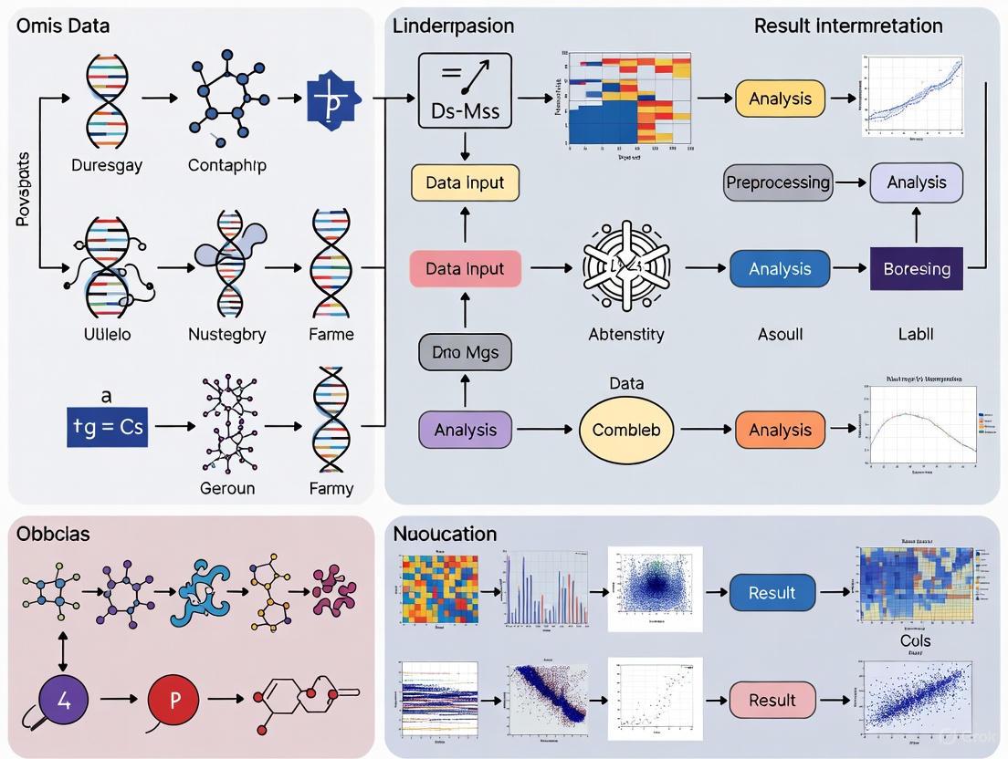
High-dimensional omics technologies provide a comprehensive, system-wide view of biological molecules, enabling researchers to move beyond studying single molecules to understanding complex interactions within cells and tissues [1].
Table 1: The Four Core Omics Disciplines
| Omics Field | Definition & Scope | Key Measurement Technologies |
|---|---|---|
| Genomics | The study of the complete sequence of DNA in a cell or organism, including genes, non-coding regions, and structural elements [1]. | Single Nucleotide Polymorphism (SNP) chips, DNA sequencing (Next-Generation Sequencing), whole-genome sequencing [1]. |
| Transcriptomics | The study of the complete set of RNA transcripts (mRNA, rRNA, tRNA, miRNA, and other non-coding RNAs) produced by the genome [1]. | Microarrays, RNA sequencing (RNA-Seq) [1]. |
| Proteomics | The study of the complete set of proteins expressed by a cell, tissue, or organism, including post-translational modifications and protein interactions [1]. | Mass spectrometry, protein microarrays, selected reaction monitoring (SRM) [1]. |
| Metabolomics | The study of the complete set of small-molecule metabolites (e.g., sugars, lipids, amino acids) found within a biological sample [1]. | Mass spectrometry, nuclear magnetic resonance (NMR) spectroscopy [1]. |
Figure 1: Omics disciplines and their primary measurement technologies.
Frequently Asked Questions (FAQs) & Troubleshooting
Q: What are the common approaches for integrating multiple omics datasets?
There are two primary approaches for multi-omics integration [2]:
1. Knowledge-Driven Integration: This approach uses prior biological knowledge from established databases (like KEGG metabolic networks, protein-protein interactions, or TF-gene-miRNA interactions) to link key features across different omics layers. This method helps identify activated biological processes but is mainly limited to model organisms and carries the bias of existing knowledge [2].
2. Data & Model-Driven Integration: This approach applies statistical models or machine learning algorithms to detect key features and patterns that co-vary across omics layers. It is not confined to existing knowledge and is more suitable for novel discovery. However, a wide variety of methods exist with no consensus approach, and each carries its own model assumptions and pitfalls [2].
Q: How should I handle missing values in my lipidomics or metabolomics data?
Missing values are common in omics data and require careful handling [3]:
- Identify the Nature of Missing Values: Determine if data are Missing Completely at Random (MCAR), Missing at Random (MAR), or Missing Not at Random (MNAR). MNAR often occurs when analyte abundance falls below the detection limit [3].
- Apply Appropriate Imputation: For MNAR data (common when concentrations are below detection limits), imputation with a percentage of the lowest concentration (half-minimum method) often works well. For MCAR/MAR data, k-nearest neighbors (kNN) or random forest imputation methods are generally recommended [3].
- Filter Before Imputation: Remove variables (lipids/metabolites) with excessive missing values (e.g., >35%) before performing imputation [3].
Q: My multi-omics data shows batch effects. How can I normalize this data effectively?
Data normalization aims to remove unwanted technical variation while preserving biological signal [3]:
- Use Quality Control (QC) Samples: Incorporate QC samples prepared from pooled aliquots of all biological samples or certified reference materials throughout your acquisition sequence [3].
- Apply Appropriate Normalization Methods:
- For scRNA-seq, scATAC-seq, and spatial transcriptomics: Log normalization is typically applied [4].
- For antibody-derived tag (ADT) data: Use centered log-ratio (CLR) transformation [4].
- For bulk RNA-seq and NanoString GeoMx data: Apply trimmed mean of M-values (TMM) normalization for dimensional reduction analysis [4].
- Leverage Visualization Tools: Use principal component analysis (PCA) and quality control trend plots to detect and correct for batch effects or systematic drift early in the preprocessing pipeline [5].
Q: Which computational tools are recommended for analyzing and visualizing omics data?
Table 2: Essential Tools for Omics Data Analysis
| Tool Name | Type | Primary Function | Key Features |
|---|---|---|---|
| R & Python | Programming Languages | Statistical analysis and visualization of omics data | Extensive packages for specialized analyses; enable reproducible research [5] [3]. |
| OmicsAnalyst | Web Platform | Data & model-driven multi-omics integration | Interactive 3D visual analytics, correlation networks, dual-heatmap viewer [2]. |
| OmnibusX | Desktop Application | Unified multi-omics analysis platform | Code-free analysis; integrates Scanpy, Seurat; privacy-focused local processing [4]. |
| MetaboAnalyst | Web Platform | Metabolomics data analysis | Comprehensive pipeline from data upload to visualization [3]. |
| GitBook Resources | Code Repository | Lipidomics/metabolomics data processing | Step-by-step R/Python notebooks for beginners [5] [3]. |
Figure 2: Omics data processing workflow with key troubleshooting checkpoints.
Q: What are the FAIR data principles and why are they important for omics research?
FAIR data principles are essential for maximizing the value and longevity of omics data [6]:
- Findable: Data and metadata should be easily locatable by both humans and computers, typically through persistent identifiers and rich descriptions.
- Accessible: Data should be retrievable using standardized protocols, even if access restrictions exist.
- Interoperable: Data should integrate with other datasets and applications through use of shared languages and vocabularies.
- Reusable: Data should have rich metadata and clear usage licenses to enable future replication and combination.
These principles extend data utility beyond original research purposes and are increasingly mandated by major funders including the NIH, NSF, and Horizon Europe [6].
The Scientist's Toolkit: Research Reagent Solutions
Table 3: Essential Research Reagents and Materials for Omics Experiments
| Reagent/Material | Function in Omics Research | Application Notes |
|---|---|---|
| NIST SRM 1950 | Certified reference material for metabolomics/lipidomics of plasma samples | Used for quality control and normalization; helps evaluate technical variability [3]. |
| Ensembl Annotation Files | Standardized gene annotations for genomic and transcriptomic data | Provides consistent gene symbols and IDs; version 111 is current standard [4]. |
| Hashtag Oligos (HTOs) | Sample multiplexing in single-cell experiments | Enables pooling of multiple samples; demultiplexing performed computationally [4]. |
| Curated Marker Sets | Cell type identification in single-cell genomics | Provides reference signatures for automated cell type prediction [4]. |
| Quality Control (QC) Samples | Pooled sample aliquots for monitoring technical variance | Critical for evaluating data quality across acquisition batches [3]. |
Data Management & Visualization Best Practices
Data Management Protocols:
- Implement Rich Metadata Capture: Document experimental steps thoroughly using relational database structures to link metadata from multi-step experiments. This practice is crucial for interoperability and correct data interpretation by future users [6].
- Ensure Data Privacy: For clinical or sensitive data, use locally executable software solutions like OmnibusX that process data on-premises without external data transfer [4].
- Adopt Standardized Gene Identifiers: Use current Ensembl releases (version 111) and map older identifiers to current standards to ensure accurate gene annotation across datasets [4].
Visualization Guidelines:
- Optimize Color Selection: Use shades of blue rather than yellow for quantitative node encoding in networks. For link colors, choose complementary colors rather than similar hues to enhance discriminability [7].
- Manage Data Volume: For interactive 3D visualizations, limit total data points to less than 5,000 to maintain responsive performance [2].
- Leverage Multiple Plot Types: Utilize volcano plots for differential analysis, PCA for quality control, heatmaps for pattern visualization, and lipid maps for class-specific trends [5] [3].
Effective management of high-dimensional omics data requires both technical expertise in specialized methodologies and strategic implementation of data management principles. By adopting the troubleshooting approaches, analytical tools, and visualization practices outlined above, researchers can navigate the complexities of multi-omics research while ensuring their data remains accessible, interpretable, and valuable for future scientific discovery.
Frequently Asked Questions (FAQs)
FAQ 1: What are the primary types of bottlenecks in modern multi-omics research? The bottleneck in omics research has shifted from the technical generation of data to the computational and analytical challenges of integration and interpretation [8]. The main bottlenecks now include:
- Data Integration: Combining heterogeneous data types (e.g., genomics, proteomics, metabolomics) with different scales, distributions, and levels of noise [8] [9].
- Interpretation: Translating integrated data into biologically meaningful insights, mechanistic understanding, and clinically actionable knowledge [10] [11].
- Workflow Efficiency: Delays caused by serial, ticket-driven data operations where researchers wait for data cleaning, access, and analysis, significantly slowing down research cycles [12].
FAQ 2: Why is multi-omics data integration so challenging from a technical perspective? Multi-omics data integration is complex due to several inherent technical characteristics of the data [8] [9]:
- High Dimensionality (HDLSS): The number of variables (e.g., genes, proteins) vastly exceeds the number of samples, increasing the risk of overfitting and spurious findings.
- Data Heterogeneity: Different omics layers have completely different data types, scales, and distributions.
- Technical Noise: Batch effects, missing values, and varying precision across measurement platforms can obscure biological signals.
- Biological Complexity: Regulatory relationships between different omics layers (e.g., how genomics influences transcriptomics) must be accounted for to achieve a holistic view.
FAQ 3: What are the main strategies for integrating multi-omics data? Integration strategies can be categorized based on when the data from different sources are combined during analysis [8] [13]:
Table 1: Multi-Omics Data Integration Strategies
| Strategy | Description | Pros | Cons |
|---|---|---|---|
| Early Integration | Raw or pre-processed data from all omics layers are concatenated into a single matrix before analysis [8] [13]. | Simple to implement [8]. | Creates a complex, high-dimensional matrix; discounts data distribution differences and can be noisy [8]. |
| Intermediate Integration | Data are transformed into new representations, and integration happens during the modeling process, often capturing joint latent structures [8] [13]. | Can reduce noise and dimensionality; captures inter-omics interactions [8]. | Requires robust pre-processing; methods can be complex [8]. |
| Late Integration | Each omics dataset is analyzed separately, and the results (e.g., model predictions) are combined at the end [8] [13]. | Circumvents challenges of assembling different data types [8]. | Fails to capture interactions between different omics layers [8]. |
FAQ 4: How can I address the "large p, small n" problem in my omics dataset? The "large p, small n" (high dimensionality, low sample size) problem can be addressed through a combination of statistical and machine learning techniques [8] [9]:
- Dimensionality Reduction: Use techniques like Principal Component Analysis (PCA) or autoencoders to project data into a lower-dimensional space while preserving key information [13].
- Feature Selection: Apply penalized regression methods like LASSO, which shrink the coefficients of non-informative features to zero, to select the most relevant variables [9] [11].
- Regularization: Incorporate penalties into model training to prevent overfitting and improve generalizability.
- Employ Specific Frameworks: Use models designed for high-dimensional data, such as DIABLO (Data Integration Analysis for Biomarker discovery using Latent cOmponents) or MOFA (Multi-Omics Factor Analysis) [9].
Troubleshooting Guides
Issue 1: Inconsistent or Inaccurate Results After Data Integration
Symptoms:
- Biomarker lists or pathway analysis results change drastically with slight changes in the data or model parameters.
- Models perform well on training data but poorly on validation data (overfitting).
- Different integration methods yield conflicting biological conclusions.
Diagnosis and Resolution:
- Check 1: Data Preprocessing
- Action: Ensure consistent and appropriate normalization has been applied to each omics dataset. For RNA-seq data, use methods like DESeq2's median-of-ratios or edgeR's TMM. For proteomics, use quantile normalization or variance-stabilizing normalization [9].
- Rationale: Technical variations like library size or ionization efficiency can confound biological signals without proper normalization.
Check 2: Batch Effect Correction
- Action: Apply batch correction algorithms such as ComBat, Limma's
removeBatchEffect(), or surrogate variable analysis (SVA) to remove non-biological technical variance [9]. - Rationale: Differences in sample processing dates, reagent batches, or sequencing lanes can introduce systematic noise that is mistaken for biological signal.
- Action: Apply batch correction algorithms such as ComBat, Limma's
Check 3: Model Validation
- Action: Implement rigorous validation using held-out test sets or resampling methods like cross-validation. Use performance metrics like AUC-ROC that are robust to class imbalance.
- Rationale: This helps identify and mitigate overfitting, a common issue in high-dimensional data, ensuring model generalizability [8] [9].
Issue 2: Poor Performance of Machine Learning Models on Integrated Omics Data
Symptoms:
- Model accuracy is no better than random chance.
- The model fails to converge during training.
- The model is unstable and produces different results on different training runs.
Diagnosis and Resolution:
- Check 1: Feature Quality and Selection
- Action: Perform rigorous feature selection to remove non-informative variables before integration and model training. Use domain knowledge or statistical filters to pre-select features.
- Rationale: High-dimensional data contains many irrelevant features that can "confuse" the model and increase noise [10] [13].
Check 2: Data Scaling
- Action: Standardize or normalize features within each omics dataset so that they are on a comparable scale (e.g., using Z-score normalization).
- Rationale: Machine learning algorithms can be sensitive to the scale of input variables. Dominant scales from one omics type can bias the model.
Check 3: Integration Strategy Suitability
- Action: Re-evaluate the choice of integration strategy. If using early integration leads to poor performance, consider an intermediate or late integration approach that better handles data heterogeneity [8].
- Rationale: The simple concatenation of data in early integration can be suboptimal for capturing complex, non-linear relationships across omics layers.
Issue 3: Difficulty in Biologically Interpreting Integrated Model Outputs
Symptoms:
- Models like deep neural networks are "black boxes," providing predictions but no mechanistic insight.
- It is challenging to link key features from the model to established biological pathways or functions.
Diagnosis and Resolution:
- Check 1: Pathway and Network Analysis
- Action: Feed the list of important features (e.g., genes, proteins) identified by your model into topology-based pathway analysis tools such as Signaling Pathway Impact Analysis (SPIA) or similar methods [11].
- Rationale: These methods consider the position, interaction type, and direction of molecules within a pathway, providing a more biologically realistic assessment of pathway dysregulation than simple enrichment analysis.
- Check 2: Use of Explainable AI (XAI) Techniques
- Action: Apply XAI methods like SHAP (SHapley Additive exPlanations) or LIME (Local Interpretable Model-agnostic Explanations) to complex models to determine which input features were most important for a given prediction [10].
- Rationale: XAI can help crack open the "black box" by quantifying feature importance, making complex model outputs more interpretable for biologists and clinicians.
Experimental Protocols
Protocol 1: A Workflow for Topology-Based Multi-Omics Pathway Activation and Drug Ranking
This protocol outlines a methodology for integrating multiple omics layers to calculate pathway activation levels and rank potential therapeutic drugs [11].
1. Objective: To integrate DNA methylation, coding RNA (mRNA), microRNA (miRNA), and long non-coding RNA (lncRNA) data to assess signaling pathway activation and compute a Drug Efficiency Index (DEI) for personalized drug ranking.
2. Materials and Reagents: Table 2: Key Research Reagent Solutions for Multi-Omics Pathway Analysis
| Item | Function / Description |
|---|---|
| Oncobox Pathway Databank (OncoboxPD) | A knowledge base of 51,672 uniformly processed human molecular pathways used for pathway activation calculations [11]. |
| SPIA Algorithm | A topology-based method that uses gene expression data and pathway structure to calculate pathway perturbation [11]. |
| Drug Efficiency Index (DEI) Software | Software that analyzes custom expression data to evaluate SPIA scores and statistically evaluate differentially regulated pathways for drug ranking [11]. |
| Normalization Reagents/Algorithms | Platform-specific reagents and software (e.g., DESeq2 for RNA-seq, quantile normalization for proteomics) to normalize raw data before integration [9]. |
3. Step-by-Step Procedure:
- Step 1: Data Generation and Preprocessing
- Generate profiles for DNA methylation, mRNA, miRNA, and lncRNA from case and control samples.
- Perform platform-specific normalization and quality control on each dataset individually [9].
Step 2: Data Transformation for Integration
- For mRNA data: Use expression values directly.
- For DNA methylation, miRNA, and lncRNA data: Treat these as repressive regulatory layers. Transform their data to reflect their negative regulatory impact on gene expression. The study calculated these SPIA values with a negative sign compared to mRNA-based values:
SPIA_methyl, ncRNA = -SPIA_mRNA[11].
Step 3: Pathway Activation Level (PAL) Calculation
- Use the SPIA algorithm to calculate pathway perturbation scores for each omics layer.
- The SPIA score combines a classical enrichment P-value with a perturbation factor that measures the propagation of expression changes through the pathway topology graph [11].
- The formula for the perturbation factor (PF) for a gene g is:
PF(g) = ΔE(g) + Σ β(g,u) * PF(u) / N_ds(u)whereΔE(g)is the normalized expression change,β(g,u)is the interaction type between gene g and its upstream gene u, andN_ds(u)is the number of downstream genes of u [11].
Step 4: Multi-Omics Data Aggregation
- Aggregate the pathway activation scores from all transformed omics layers (mRNA, methylation, miRNA, lncRNA) into a unified platform.
Step 5: Drug Efficiency Index (DEI) Calculation and Ranking
- Use the DEI software to analyze the integrated pathway activation profiles from your sample against a built-in set of controls.
- The DEI algorithm ranks drugs based on their predicted ability to reverse the disease-specific pathway activation signature toward a normal state [11].
4. Visualization of Workflow: The following diagram illustrates the logical flow of the multi-omics integration and analysis protocol.
Protocol 2: A Deep Learning Framework for Multi-Omics Integration (SynOmics)
This protocol describes using a graph-based deep learning model, SynOmics, to integrate multi-omics data for biomedical classification tasks [14].
1. Objective: To construct a graph convolutional network that models both within-omics and cross-omics feature dependencies for enhanced predictive performance.
2. Materials:
- Software: Python with deep learning libraries (e.g., PyTorch, TensorFlow).
- Omics Data: Matched multi-omics datasets (e.g., genomics, transcriptomics, proteomics) with sample labels.
3. Step-by-Step Procedure:
- Step 1: Data Preprocessing
- Clean, normalize, and scale each omics dataset individually. Handle missing values through imputation or removal.
Step 2: Network Construction
- Construct two types of networks in the feature space:
- Intra-omics Networks: Capture relationships between features within the same omics layer.
- Cross-omics Bipartite Networks: Capture regulatory and interaction relationships between features from different omics layers [14].
- Construct two types of networks in the feature space:
Step 3: Model Training with Graph Convolutional Networks (GCN)
- The SynOmics framework uses a parallel learning strategy where both intra-omics and inter-omics graphs are processed simultaneously at each layer of the GCN.
- The model learns node embeddings that incorporate both the local structure of one omics type and the global structure from cross-omics interactions [14].
Step 4: Model Validation and Interpretation
- Validate the model on held-out test sets for classification accuracy.
- Use feature importance and graph analysis techniques to interpret which intra- and inter-omics interactions were most informative for the prediction.
4. Visualization of Framework: The following diagram outlines the core architecture of the SynOmics integration model.
Statistical and Data Handling Reference
Summary of Common Statistical Challenges and Solutions in Multi-Omics Research Table 3: Statistical Pitfalls and Remedial Strategies for High-Dimensional Omics Data
| Statistical Challenge | Potential Impact on Research | Recommended Solutions & Methods |
|---|---|---|
| High Dimensionality (HDLSS) | Overfitting, spurious associations, reduced model generalizability [8] [9]. | Dimensionality reduction (PCA, Autoencoders), feature selection (LASSO), penalized regression [9] [13]. |
| Batch Effects | False positives/negatives, technical variance mistaken for biological signal [9]. | Batch correction algorithms (ComBat, Limma), study design randomization, SVA [9]. |
| Data Heterogeneity | Inability to directly compare or integrate datasets, leading to biased or incomplete models [8]. | Use of integration frameworks designed for heterogeneity (e.g., MOFA, DIABLO), late or intermediate integration strategies [8] [9]. |
| Missing Values | Reduced sample size, biased parameter estimates if not handled correctly [8]. | Imputation methods (e.g., k-nearest neighbors, matrix completion), or model-based approaches that account for missingness [8]. |
Frequently Asked Questions (FAQs)
Q1: What are the main computational challenges when analyzing high-dimensional single-cell data, such as from CyTOF or scRNA-seq?
The primary challenges stem from the data's high dimensionality and complex nature. Traditional analysis methods like manual gating become inefficient and biased when dealing with 50+ parameters per cell [15]. Key issues include:
- Dimensionality Explosion: The number of required bivariate plots increases quadratically with the number of measured parameters, making manual analysis laborious and confounded [15].
- Noire: The presence of technical and biological noise can obscure true signals, complicating the identification of genuine cell populations and biomarkers [16].
- Cellular Heterogeneity and Complexity: High-resolution technologies reveal continuous cellular differentiation trajectories and complex subset relationships that are difficult to capture with manual, discrete gating strategies [15].
- Data Scale and Integration: Multiomics studies generate massive, disparate datasets with varying formats, scales, and biological contexts. Integrating these data requires sophisticated computational tools and harmonization efforts [17].
Q2: How can I visualize high-dimensional data to better understand cellular heterogeneity and transitions?
Non-linear dimensionality reduction techniques are essential for visualizing high-dimensional data in two or three dimensions. The table below compares common methods:
| Method | Description | Key Advantages | Considerations |
|---|---|---|---|
| t-SNE [15] | t-stochastic neighbor embedding; maps cells to a lower-dimensional space based on pairwise similarities. | Provides intuitive clustering of similar cells; excellent for revealing local structure and distinct populations. | Can be stochastic (results vary per run); less effective at preserving global data structure; perplexity parameter requires tuning. |
| UMAP [15] | Uniform Manifold Approximation and Projection; a novel manifold learning technique. | Better preservation of global data structure than t-SNE; faster and more scalable; good resolution of rare and transitional cell types [15]. | |
| PHATE [16] | Potential of Heat Diffusion for Affinity-based Transition Embedding; encodes local and global data structure using a potential distance. | Robust to noise; particularly effective for identifying patterns like branching trajectories (e.g., cell differentiation) [16]. | |
| HSNE [15] | Hierarchical Stochastic Neighbor Embedding; constructs a hierarchy of non-linear similarities. | Enables interactive exploration of large datasets from an overview down to single-cell details; effective for rare cell type identification [15]. |
Q3: Our multi-omics data comes from different cohorts and labs, leading to integration issues. How can this be addressed?
Harmonizing disparate data sources is a central challenge in multi-omics. An optimal approach involves:
- Pre-Integration Harmonization: Collecting multiple omics datasets on the same set of samples where possible [17].
- Integrated Data Processing: Integrating data signals from each omics layer prior to higher-level statistical analysis, rather than analyzing datasets individually and correlating results post-hoc [17].
- Network Integration: Mapping multiple omics datasets onto shared biochemical networks (e.g., connecting transcription factors to their target transcripts or enzymes to their metabolites) to improve mechanistic understanding [17].
- AI and Machine Learning: Leveraging advanced computational tools to detect intricate patterns and interdependencies across the integrated data modalities [17].
Q4: What are the best practices for identifying cell populations in an unbiased way in high-dimensional cytometry data?
Unsupervised clustering methods are recommended to overcome the bias of manual gating [15]. The following table outlines key algorithms:
| Method | Type | Description | Key Utility |
|---|---|---|---|
| FlowSOM [15] | Clustering | Uses self-organizing maps trained to detect cell populations. | Fast, scalable method for automatic cell population identification. |
| SPADE [15] | Clustering & Visualization | Creates a hierarchical branched tree representation of cell relationships. | Helps in understanding cellular hierarchy and relationships between subsets. |
| PAGA [15] | Trajectory Inference & Graph Abstraction | Reconstitutes topological information from single-cell data into a graph of cellular relationships. | Provides an interpretable graph-based map of cellular dynamics, such as differentiation trajectories. |
Q5: How can I infer dynamic processes, like cellular differentiation, from static snapshot single-cell data?
Trajectory inference algorithms can reconstruct dynamic temporal ordering from static data. Diffusion Pseudotime (DPT) is a method that investigates continuous cellular differentiation trajectories, allowing researchers to order cells along a pseudo-temporal continuum based on their expression profiles [15]. This is particularly powerful for understanding processes like immune cell differentiation or stem cell development from a single snapshot sample.
Troubleshooting Guides
Issue: Poor Cell Population Separation in Dimensionality Reduction Plots
Problem: Your t-SNE or UMAP plot appears as a single, unresolved blob, making it difficult to distinguish distinct cell populations.
Solution:
- Check Data Preprocessing: Ensure data has been properly transformed (e.g., arcsinh for CyTOF) and normalized. Technical noise can mask biological signals.
- Adjust Algorithm Parameters:
- For t-SNE, try increasing the
perplexityparameter (values between 5-50 are common) and the number ofiterations[15]. Run multiple times to ensure stability. - For UMAP, adjust the
n_neighborsparameter. A lower value emphasizes local structure, while a higher value captures more global structure.
- For t-SNE, try increasing the
- Consider Alternative Methods: If non-linear methods fail, try a different visualization algorithm like PHATE, which is specifically designed to capture continuous trajectories and can be more robust to noise [16].
- Re-evaluate Clustering: The clusters used to color the plot may not be optimal. Experiment with different clustering algorithms (e.g., FlowSOM) or resolution parameters.
Issue: Integrating Disparate Multi-Omics Data Types
Problem: You have genomic, transcriptomic, and proteomic data from the same biological system, but cannot effectively combine them for a unified analysis.
Solution:
- Data Harmonization: Before integration, ensure all datasets are reprocessed with standardized metadata and normalized to account for technical batch effects [17].
- Use Multi-Modal Integration Tools: Employ computational frameworks designed for multi-omics integration rather than analyzing each data type in a siloed workflow [17].
- Leverage Network-Based Integration: Map your various omics datasets (e.g., genes, transcripts, proteins) onto shared biochemical networks. This connects analytes based on known interactions, providing a mechanistic framework for integration [17].
- Apply AI/ML Models: Utilize artificial intelligence and machine learning methods, such as variational autoencoders (VAEs) or graph convolutional networks (GCNs), which are advanced for extracting features and integrating multiple data modalities into a cohesive model [18] [17].
The Scientist's Toolkit: Research Reagent Solutions
This table details key reagents and materials used in high-dimensional single-cell and multi-omics research.
| Item | Function |
|---|---|
| Antibodies Labeled with Metal Isotopes | For mass cytometry (CyTOF); enables simultaneous measurement of >40 protein markers per cell without spectral overlap found in fluorescence-based flow cytometry [15]. |
| Heavy Metal Isotopes | The labels for antibodies in CyTOF; their detection via time-of-flight mass spectrometry allows for high-parameter single-cell proteomic profiling [15]. |
| Single-Cell Multi-Omics Assay Kits | Commercial kits that enable correlated measurements of genomic, transcriptomic, and epigenomic information from the same single cells [17]. |
| Cell Hash Tagging Antibodies | Antibodies conjugated to oligonucleotides that allow sample multiplexing, reducing batch effects and reagent costs in single-cell sequencing experiments. |
| Viability Stain (e.g., Cisplatin) | A cell membrane-impermeant metal chelator used in CyTOF to identify and exclude dead cells during analysis. |
Experimental Protocols
Protocol 1: Standard Workflow for High-Dimensional CyTOF Data Analysis
This protocol outlines a standard computational pipeline for analyzing CyTOF data, from raw files to biological insights [15].
Detailed Methodology:
Data Pre-processing & Normalization:
- Load the raw data (FCS files) into your analysis platform (e.g., R, Python, or specialized software like Cytobank).
- Normalize signal intensity across samples to correct for instrument drift and variation using bead-based or standard normalization algorithms.
- Apply an arcsinh transformation with a cofactor (e.g., 5) to stabilize the variance of the measured markers and bring the data to a more Gaussian-like distribution.
Dimensionality Reduction:
- Select the parameters (markers) for analysis, typically those defining cell phenotype and function.
- Run a non-linear dimensionality reduction algorithm such as UMAP or t-SNE on the transformed data to visualize the high-dimensional data in 2D. This step helps reveal the underlying structure and heterogeneity of the cell population.
- Optional: Use PCA as an initial step to reduce computational time for subsequent analyses.
Unsupervised Clustering:
- Apply an automated clustering algorithm like FlowSOM or PhenoGraph to identify distinct cell populations in an unbiased manner. These algorithms group cells based on the similarity of their marker expression profiles.
- Overlay the cluster identities onto the UMAP/t-SNE plot to visualize the results.
Differential Analysis & Biomarker Identification:
- Compare marker expression levels (using statistical tests like t-tests or Mann-Whitney U tests) between clusters to identify defining features (biomarkers) for each population.
- Compare the relative abundances of cell populations between different experimental conditions (e.g., healthy vs. diseased).
Trajectory Inference (if applicable):
- For data suggesting continuous processes (e.g., differentiation), use a trajectory inference algorithm like Diffusion Pseudotime (DPT) [15] to reconstruct the cellular progression and order cells along a pseudo-temporal trajectory.
Protocol 2: Integrated Multi-Omics Analysis for Biomarker Discovery
This protocol describes a strategy for integrating multiple omics datasets to discover robust biomarkers and therapeutic targets [17].
Detailed Methodology:
Sample & Data Collection:
- Collect multiple omics datasets (e.g., genomics, transcriptomics, proteomics) from the same set of patient samples to ensure direct comparability.
Data Harmonization & Pre-processing:
- Independently pre-process each omics dataset using established pipelines (e.g., alignment for sequencing data, normalization for proteomics data).
- Perform batch effect correction and data harmonization to account for technical variations across different processing batches or platforms.
Integrated Data Analysis:
- Early Integration: Combine the normalized data matrices from the different omics layers into a single, unified dataset prior to running statistical or machine learning models. This approach allows the model to learn from the combined signal of all modalities simultaneously [17].
- Network Integration: As an alternative or complementary approach, map the analytes from each omics dataset (e.g., differentially expressed genes, proteins) onto a shared interaction network (e.g., protein-protein interaction, metabolic pathway). This contextualizes findings within known biology and can reveal dysregulated network modules.
AI/ML-Based Pattern Recognition:
- Apply machine learning or deep learning models (e.g., autoencoders, graph neural networks) to the integrated data. These models are adept at detecting complex, non-linear patterns that would be impossible to find by analyzing each data type individually [18] [17].
- Use these models for tasks like patient stratification, prediction of disease progression, or identification of master regulatory nodes in the integrated network.
Validation:
- Validate the discovered biomarkers or targets using an independent cohort of samples or through functional experimental studies.
Technical Support Center
Troubleshooting Guides
Guide 1: Troubleshooting High-Dimensional Data Analysis
Reported Issue: Model overfitting and poor generalizability on new datasets.
- Potential Cause: High-dimension low sample size (HDLSS) problem where variables significantly outnumber samples [8].
- Solution:
Reported Issue: Inability to integrate multiple omics data types.
- Potential Cause: Data heterogeneity across different omics platforms and modalities [8].
- Solution:
- Apply data transformation and normalization specific to each omics modality [8].
- Choose appropriate integration strategy (Early, Mixed, Intermediate, Late, or Hierarchical) based on research goals [8].
- Use tools like Multi-Omics Factor Analysis (MOFA) for pattern recognition across omics layers [19].
Guide 2: Troubleshooting Computational Infrastructure Limitations
Reported Issue: Insufficient computing power for population-scale omics analysis.
- Potential Cause: Local computational resources cannot handle datasets with thousands of samples and millions of data points [20] [21].
- Solution:
Reported Issue: Long processing times for genome-wide association studies.
- Potential Cause: Standard workstations insufficient for datasets with 1000 individuals and 100,000 SNPs [20].
- Solution:
Frequently Asked Questions (FAQs)
Q: What strategies can help overcome the high computational costs of omics analysis? A: Several cost-management strategies can improve accessibility:
- Implement pay-as-you-go cloud computing models to avoid large capital investments [21].
- Use data compression algorithms to reduce storage requirements [19].
- Leverage public data repositories like TCGA that offer cloud-based access to minimize data transfer costs [22].
- Apply efficient imputation methods for missing values rather than repeating expensive experiments [8] [19].
Q: How can researchers with limited bioinformatics training analyze complex omics datasets? A: Multiple user-friendly solutions are available:
- Utilize graphical interfaces like EasyOmics that enable point-and-click analysis without coding [20].
- Implement platforms like Omics Playground for interactive visualization and analysis [23].
- Access pre-built workflows on cloud platforms (Terra, DNAnexus) that automate complex analyses [21].
- Employ multivariate data analysis software like SIMCA with specialized omics interfaces [24].
Q: What are the best practices for ensuring statistical rigor in high-dimensional omics studies? A: Follow these established methodologies:
- Apply multiple testing corrections (e.g., Benjamini-Hochberg) to control false discovery rates [19].
- Implement rigorous quality control measures including control samples and replicates [19].
- Use cross-validation and held-out datasets to verify predictions [25].
- Correct for batch effects using methods like quantile normalization and ComBat [19].
Q: How can we effectively integrate multi-omics data from different technological platforms? A: Successful integration requires:
- Standardization using FAIR (Findable, Accessible, Interoperable, Reusable) data principles [19].
- Choosing integration methods based on data structure (horizontal vs. vertical integration) [8].
- Using specialized tools for simultaneous visualization of multiple omics types on metabolic networks [26].
- Applying novel approaches like the HYFT framework that tokenizes biological data to a common language [8].
Quantitative Data Tables
Computational Requirements for Omics Analysis
Table 1: Cloud Computing Costs for Different Omics Data Types (Approximate)
| Omics Type | Platform | Data Size | Analysis Cost |
|---|---|---|---|
| Genome [22] | DNA sequencing | >100 GB | $40-$66 per test |
| Transcriptome [22] | RNA-seq | >2000 samples | $1.30 per sample |
| Proteome [22] | Protein mass spectrometry | Standard mix dataset | >$1 per database search |
| Metabolite [22] | Metabolite mass spectrometry | ~1 GB | $11 per processing |
| Microbiome [22] | rRNA gene sequencing | >90 GB | ~$8 per GB + $400 prep |
Table 2: Computational Performance Metrics for Omics Analysis
| Analysis Type | Dataset Size | Runtime | RAM Usage |
|---|---|---|---|
| GWAS [20] | 1,000 individuals, 100,000 SNPs | <2 minutes | <16 GB |
| GWAS [20] | 1,000 individuals, 10,000,000 SNPs | ~110 minutes | <16 GB |
| OmicQTL [20] | 1,000 individuals, 10M SNPs, 20K features | Hours | ≤15 GB |
| Multi-omics Visualization [26] | 3,209 reactions, 1,796 compounds, 20 timepoints | 20 seconds | Moderate |
Experimental Protocols
Protocol 1: Multi-Omics Data Integration and Analysis
Objective: To integrate and analyze datasets from multiple omics platforms (genomics, transcriptomics, proteomics, metabolomics) for comprehensive biological insight.
Methodology:
- Data Pre-processing
Data Integration Strategy Selection
- Choose based on research question:
- Early Integration: Concatenate all datasets into single matrix [8].
- Mixed Integration: Separately transform each dataset then combine [8].
- Intermediate Integration: Simultaneous integration producing common and omics-specific representations [8].
- Late Integration: Analyze each omics separately then combine predictions [8].
- Hierarchical Integration: Include prior regulatory relationships between omics layers [8].
- Choose based on research question:
Multivariate Data Analysis
Protocol 2: Population-Scale Omics Association Analysis
Objective: To identify genetic variations associated with complex traits using population-scale omics data.
Methodology:
- Data Quality Control and Preparation
Genome-Wide Association Scan
- Implement mixed linear model to detect genetic variations [20].
- Apply Bonferroni-corrected threshold for significance [20].
- Generate Manhattan plots and quantile-quantile (QQ) plots automatically [20].
- Prioritize association signals by statistical significance, physical distance, and linkage disequilibrium [20].
Validation and Interpretation
Visualizations
Multi-Omics Data Integration Workflow
Omics Data Analysis Challenges and Solutions
Research Reagent Solutions
Table 3: Essential Tools and Platforms for Omics Research
| Tool Category | Specific Solutions | Function |
|---|---|---|
| Data Analysis Platforms | EasyOmics [20] | User-friendly graphical interface for association analysis without coding |
| Omics Playground [23] | Interactive visualization and analysis platform | |
| SIMCA [24] | Multivariate data analysis software with specialized omics capabilities | |
| Cloud Computing Platforms | Terra [21] | Cloud platform optimized for genomic workflows |
| AWS HealthOmics [21] | Amazon's specialized service for healthcare omics data | |
| Google Cloud Life Sciences [21] | Google's solution for life sciences data analysis | |
| Visualization Tools | Pathway Tools Cellular Overview [26] | Simultaneous visualization of up to four omics data types on metabolic networks |
| Cytoscape [26] | Network visualization and analysis | |
| Escher [26] | Manual creation of pathway diagrams with data overlay | |
| Statistical Analysis | MOFA [19] | Multi-Omics Factor Analysis for identifying patterns across omics layers |
| iCluster [19] | Tool for integrated clustering of multiple omics data types |
Computational Tools and Integration Strategies for Multi-Omics Analysis
In multi-omics research, data integration is the computational process of combining multiple layers of biological information (such as genomics, transcriptomics, proteomics, and epigenomics) to gain a unified and comprehensive understanding of a biological system. [27] [28]
The core challenge is that each omics layer has a unique data scale, noise ratio, and preprocessing steps, making integration a complex task without a universal one-size-fits-all solution. [27] [29] The choice of integration strategy is primarily determined by how the data was collected—specifically, whether different omics layers were measured from the same cells or from different samples. [27] This article classifies these approaches into three main types: Matched, Unmatched, and Mosaic integration, providing a troubleshooting guide to help you select and successfully apply the correct method for your research.
Comparison of Multi-Omics Integration Types
The following table summarizes the key characteristics, typical use cases, and popular tools for the three primary integration types.
| Integration Type | Data Source & Anchors | Primary Challenge | Example Tools & Methods |
|---|---|---|---|
| Matched (Vertical) [27] [28] | Data from different omics layers profiled from the same cell or sample. The cell itself is the anchor. [27] | Managing different data scales and noise profiles from multiple modalities measured on the same cell. [27] [29] | Seurat v4 [27], MOFA+ [27] [28], totalVI [27], DCCA [27] |
| Unmatched (Diagonal) [27] [28] | Data from different omics layers profiled from different cells or samples. Requires a computational anchor. [27] | Finding commonality between cells without a biological anchor; often requires projecting cells into a co-embedded space. [27] | GLUE [27], LIGER [27] [30], Pamona [27], Seurat v3 (CCA) [27] [30] |
| Mosaic [31] | Multiple datasets with varying combinations of omics layers. Requires sufficient overlapping features or datasets to connect them. [31] | Integrating datasets where some pairs do not share any direct features ("multi-hop" integration). [31] | StabMap [27] [31], Cobolt [27] [31], MultiVI [27] [31], Bridge Integration [27] [30] |
Troubleshooting Guide: FAQs and Solutions
Matched (Vertical) Integration
Q: My matched RNA and protein data show poor correlation for key markers. What could be wrong? A: This is a common occurrence, not necessarily an error. A weak correlation can reflect real biology, such as post-transcriptional regulation. Before concluding, troubleshoot the following:
- Check for Sample Misalignment: Ensure the RNA and protein data are truly from the same set of cells and that no sample mix-up occurred. [29]
- Verify Normalization: Confirm that each modality has been properly and independently normalized (e.g., RNA by library size, protein counts by CLR) before integration. Naively concatenating raw counts will skew results. [29]
- Investigate Biology: Use the discordance as a discovery tool. A highly expressed gene with low protein abundance may be subject to rapid degradation or strong microRNA regulation. [29]
Q: When I integrate my matched scRNA-seq and scATAC-seq data, the chromatin accessibility signal dominates the clustering. Why? A: This is often a normalization or scaling issue.
- Cause: The integration method may not be weighting the modalities equally. If one dataset has higher overall variance or was not scaled to a comparable range, it can dominate the shared low-dimensional space. [29]
- Solution: Use integration-aware tools like MOFA+ or Seurat v4 that are designed to handle the distinct statistical properties of each modality. [27] [19] Ensure you follow the tool-specific guidelines for normalizing each data type individually before running the integration workflow. [29]
Unmatched (Diagonal) Integration
Q: I am trying to integrate scRNA-seq from one experiment with scATAC-seq from another, but the cell types won't align. What anchors should I use? A: With unmatched data, the "anchor" is not biological but computational.
- Cause: The algorithm may be failing to find a valid common latent space, often due to strong batch effects or insufficient overlapping biological signal. [27]
- Solution:
- Leverage Manifold Alignment: Use methods like GLUE or Pamona, which project cells from both modalities onto a non-linear manifold to find commonality. [27]
- Incorporate Prior Knowledge: Tools like GLUE can use prior biological knowledge (e.g., gene-to-peak links) to guide the alignment and create a more accurate anchor. [27]
- Apply Batch Correction: Perform batch effect correction within each modality before attempting cross-modal integration to ensure technical differences are minimized. [29]
Q: How can I validate an unmatched integration result if I don't have ground truth? A: While challenging, you can assess integration quality using:
- Label Transfer Accuracy: If you have cell type labels for both datasets, train a classifier on one dataset and see how well it predicts labels in the other, using the integrated space. [31]
- Conservation of Structure: Check if the local neighborhood structure of cells from the same cell type is preserved in the integrated embedding. Good integration should group similar cell types together, regardless of their omics source. [31]
Mosaic Integration
Q: What is "multi-hop" integration, and when is it necessary? A: Multi-hop integration is a specific capability of mosaic integration tools.
- Scenario: You have three datasets: Dataset A (RNA+Protein), Dataset B (RNA+ATAC), and Dataset C (ATAC+Protein). There is no single feature shared by all three, but each pair has an overlap.
- Solution: Mosaic tools like StabMap or Bridge Integration can use Dataset B (RNA+ATAC) as a "bridge" to connect Dataset A and Dataset C. The method traverses the shortest path along this "mosaic data topology" to integrate all datasets into a common space, even without a globally shared feature. [31]
Q: My mosaic integration produces a fragmented embedding where datasets don't mix well. How can I improve stability? A: Fragmentation often occurs when the connections (shared features) between datasets are too weak or few.
- Cause: The mosaic data topology is unstable. If the shared feature set between two key datasets is small or non-informative, the mapping between them becomes unreliable. [31]
- Solution: Use a method like StabMap, which is explicitly designed to stabilize mapping by exploiting both shared and non-overlapping features across the entire topology of datasets, leading to a more robust and coherent integration. [31]
Workflow Diagrams for Integration Strategies
The following diagrams illustrate the logical decision process and core mechanisms for each integration type.
Decision Workflow for Multi-Omics Integration Types
Mechanisms of Matched, Unmatched, and Mosaic Integration
The Scientist's Toolkit: Key Research Reagents and Computational Solutions
The following table lists essential computational tools and conceptual "reagents" crucial for successful multi-omics integration.
| Tool / Solution | Function | Primary Integration Type |
|---|---|---|
| Seurat (v4/v5) [27] [30] | A comprehensive toolkit for single-cell analysis. Provides weighted nearest neighbors (WNN) for matched integration and bridge integration for complex mosaic scenarios. [27] | Matched, Mosaic |
| MOFA+ [27] [28] | A factor analysis model that infers a small number of latent factors that capture the shared and unique sources of variation across multiple omics layers. [27] [28] | Matched |
| StabMap [27] [31] | A mosaic data integration method that projects cells onto a reference by traversing shortest paths along a dataset topology, enabling "multi-hop" integration. [31] | Mosaic |
| GLUE (Graph-Linked Unified Embedding) [27] | A variational autoencoder-based method that uses a prior knowledge graph to link different omics layers, guiding the integration of unmatched data. [27] | Unmatched |
| LIGER (Linked Inference of Genomic Experimental Relationships) [27] [30] | Uses integrative non-negative matrix factorization (iNMF) to identify shared and dataset-specific factors, effective for aligning datasets from different modalities or technologies. [27] [30] | Unmatched |
| Integration Anchors (Conceptual) [27] [29] | The shared features or cells used to align datasets. Correctly identifying and using anchors is critical. These can be biological (the cell) or computational (shared features/latent space). [27] [29] | All Types |
| Cross-Modality Normalization (Conceptual) [32] [29] | The process of scaling different omics data types (e.g., RNA counts, protein counts, ATAC peaks) to a comparable range to prevent one modality from dominating the integrated analysis. [29] | All Types |
The advancement of high-throughput technologies has moved biomedical research into the age of omics, enabling scientists to track molecules such as DNAs, RNAs, proteins, and metabolites for a better understanding of human diseases [33]. However, translating large volumes of omics data into knowledge presents significant challenges, including missing observations, batch effects, and the complexity of choosing appropriate statistical models [33]. Multi-omics characterization of individual cells offers remarkable potential for analyzing the dynamics and relationships of gene regulatory states across millions of cells, but how to effectively integrate multimodal data remains an open problem [34]. This technical support center addresses specific issues researchers encounter when working with state-of-the-art tools for managing high-dimensional omics data, providing practical troubleshooting guides and FAQs framed within the broader context of omics data research management.
MOFA+ Troubleshooting Guide
Frequently Asked Questions
Q: I get the following error when running run_mofa in R: AttributeError: 'module' object has no attribute 'core.entry_point' or ModuleNotFoundError: No module named 'mofapy2'
A: This error typically indicates a Python configuration issue. First, restart R and try again. If the error persists, either the mofapy2 Python package is not installed, or R is detecting the wrong Python installation. Specify the correct Python interpreter at the beginning of your R script:
Alternatively, use use_conda() if you work with conda environments. You must install the mofapy2 Python package following the official installation instructions [35].
Q: I encounter installation errors for the MOFA2 R package with messages about unavailable dependencies
A: This occurs when trying to install Bioconductor dependencies using install.packages(). Instead, install these packages directly from Bioconductor:
Replace "DEPENDENCY_NAME" with the specific missing dependencies mentioned in the error message [35].
Q: My R version is older than 4.0. Can I still use MOFA2?
A: Yes, MOFA2 works with R versions 3 and above. You need to clone the repository and edit the DESCRIPTION file:
Edit the Depends option in the DESCRIPTION file to match your R version, then install using:
MOFA-FLEX Experimental Protocol
MOFA-FLEX represents a framework for factor analysis of multimodal data, with a focus on single-cell omics, modeling an observed data matrix as a product of low-rank factor and weight matrices [36]. Below is a detailed methodology for analyzing PBMC multiome data:
1. Data Import and Setup
2. Preprocessing for RNA Modality
3. Preprocessing for ATAC Modality
4. Model Fitting MOFA-FLEX automatically fits the model upon object creation. For normalized data, use a Normal (Gaussian) likelihood, while negative binomial likelihood is more appropriate for unnormalized count data [36].
MOFA+ Integration Workflow
Seurat Troubleshooting Guide
Frequently Asked Questions
Q: After merging multiple samples using the merge() function in Seurat v5.0, I get an error with GetAssayData()
A: In Seurat v5.0, the merge() function creates separate count layers for each sample by default, which prevents GetAssayData() from extracting the matrix. Resolve this by joining the layers before using GetAssayData():
After joining the layers, you can use GetAssayData() without errors. Note that this issue does not occur in Seurat v4 and earlier versions [37].
Q: I experience crashes when running Python-based functions like scVelo or Palantir on macOS
A: This issue differs between Intel and Apple Silicon Macs:
Intel Macs: When using R Markdown in RStudio with Python tools, the R session may crash unexpectedly. Use regular .R script files instead of R Markdown files.
Apple Silicon (M1/M2/M3/M4): If you load R objects before calling Python functions, the R session may crash due to memory management issues. Always initialize the Python environment BEFORE loading any R objects:
Q: I get a GLIBCXX version error when running scVelo-related functions in RStudio on Linux
A: This error occurs because RStudio cannot find the correct shared library 'libstdc++'. Check the library paths with Sys.getenv("LD_LIBRARY_PATH"). Copy the following files from your conda environment lib directory to one of the paths in LD_LIBRARY_PATH:
- libR.so
- libstdc++.so
- libstdc++.so.6
- libstdc++.so.6.0.32
After copying these files, restart your R session [37].
Seurat Data Preprocessing Protocol
1. Data Loading and Initialization
2. Quality Control Metrics
3. Quality Control Understanding
- nFeature_RNA: Number of genes detected per cell (low counts indicate dying cells)
- nCount_RNA: Total molecules detected per cell (high counts may indicate doublets/multiplets)
- percent.mt: Percentage of mitochondrial reads (high percentage indicates poor-quality cells or contamination) [38]
4. Data Filtering and Normalization
Seurat Analysis Workflow
Multi-omics Integration Tools Comparison
Performance Benchmarking of Integration Methods
Recent benchmarking studies evaluate multi-omics integration methods across multiple datasets and performance metrics. The following tables summarize quantitative comparisons based on studies of paired and unpaired single-cell multi-omics data.
Table 1: Performance Comparison on Paired 10x Multiome Data
| Method | Cell-type ASW | Batch ASW | FOSCTTM | Seurat Alignment Score |
|---|---|---|---|---|
| scHyper | High | High | Lowest | Better |
| scJoint | Moderate | Moderate | Moderate | Moderate |
| Seurat | Low | Low | High | Low |
| Liger | Low | Low | High | Low |
| Harmony | Low | Moderate | High | Low |
| GLUE | Low | Low | High | Low |
Table 2: Performance on Unpaired Mouse Atlas Data
| Method | Label Transfer Accuracy | Cell-type Silhouette | Batch ASW | FOSCTTM |
|---|---|---|---|---|
| scHyper | 85% | High | High | Lowest |
| GLUE | 77% | Moderate | Moderate | Low |
| scJoint | 72% | High | Moderate | Moderate |
| Conos | 67% | Low | Low | High |
| Harmony | 68% | Low | Moderate | High |
| Seurat | 56% | Low | Low | High |
Table 3: Performance on Multimodal PBMC Data (CITE-seq + ASAP-seq)
| Method | Label Transfer Accuracy | Cell-type Silhouette | Integration Quality |
|---|---|---|---|
| scHyper | 86% | High | High |
| scJoint | 84% | Moderate | Moderate |
| GLUE | 80% | Moderate | Moderate |
| Seurat | 75% | Low | Low |
Performance metrics explanation:
- ASW (Average Silhouette Width): Measures separation between cell types (higher is better)
- FOSCTTM (Fraction Of Samples Closer Than The True Match): Measures preservation of biological variation (lower is better)
- Seurat Alignment Score: Measures dataset integration (higher indicates better alignment) [34]
scHyper Integration Protocol
scHyper is a deep transfer learning model for paired and unpaired multimodal single-cell data integration that uses hypergraph convolutional encoders to capture high-order data associations across multi-omics data [34].
Experimental Workflow:
Hypergraph Construction: Create a hypergraph for each modality individually, forming multi-omics hypergraph topology by combining modality-specific hyperedges.
Feature Encoding: Use hypergraph convolutional encoder to capture high-order data associations across multi-omics data.
Transfer Learning: Apply efficient transfer learning strategy for large-scale atlas data integration.
Integration Evaluation: Assess using cell-type silhouette coefficient, ASW for cell types and omics layers, Seurat Alignment Score, and FOSCTTM values.
Key Advantages:
- Effectively handles both paired and unpaired multimodal data
- Achieves high accuracy in label transfer (85% on mouse atlas data)
- Maintains balance between reducing technical variations and preserving cell-type signals
- Demonstrates scalability to large atlas-level datasets [34]
Multi-omics Integration Decision Framework
Essential Research Reagent Solutions
Table 4: Key Computational Tools for Multi-omics Analysis
| Tool/Resource | Function | Application Context |
|---|---|---|
| MOFA2/MOFA-FLEX | Factor analysis for multimodal data | Multi-omics integration, dimensionality reduction |
| Seurat | Single-cell RNA-seq analysis | Clustering, visualization, differential expression |
| scHyper | Deep transfer learning for multi-omics | Paired and unpaired data integration |
| GLUE | Graph-linked unified embedding | Multi-omics integration, regulatory inference |
| Scanpy | Single-cell analysis in Python | Preprocessing, visualization, clustering |
| MuData | Multimodal data container | Standardized format for multi-omics data |
| AnnData | Annotated data matrix | Single-cell data representation |
| scVelo | RNA velocity analysis | Cell fate determination, dynamics |
The integration of multi-omics data remains a complex challenge in single-cell research, with various tools offering different strengths and limitations. As demonstrated by the benchmarking results, newer methods like scHyper show promising performance in both paired and unpaired data integration scenarios, particularly for large-scale atlas data [34]. The field continues to evolve with emerging approaches that better balance the reduction of technical variations with the preservation of biological signals.
Successful management of high-dimensional omics data requires not only selecting appropriate tools but also understanding their specific troubleshooting requirements, configuration dependencies, and optimal application contexts. The protocols and troubleshooting guides provided here offer researchers practical solutions for common challenges encountered when working with state-of-the-art multi-omics analysis tools.
As the volume and complexity of omics data continue to grow, developing robust, scalable, and user-friendly integration methods will remain crucial for extracting meaningful biological insights and advancing biomedical research.
Machine Learning and Deep Learning Approaches for Data Fusion
Troubleshooting Guides
Troubleshooting Scenario 1: Handling Missing or Noisy Multi-Omics Data
- Problem: Your deep learning model for cancer subtyping fails to converge or shows poor performance. Diagnostics reveal your multi-omics dataset (e.g., from TCGA) has missing values in the methylation data and noisy segments in the transcriptomic data.
- Background: High-dimensional omics data is often sparse and heterogeneous. Missing values can arise from experimental limitations, while noise can stem from technical artifacts [39] [40].
- Solution:
- Data Audit: Use summary statistics and visualization (e.g., heatmaps of missingness) to identify patterns. Determine if data is Missing Completely at Random (MCAR) or not.
- Imputation: Apply advanced imputation techniques.
- Robust Modeling: If missingness is significant, employ modeling strategies that are inherently robust, such as:
Troubleshooting Scenario 2: Model Interpretability in a Translational Study
- Problem: You have developed a high-accuracy deep learning model using
Flexynesisto predict drug response from genomic and proteomic data. However, for a publication on precision oncology, reviewers demand insights into the biological mechanisms and features driving the predictions. - Background: A key challenge of deep learning models is their "black box" nature, which limits clinical adoption as professionals require understandable rationale for decisions [43] [41].
- Solution:
- Attention Mechanisms: If your model uses an attention layer, visualize the attention weights to see which features (e.g., specific genes or proteins) the model "attendsto" for each prediction [43] [40].
- Post-hoc Interpretation:
- SHAP (SHapley Additive exPlanations): Use this method on your trained model to compute the contribution of each input feature to the final prediction for a specific sample.
- Marker Discovery: Utilize built-in functions in frameworks like
Flexynesisthat aid in biomarker discovery from the trained model's weights or through permutation feature importance [41].
- Pathway Enrichment Analysis: Take the top features identified as important for your model's predictions and perform over-representation analysis using databases like KEGG or GO. This translates a list of genes/proteins into biologically meaningful pathways.
Troubleshooting Scenario 3: Inconsistent Multi-Omics Integration in a Single-Cell Study
- Problem: When integrating single-cell RNA-seq and ATAC-seq data to study cellular heterogeneity, the integrated latent space shows poor alignment of modalities, and clusters do not correspond to known cell types.
- Background: Single-cell multi-omics data presents unique challenges in correlating genomic, transcriptomic, and/or epigenomic changes within the same cells. Effective integration requires specialized algorithms to determine which changes co-occur [17].
- Solution:
- Check Data Preprocessing: Ensure proper normalization for each modality and that features are scaled appropriately.
- Algorithm Selection: Move beyond simple correlation-based methods.
- Employ methods designed for unpaired or mosaic data integration, such as UINMF, which can handle features present in only a subset of omics datasets [39].
- Leverage neural network architectures with contrastive learning objectives. These are powerful for creating integrated embeddings by pulling together data points from the same cell while pushing apart those from different cells [39] [40].
- Benchmarking: Use a toolkit that allows for flexible architecture swapping. Benchmark deep learning approaches against classical methods like Random Forests or Survival Models, as they can sometimes outperform more complex models on specific tasks [41].
Frequently Asked Questions (FAQs)
FAQ 1: What is the fundamental difference between early, intermediate, and late fusion strategies, and when should I use each?
The choice of fusion strategy is critical and depends on your data alignment and the goal of your analysis [42] [40].
Table: Comparison of Data Fusion Strategies
| Fusion Strategy | Description | Best Use Cases | Advantages | Limitations |
|---|---|---|---|---|
| Early Fusion | Raw or pre-processed data from different modalities are combined into a single input vector before being fed into the model [42] [40]. | Modalities are perfectly aligned and have the same dimensionalities (e.g., multi-omics data from the same set of patient samples). | Allows the model to learn complex, low-level interactions between modalities directly from the data. | Requires precise data alignment; highly sensitive to noise and missing data in any single modality. |
| Intermediate Fusion | Features are extracted separately for each modality and then combined in an intermediate layer of the model (e.g., via concatenation or attention) [42] [40]. | The most common and flexible approach. Suitable when modalities have different representations but are related. | Balances modality-specific processing with joint representation learning; can capture complex cross-modal interactions. | Model architecture becomes more complex; requires careful tuning to balance learning across modalities. |
| Late Fusion | Each modality is processed by a separate model, and the final predictions (or decisions) are combined, for example, by averaging or voting [42] [40]. | Modalities are asynchronous, have different sampling rates, or are prone to missing data. | Highly flexible and robust to missing modalities; allows use of best model for each data type. | Cannot model cross-modal interactions at the feature level; may miss synergistic information. |
FAQ 2: Which deep learning architecture is best for integrating bulk multi-omics data for a classification task like cancer subtyping?
There is no single "best" architecture, as performance is highly task-dependent [41]. However, several architectures have proven effective:
- Multi-Layer Perceptrons (MLPs) with Encoders: A common approach involves using separate MLP encoders for each omics type to create a lower-dimensional representation, which is then fused and fed into a classifier [41]. This is a core component of toolkits like
Flexynesis. - Graph Neural Networks (GNNs): If your biological question can be framed as a network (e.g., a protein-protein interaction network), GNNs are powerful for capturing topological information and integrating node features from multiple omics [43].
- Transformers with Attention: Transformer models excel at weighing the importance of different features. They can be adapted for omics data to model long-range dependencies and identify key biomarkers across different data types [43] [40].
Recommendation: Start with a flexible toolkit like Flexynesis [41], which allows you to benchmark multiple deep learning architectures (and classical machine learning models) on your specific dataset to determine the best performer.
FAQ 3: How can I handle the computational complexity and resource demands of deep learning for large-scale multi-omics studies?
This is a common challenge given the high-dimensional nature of omics data.
- Utilize Cloud Computing: Platforms like Google Cloud, AWS, and Azure offer scalable computing resources, including GPUs, which are essential for training deep learning models efficiently.
- Dimensionality Reduction: Perform an initial feature selection step to reduce the dimensionality of your omics data before model training. This can be done using domain knowledge (e.g., selecting pathway genes) or algorithmic methods (e.g., variational autoencoders) [39].
- Transfer Learning: Consider using pre-trained models on large, public multi-omics datasets (like TCGA) and fine-tune them on your specific, smaller dataset. This can significantly reduce training time and data requirements.
- Optimized Toolkits: Use tools that are designed with efficiency in mind. For example,
Flexynesisstreamlines data processing, feature selection, and hyperparameter tuning, reducing unnecessary computational overhead [41].
Experimental Protocols & Methodologies
Protocol 1: Implementing a Multi-Task Learning Experiment with Flexynesis
This protocol outlines how to use the Flexynesis toolkit to build a model that predicts multiple clinical outcomes simultaneously from multi-omics data [41].
- Input Data Preparation: Format your multi-omics data (e.g., gene expression, methylation) into matrices where rows are samples and columns are features. Prepare corresponding outcome variables (e.g., drug response as a continuous value, cancer subtype as a class label, and survival data).
- Toolkit Configuration: Install
Flexynesis(available via PyPi, Bioconda, or Galaxy Server). Choose a multi-task architecture where separate Multi-Layer Perceptrons (MLPs) for regression, classification, and survival are attached to the encoder network. - Model Training:
- The encoder network (e.g., a fully connected or graph-convolutional encoder) learns a joint latent representation from the input omics data.
- Each supervisor MLP uses this joint representation to learn its specific task (e.g., Cox Proportional Hazards loss for survival, cross-entropy for classification).
- The training process jointly optimizes the weights of the encoder and all supervisors, allowing the latent space to be informed by all tasks.
- Output and Analysis: The model outputs predictions for each task. Analyze the latent space embeddings to see if samples cluster by known biological groups and use the model's interpretability features to identify key cross-omics features driving the predictions.
Protocol 2: Benchmarking Fusion Strategies for Physiological Time Series Data
This protocol describes a methodology for comparing early, intermediate, and late fusion when integrating heterogeneous biomedical time series (e.g., EEG, ECG) with clinical records [40].
- Data Preprocessing & Alignment:
- Time Series: Filter noise, extract features (e.g., heart rate variability from ECG), or use raw signals. Segment data into uniform time windows.
- Clinical Data: Normalize numerical values and encode categorical variables.
- Temporal Alignment: Ensure all data streams are synchronized to a common timeline.
- Model Architecture Design:
- Early Fusion: Concatenate the raw/preprocessed time-series features and clinical data into a single input vector. Feed into a classifier (e.g., an MLP).
- Intermediate Fusion: Use a hybrid model (e.g., CNN-RNN) to extract features from the time series. At an intermediate layer, concatenate these features with the encoded clinical data, then pass through a final classifier.
- Late Fusion: Train a separate model (e.g., a 1D-CNN) on the time-series data and another model (e.g., an MLP) on the clinical data. Combine their final prediction probabilities via a meta-classifier (e.g., a logistic regressor) or weighted averaging.
- Evaluation: Evaluate all models on a held-out test set using metrics like Accuracy, F1-Score, and AUC-ROC. The best-performing strategy will depend on the specific interdependence of the modalities in your dataset.
Data Presentation
Quantitative Performance of Multi-Omics Integration Methods
Table: A summary of model performances on common tasks, as demonstrated in the reviewed literature. Performance is task-specific and these values are for illustrative comparison.
| Model/Tool | Data Types | Task | Reported Performance | Key Characteristics |
|---|---|---|---|---|
| Flexynesis [41] | Gene Expression, Copy Number Variation | Drug Response (Lapatinib) Prediction | High correlation on external test set (GDSC2) | Flexible, multi-task; supports regression, classification, survival. |
| Flexynesis [41] | Gene Expression, Promoter Methylation | Microsatellite Instability (MSI) Classification | AUC = 0.981 | Demonstrates high accuracy without using mutation data. |
| Adaptive Multimodal Fusion Network (AMFN) [40] | Physiological Signals, EHRs | Biomedical Time Series Prediction | Outperformed state-of-the-art baselines | Uses attention-based alignment and graph-based learning. |
| DIABLO [39] | Multiple Omics | Supervised Classification & Biomarker Discovery | Effective for selecting co-varying modules | A supervised extension of sGCCA; good interpretability. |
Workflow Visualization
Diagram: Multi-Omics Data Fusion Workflow
This diagram illustrates a generalized computational workflow for integrating multi-omics data using deep learning, from raw data to biological insight.
Diagram: Multimodal Fusion Strategies
This diagram provides a visual comparison of the three core data fusion strategies: Early, Intermediate, and Late Fusion.
The Scientist's Toolkit
Essential Research Reagent Solutions for Computational Multi-Omics
Table: A list of key software tools, libraries, and data resources essential for conducting machine learning and deep learning-based data fusion research.
| Tool/Resource Name | Type | Primary Function | Application in Data Fusion |
|---|---|---|---|
| Flexynesis [41] | Software Toolkit | An accessible deep learning framework for bulk multi-omics integration. | Streamlines data processing, model building (classification, regression, survival), and benchmarking for precision oncology and beyond. |
| The Cancer Genome Atlas (TCGA) [39] | Data Repository | A comprehensive public database containing genomic, epigenomic, transcriptomic, and proteomic data from thousands of cancer patients. | Provides standardized, multi-modal datasets that are the benchmark for developing and validating new data fusion algorithms. |
| Cancer Cell Line Encyclopedia (CCLE) [41] | Data Repository | A compilation of gene expression, chromosomal copy number, and sequencing data from human cancer cell lines. | Used for pre-clinical research, e.g., building models to predict drug response from multi-omics profiles of cell lines. |
| Variational Autoencoders (VAEs) [39] | Algorithm / Model | A class of deep generative models that learn a latent, low-dimensional representation of complex input data. | Used for multi-omics data imputation, denoising, augmentation, and creating joint embeddings for downstream tasks. |
| Canonical Correlation Analysis (CCA) [39] | Algorithm / Method | A classical statistical method that finds relationships between two sets of variables. | Foundation for methods like sGCCA and DIABLO, used for supervised and unsupervised integration to find correlated features across omics. |
| Transformer/Attention Mechanisms [43] [40] | Neural Network Component | A mechanism that allows models to dynamically weigh the importance of different parts of the input data. | Enables fine-grained cross-modal interaction in fusion models, improving both performance and interpretability by highlighting salient features. |
Frequently Asked Questions (FAQs) for High-Dimensional Omics Data Research
Q1: What are the primary challenges in identifying robust biomarkers from high-dimensional omics data? A primary challenge is the "garbage in, garbage out" (GIGO) principle, where the quality of results is directly determined by the quality of the input data [44]. Issues like sample mislabeling, batch effects, and technical artifacts can severely distort key outcomes like transcript quantification and differential expression analyses [44]. Furthermore, in fields like hepatocellular carcinoma (HCC) research, a lack of homogenous driver mutations and limited access to tumor tissue samples add to the complexity [45].
Q2: How can I ensure the quality of my data throughout the bioinformatics pipeline? Ensuring data quality requires a multi-layered approach:
- Standardized Protocols: Implement detailed Standard Operating Procedures (SOPs) for every step, from sample collection to data generation [44].
- Quality Control Metrics: Establish QC checkpoints at each stage. For sequencing data, this includes monitoring Phred scores, read length distributions, and GC content using tools like FastQC [44].
- Data Validation: Check that data makes biological sense by comparing gene expression profiles to known tissue types or using cross-validation methods like qPCR to confirm RNA-seq findings [44].
- Version Control: Use systems like Git to track changes to datasets and analysis workflows, creating an audit trail [44].
Q3: What computational methods are effective for biomarker discovery from high-dimensional datasets? Regularization methods that perform automatic feature selection are highly effective for robust biomarker identification. These include:
- LASSO (Least Absolute Shrinkage and Selection Operator): Applies a penalty to the absolute size of regression coefficients (L1 norm), driving coefficients of non-informative features to zero [46].
- Elastic Net: A hybrid of LASSO and Ridge regression that is particularly useful when dealing with highly correlated variables, as it allows for their grouped selection or de-selection [46]. A robust pipeline should incorporate these methods alongside data pre-processing, cross-validation, and combinatorial analysis to select the best-performing multivariate model [46].
Q4: Why are biomarkers especially critical in the context of rare diseases? For rare diseases, small patient populations make it difficult to determine a therapy's significant effect. Identifying predictive biomarkers allows for the enrollment of patients based on their molecular profile, regardless of disease subtype. This enables patients with rare diseases to join larger studies, accelerating access to therapies and improving the statistical power of trials [47].
Q5: How does tumor microenvironment (TME) complexity impact drug development and biomarker discovery? The TME, particularly in HCC, plays a key role in treatment response and resistance. For instance, hypoxia in the TME can induce immune suppression [45]. The concept of "vascular normalization" suggests that the dosage of anti-angiogenic drugs is critical; lower doses may improve T cell trafficking and function, while higher doses may paradoxically increase hypoxia and promote immune suppression [45]. This complexity makes it essential to understand the biologically effective dosage of targeted agents.
Troubleshooting Guides
Problem: Inconsistent Biomarker Identification Across Study Batches Potential Cause: Batch effects are a common pitfall, where non-biological factors (e.g., processing time, reagent lot) introduce systematic errors [44]. Solution:
- Experimental Design: Randomize samples across batches whenever possible.
- Detection: Use unsupervised learning methods like Principal Component Analysis (PCA) to visualize whether samples cluster by batch instead of biological group.
- Correction: Apply statistical methods like ComBat to remove batch effects while preserving biological signal.
Problem: High-Dimensional Data Model Suffers from Overfitting Potential Cause: The model is too complex and has learned noise from the training data instead of the underlying biological signal. Solution:
- Apply Regularization: Use feature selection methods like LASSO or Elastic Net, which penalize model complexity and provide a sparse solution of the most important features [46].
- Robust Validation: Always split data into training and test sets. Use k-fold cross-validation on the training set to tune parameters, and use the held-out test set for a final, unbiased assessment of model performance [46].
- Stability Analysis: Run the feature selection process over many iterations (e.g., 100x with random data splits) to identify biomarkers that are consistently selected [46].
Problem: Translational Research Findings Fail in Clinical Validation Potential Cause: A significant gap often exists between pre-clinical models and human disease. For example, mouse models may not fully recapitulate the clinical features of a human disease emerging from an inflammatory background [45]. Solution:
- Model Selection: Invest in developing more sophisticated pre-clinical models (e.g., patient-derived organoids) that better mimic human disease physiology.
- Mechanistic Exploration: Use multi-omics analyses (genomics, transcriptomics, proteomics) to enhance the understanding of the underlying mechanisms of therapy and characterize patient subgroups more effectively [45].
- Pharmacodynamic Biomarkers: Develop and validate biomarkers early on to monitor the extent of target engagement and biological effect in clinical trials [45].
Experimental Protocols & Methodologies
Protocol 1: A Computational Pipeline for Robust Biomarker Identification
This protocol is adapted from a pipeline designed to identify clinically relevant biomarkers from various -omics datasets [46].
1. Input Data and Pre-processing:
- Input: Collect your outcome variable and independent variables (e.g., gene expression matrix from transcriptomics).
- Normalization: Apply appropriate normalization to correct for technical variation.
- Missing Values: Impute or remove missing values using statistically sound methods.
- Data Splitting: Randomly divide samples into a training set (e.g., 70%) and a test set (e.g., 30%).
2. Feature Selection with Regularization:
- Apply Methods: Use the training set to apply LASSO and Elastic Net feature selection.
- Optimize Parameters: Use 10-fold cross-validation on the training set to optimize the penalization parameters (λ for LASSO; λ and α for Elastic Net).
3. Model Validation and Assessment:
- Iterate: Repeat the process of data splitting and feature selection for 100 iterations.
- Combinatorial Analysis: Analyze the results from all iterations to select the best-performing multivariate model and identify the most stable features (biomarkers).
- Evaluate Performance: Assess the final model's quality using the Area Under the Curve (AUC) of the Receiver Operating Characteristic (ROC) curve on the permuted and real test set data over 1000 randomizations.
Workflow Diagram: The following diagram illustrates the key steps in this computational pipeline.
Protocol 2: Assessing Treatment Response and Resistance in the Tumor Microenvironment
This methodology focuses on investigating the complex interactions within the TME, particularly relevant for immunotherapy and anti-angiogenic therapy [45].
1. Pre-clinical Model Dosing Strategy:
- Objective: Determine the biologically effective dosage, considering the vascular normalization theory.
- Method: In animal models, use lower doses of anti-angiogenic multi-kinase inhibitors (e.g., regorafenib at 5 mg/kg/day) to mimic a human-equivalent dose that may promote vascular normalization, reduce hypoxia, and improve anti-tumor immunity, as opposed to the maximum tolerated dose [45].
2. Multi-omics Analysis of the TME:
- Sample Processing: Collect tumor tissue post-treatment and perform single-cell RNA sequencing (scRNA-seq) or spatial transcriptomics.
- Computational Analysis:
- Cell Type Deconvolution: Use computational biology tools to characterize immune cell populations (e.g., effector T cells, tumor-associated macrophages) within the TME.
- Pathway Analysis: Identify signaling pathways that are differentially activated in responding versus non-responding models.
- Hypoxia Signature: Apply gene expression signatures to assess levels of hypoxia within the TME.
3. Correlate with Pharmacodynamic Biomarkers:
- Circulating Biomarkers: Measure levels of circulating cytokines (e.g., VEGF) or angiogenic progenitor cells in blood samples as potential pharmacodynamic markers [45].
- Functional Imaging: Utilize DCE-MRI to assess changes in tumor vasculature and perfusion in response to therapy [45].
Signaling Pathway Diagram: The diagram below summarizes key pathways and interactions in the tumor microenvironment that influence therapy response.
Summarized Quantitative Data
Table 1: Common Data Quality Metrics and Recommended Thresholds for Sequencing Data [44]
| Quality Control Metric | Measurement Tool | Recommended Threshold | Purpose |
|---|---|---|---|
| Base Call Quality (Phred Score) | FastQC | Q ≥ 30 (99.9% accuracy) | Ensures accuracy of individual base calls in sequencing reads. |
| Alignment Rate | SAMtools, Qualimap | > 70-90% (depends on organism) | Measures the proportion of reads that successfully map to the reference genome. |
| Coverage Depth | SAMtools, Qualimap | Varies by application (e.g., 30x for WGS) | Ensures sufficient sequencing reads cover each genomic region for reliable variant calling. |
| RNA Integrity Number (RIN) | Bioanalyzer | RIN ≥ 8 for most RNA-seq | Assesses the quality and degradation level of RNA samples. |
Table 2: Comparison of Regularization Methods for Feature Selection [46]
| Method | Penalty Function | Key Characteristic | Best Use Case |
|---|---|---|---|
| LASSO | L1 Norm (Absolute value) | Creates a sparse model by forcing some coefficients to exactly zero. | When you expect only a small number of features to be strong predictors. |
| Elastic Net | L1 + L2 Norms | Groups correlated variables together, selecting or de-selecting them as a group. | When features are highly correlated or when the number of features (p) is much larger than samples (n). |
| Ridge Regression | L2 Norm (Squared value) | Shrinks coefficients but never sets them to zero; all features remain in the model. | When the goal is prediction accuracy and not feature interpretation. |
The Scientist's Toolkit: Essential Research Reagents & Materials
Table 3: Key Research Reagent Solutions for Translational Omics Research
| Item | Function | Example/Note |
|---|---|---|
| Next-Generation Sequencer | Generates high-throughput genomic, transcriptomic, or epigenomic data. | Platforms from Illumina, PacBio, or Oxford Nanopore. |
| Laboratory Information Management System (LIMS) | Tracks samples and associated metadata throughout the experimental workflow, preventing mislabeling [44]. | Commercial or open-source systems. |
| Quality Control Software | Provides initial assessment of raw sequencing data quality. | FastQC is a standard tool for this purpose [44]. |
| Variant Calling Software | Identifies genetic variants (SNPs, indels) from sequencing data. | Genome Analysis Toolkit (GATK) provides best-practice pipelines [44]. |
| scRNA-seq Kit | Enables profiling of gene expression at the single-cell level to deconvolute the tumor microenvironment [45]. | Kits from 10x Genomics, Parse Biosciences. |
| Statistical Computing Environment | Provides the platform for data pre-processing, analysis, and visualization. | R or Python are the most common environments [46]. |
Overcoming Practical Hurdles: A Guide to Robust Multi-Omics Workflows
Ten Quick Tips for Avoiding Common Pitfalls in Data Integration
Troubleshooting Guides
Q1: My integrated resource is underutilized by other researchers. How can I make it more user-friendly?
This common pitfall often occurs when resources are designed from a data curator's perspective rather than the end-user's.
- Diagnosis: The bioinformatics resource has been optimized for the people who built it, making it difficult for the broader research community to use effectively, leading to a waste of effort and funds [32].
- Solution: Design your integrated data resource from the perspective of the users, not the data curators [32].
- Actionable Steps:
- Define Use Cases: Pretend you are an analyst needing to solve a specific biomedical problem. Map out every step you would take using your resource [32].
- Identify Requirements: Ask critical questions: What data would you need? What functionalities are missing? What is difficult to do? What could be improved? [32]
- Apply Existing Methods: Test the user experience by running existing integrative methods (e.g., mixOmics in R or INTEGRATE in Python) on your data [32].
- Actionable Steps:
- Prevention: Follow the example of successful projects like ENCODE, which is designed from the user's perspective, making it a popular and well-documented resource [32].
Q2: I have combined multiple omics datasets, but the analysis results are inconsistent and noisy. What went wrong?
This issue typically stems from inadequate data preprocessing before integration.
- Diagnosis: Omics data from different technologies (genomics, proteomics, etc.) have unique characteristics, measurement units, and technical biases. Without proper standardization, they become incompatible [32].
- Solution: Preprocess your data thoroughly by standardizing and harmonizing it [32].
- Actionable Steps:
- Normalize Data: Account for differences in sample size, concentration, and scale to make datasets comparable [32].
- Remove Technical Biases: Correct for batch effects and remove technical artifacts or outliers [32] [8].
- Handle Missing Values: Use imputation processes to infer missing values in incomplete datasets, which are common in omics data and can hamper analysis [8].
- Store Raw Data: For full reproducibility, always store and provide access to the raw instrumentation data [32].
- Actionable Steps:
- Prevention: Always describe your preprocessing and normalization techniques precisely in your project documentation. If authorized, release both raw and preprocessed data in public repositories [32].
Q3: My multi-omics data matrices are too large and complex, causing machine learning models to overfit. How can I manage this?
This is known as the high-dimension low sample size (HDLSS) problem, where variables vastly outnumber samples [8].
- Diagnosis: A simple "early integration" approach—concatenating all datasets into one large matrix—creates a complex, noisy, and high-dimensional matrix that causes ML algorithms to overfit and lose generalizability [8].
- Solution: Choose an integration strategy that reduces dimensionality and accounts for data structure.
- Actionable Steps: Select from these five vertical data integration strategies [8]:
- Mixed Integration: Separately transform each omics dataset into a new, lower-dimensional representation before combining them for analysis.
- Intermediate Integration: Simultaneously integrate datasets to output both common and omics-specific representations.
- Hierarchical Integration: Include prior knowledge about regulatory relationships between different omics layers in the analysis.
- Actionable Steps: Select from these five vertical data integration strategies [8]:
- Prevention: Avoid simple early integration for complex datasets. Invest time in understanding the different conceptual approaches to multi-omics data integration [8].
Q4: I'm getting validation errors in my data integration project. What are the most common causes?
Validation errors often occur due to issues in project setup or data mapping.
- Diagnosis: Common reasons include an incorrect company/business unit selected during project creation, missing mandatory columns, incomplete or duplicate mapping, and field type mismatches [48].
- Solution: Systematically check your project configuration and mappings.
- Actionable Steps:
- Verify Organization Settings: Ensure the correct legal entity or business unit is specified [48].
- Check Mandatory Columns: Confirm that all required data columns are present and populated [48].
- Inspect Mappings: Look for and eliminate duplicate field mappings or incorrect field assignments (e.g., a "fax" field incorrectly mapped to "ADDRESSCITY") [48].
- Actionable Steps:
- Prevention: Thoroughly validate your data integration project before execution. Use the platform's validation features to identify issues early [48].
Q5: How can I ensure my integrated data remains usable and interpretable for the long term?
The long-term value of data is dependent on high-quality, descriptive metadata.
- Diagnosis: Data without sufficient descriptive metadata cannot be used broadly by the scientific community. Just like a photo without metadata (like date or camera settings) loses context, biological data without metadata loses scientific value [32].
- Solution: Value your data with comprehensive metadata [32].
- Actionable Steps:
- Document Extensively: Generate replicates, documentation, and project metadata alongside the primary data [32].
- Use Standardized Formats: Leverage domain-specific ontologies or other standardized data formats to describe your data [32].
- Include Full Descriptions: For preprocessed data, include complete descriptions of the samples, equipment, and software used [32].
- Actionable Steps:
- Prevention: Integrate metadata collection into every step of your data generation and integration workflow, not as an afterthought [32].
Data Integration Standards and Requirements
Table 1: WCAG 2.1 Color Contrast Requirements for Data Visualizations
Ensuring sufficient color contrast in diagrams and charts is critical for accessibility and interpretability. The following standards should be applied to all visualizations [49].
| Element Type | Minimum Ratio (AA) | Enhanced Ratio (AAA) | Notes |
|---|---|---|---|
| Body Text | 4.5:1 | 7:1 | Applies to most text in visuals. |
| Large-Scale Text | 3:1 | 4.5:1 | Text 18pt+ or 14pt+ bold. |
| UI Components & Graphical Objects | 3:1 | Not Defined | Graphs, icons, and interface elements. |
Table 2: Multi-Omics Data Integration Methods
Choosing the right integration method is crucial. Below is a comparison of common approaches [8] [28].
| Method | Integration Type | Key Characteristic | Best For |
|---|---|---|---|
| Early Integration | Vertical | Concatenates all datasets into a single matrix. | Simple, quick projects with low-dimensional data. |
| Mixed Integration | Vertical | Transforms datasets before combination. | Reducing noise and dimensionality. |
| Intermediate Integration | Vertical | Outputs common and dataset-specific representations. | Capturing shared and unique signals. |
| Late Integration | Vertical | Analyzes datasets separately, combines predictions. | When datasets are very heterogeneous. |
| Hierarchical Integration | Vertical | Includes prior regulatory relationships. | Modeling known biological interactions. |
| MOFA | Unsupervised | Bayesian factor analysis to find latent sources of variation. | Exploratory analysis of matched samples. |
| DIABLO | Supervised | Uses phenotype labels to guide integration and feature selection. | Biomarker discovery and classification. |
| SNF | Network-based | Fuses sample-similarity networks from each data type. | Identifying sample clusters across omics layers. |
Experimental Protocols
Protocol 1: Standardized Workflow for Multi-Omics Data Preprocessing
This protocol is adapted from best practices in the field to ensure data compatibility before integration [32].
Data Collection & Storage
- Collect data with a sample size that provides enough statistical power.
- Generate biological and technical replicates.
- Store raw data in its original format to ensure full reproducibility.
Normalization
Data Cleansing
- Filter data to remove low-quality data points and outliers.
- Address the problem of missing values using appropriate imputation methods [8].
Format Unification
Harmonization
- Align data from different sources using a common reference, such as genomic coordinates.
- Use domain-specific ontologies to map metadata onto a standardized scale [32].
Documentation & Release
- Precisely document all preprocessing and normalization steps.
- If authorized, release both raw and preprocessed data into a public repository [32].
Protocol 2: Implementing a Similarity Network Fusion (SNF) Analysis
SNF is a powerful method for integrating horizontal or heterogeneous multi-omics datasets [8] [28].
Input Data Preparation: Start with multiple omics data matrices (e.g., mRNA expression, DNA methylation) collected from the same set of samples. Ensure each dataset is preprocessed and normalized individually.
Similarity Network Construction:
- For each omics data type, construct a sample-similarity network.
- In this network, nodes represent individual samples (e.g., patients).
- Edges between nodes represent the pairwise similarity between samples, typically calculated using a Euclidean distance kernel or other appropriate metric.
Network Fusion:
- Fuse the individual similarity networks from each omics layer through an iterative, non-linear process.
- This step propagates information across the networks, so that strong similarities in one data type can strengthen similarities in another.
Output and Analysis:
- The result is a single, fused network that captures the complementary information from all input omics datasets.
- This fused network can then be used for downstream analyses like clustering to identify disease subtypes that are consistent across multiple molecular layers.
The Scientist's Toolkit
Table 3: Essential Research Reagents & Solutions for Multi-Omics Data Integration
This table lists key computational tools and resources that function as the "reagents" for successful data integration projects.
| Item / Tool | Function | Application Context |
|---|---|---|
| mixOmics (R package) [32] | Provides a wide range of multivariate methods for the integration of multi-omics datasets. | General purpose vertical data integration. |
| INTEGRATE (Python) [32] | A Python-based tool for integrating biological data from different sources. | General purpose vertical data integration. |
| MOFA [28] | An unsupervised Bayesian method that infers latent factors capturing shared and specific variations across omics layers. | Exploratory analysis of matched multi-omics samples. |
| DIABLO [28] | A supervised integration method that uses phenotype labels to identify discriminative features across omics datasets. | Biomarker discovery and classification tasks. |
| SNF (Similarity Network Fusion) [28] | A network-based method that fuses sample-similarity networks from different data types. | Clustering of samples using horizontal or heterogeneous data. |
| TCGA2BED [32] | A standardization tool that converts public data (e.g., from TCGA) into a uniform BED format. | Data harmonization and preprocessing. |
| Conditional Variational Autoencoders [32] | A deep learning approach for data harmonization, such as for RNA-seq data. | Removing batch effects and technical variation. |
| HYFTs (MindWalk Platform) [8] | A framework that tokenizes biological sequences into a common language for one-click normalization and integration. | Large-scale integration of proprietary and public omics data. |
Workflow Visualization
Multi-Omics Integration Workflow
SNF Method Diagram
FAQs: Identifying and Solving Batch Effect Problems
Q1: What is a batch effect, and why is it a critical issue in omics research?
A: A batch effect is a technical source of variation introduced when samples are processed or measured in different batches (e.g., on different days, by different technicians, using different sequencing platforms or reagent lots) [50] [51]. It constitutes a systematic bias that is unrelated to the biological variables of interest.
In high-dimensional omics research, where sensitive detection of subtle biological signals is paramount, batch effects can have severe consequences [51]. They can:
- Obscure true biological differences by introducing variation that is larger than the effect under study.
- Create false positives by causing non-biological clusters that can be misinterpreted as biologically significant.
- Compromise data integration, making it invalid to combine datasets from different sources without proper correction.
A seminal example comes from a PNAS study comparing transcriptional landscapes between human and mouse tissues, where initial results showed clustering by species rather than tissue type. This was later attributed to a strong batch effect; after correction with the ComBat method, the expected clustering by tissue type emerged [50].
Q2: How can I quickly diagnose if my dataset has a batch effect?
A: Several visualization and statistical techniques are commonly employed to diagnose batch effects. The table below summarizes the primary methods [50] [52]:
| Method | Description | What to Look For |
|---|---|---|
| Principal Component Analysis (PCA) | A dimensionality reduction technique that projects data onto axes of greatest variance. | Samples clustering strongly by batch (e.g., platform, lab, date) instead of by biological group in the first few principal components [50] [52]. |
| Hierarchical Clustering | An unsupervised method that groups samples based on the similarity of their expression profiles. | Samples from the same batch forming distinct clusters separate from samples of the same biological type from other batches [50] [52]. |
| Data Distribution Plots | Viewing the overall distribution of expression values (e.g., density plots, boxplots) across samples. | Clear shifts in the median or shape of the distribution between batches [52]. |
The following diagram illustrates the logical workflow for diagnosing a batch effect using these methods:
Q3: What are the main statistical methods for correcting batch effects?
A: Correction methods range from simple linear adjustments to advanced Bayesian approaches. The choice depends on your experimental design and whether the batch information is known.
| Method | Principle | Use Case | Key Tool(s) |
|---|---|---|---|
| Linear Models (in Differential Expression Tools) | Incorporates batch as a covariate directly in the model used for identifying differentially expressed genes. | When you have known batches and are performing a differential expression analysis. | DESeq2 [50] [53] |
| Empirical Bayes (ComBat) | Uses a Bayesian framework to shrink the batch-effect estimates towards the overall mean, making it powerful for small sample sizes. It can preserve biological variation by including a model of the conditions of interest [54]. | Correcting for known batch effects in gene expression or methylation array data. | sva R package [51] [55] |
| Remove Batch Effect (Linear) | Fits a linear model to the data and removes the component that can be attributed to the batch. | When a corrected matrix is needed for downstream analyses like clustering or visualization, but not for direct differential testing. | limma R package [55] [54] |
| Surrogate Variable Analysis (SVA) | Identifies and estimates unmodeled sources of variation (unknown batches or other confounders) from the data itself. | When batch effects are unknown or unrecorded. | sva R package [56] |
Q4: Can you provide a step-by-step protocol for correcting known batch effects with ComBat?
A: Yes. This protocol uses the ComBat function from the sva package in R and assumes you have a normalized expression matrix (e.g., from microarray or RNA-seq).
Experimental Protocol: Known Batch Correction with ComBat
Preparation: Install and load the required R packages.
Load Data: Read your data into R. You need:
expr_mat: A matrix of normalized expression values, with rows as features (genes) and columns as samples.metadata: A data frame with row names matching the columns ofexpr_matand columns indicating thebatchand the biologicalcondition.
Define Model Matrices: Create a model matrix for the biological condition you wish to preserve.
Run ComBat: Execute the
ComBatfunction with the expression matrix, batch vector, and biological model.dat: The normalized expression matrix.batch: A factor vector specifying the batch for each sample.mod: (Optional but recommended) The model matrix for biological conditions. Including this helps prevent ComBat from removing biological signal along with the batch effect [54].
Validation: Always validate the correction by repeating the PCA from Q2 on the
corrected_matrixto confirm that batch clustering has been diminished.
Q5: How do I handle batch effects when the batch information is unknown?
A: For unknown batches or unmeasured confounders, you can use Surrogate Variable Analysis (SVA). The workflow integrates with differential expression analysis in tools like limma.
Experimental Protocol: Unknown Batch Correction with SVA
Preparation: Load the
svapackage.Define Models: Create two model matrices: a full model with your biological variable of interest (
mod1), and a null model without it (mod0).Estimate Surrogate Variables (SVs):
Incorporate SVs in Downstream Analysis: Add the identified surrogate variables to your linear model in
limmato adjust for the hidden batch effects during differential expression.
The following diagram outlines the core decision-making process for selecting a batch effect correction strategy:
Q6: What are common pitfalls in batch effect correction, and how can I avoid them?
A: Troubleshooting is essential for effective correction.
| Problem | Potential Cause | Solution |
|---|---|---|
| Loss of Biological Signal | Over-correction when the batch is confounded with the biological condition (e.g., all controls in one batch and all treatments in another) [50] [54]. | This is primarily an experimental design flaw. If present, correction is risky. Always strive for a balanced design where each batch contains a mix of all biological groups [50]. |
| "Error: Design matrix is not full rank" | High multicollinearity between the batch variable and the biological variable in the model, often due to confounding [54]. | Re-check your design for confounding. If severe, correction may not be possible, highlighting the need for proper experimental design. |
| Poor Correction Performance | The chosen method or its parameters are unsuitable for the data. | Ensure data is properly normalized before batch correction. For complex multi-omics data, consider specialized methods like MultiBaC [57]. |
The Scientist's Toolkit: Key Research Reagent Solutions
The following table details essential software tools and their functions in the battle against batch effects.
| Tool / Reagent | Function in Batch Effect Management |
|---|---|
| DESeq2 [53] | An R package for differential analysis of RNA-seq data. It can account for known batch effects by including them as factors in its design formula, preventing them from inflating the model's error. |
| sva Package [55] [56] | Contains the ComBat function for known batch correction and the sva function for identifying unknown batches and surrogate variables. A cornerstone for advanced correction. |
| limma Package [55] [54] | Provides the removeBatchEffect function, which is useful for creating corrected expression matrices for visualization and clustering (though not for direct differential testing). |
| PVCA (Principal Variance Component Analysis) [56] | A method to quantify the proportion of variance in the dataset attributable to different factors (e.g., batch, condition), helping to objectively assess batch effect strength. |
The Critical Role of High-Quality Metadata
Frequently Asked Questions (FAQs)
1. What exactly is metadata in the context of omics research? Metadata is "data about data." In omics studies, it provides the critical context for your biological samples and experiments. This includes information about when and where a sample was collected, what the experimental conditions were, demographic details of participants, and technical methods used for sample processing and analysis [58] [59]. It is the essential information that makes your genomic, proteomic, or metabolomic data interpretable and reusable.
2. Why is high-quality metadata critical for my multi-omics study? High-quality metadata is the foundation for meaningful and reproducible biological insights. It enables you to:
- Integrate diverse data types (genomics, transcriptomics, proteomics) by providing a common framework [8] [60].
- Ensure analytical integrity by correctly aligning samples with their experimental conditions, preventing biases and errors in downstream analysis [58].
- Facilitate secondary use of your data, maximizing its long-term value for future research and meta-analyses, including machine learning applications [61] [32].
3. What are the most common pitfalls in metadata management? Researchers often encounter several key challenges:
- Missing Values: Incomplete metadata for samples, which can hamper integrative analyses [61] [8].
- Inconsistent Formatting: A lack of standardized terms and formats (e.g., "USA" vs. "United States," or different date formats) makes data integration and searching difficult [59] [62].
- Inadequate Descriptions: Metadata that lacks sufficient detail for others (or yourself in the future) to understand the experimental context [61].
- Misalignment with Omics Data: Simple typos or inconsistencies in sample IDs can break the link between metadata and the associated omics data files [58].
4. Which metadata standards should I use for my project? Adhering to community-accepted standards is a best practice. Key standards include:
- MIxS (Minimum Information about any (x) Sequence): A widely used checklist for nucleotide sequences, encompassing standards for genomes (MIGS), metagenomes (MIMS), and marker genes (MIMARKS) [59] [62].
- Domain-specific Standards: For environmental data, standards like Darwin Core for biodiversity are highly recommended [59]. Always consult the specific requirements of the data repository you plan to submit to.
Table: Common Metadata Standards and Their Applications
| Standard/Acronym | Full Name | Primary Application |
|---|---|---|
| MIxS | Minimum Information about any (x) Sequence | Umbrella framework for genomic, metagenomic, and marker gene sequences [62] |
| MIGS | Minimum Information about a Genome Sequence | Isolated genome sequences [62] |
| MIMS | Minimum Information about a Metagenome Sequence | Metagenome sequences [62] |
| MIMARKS | Minimum Information about a Marker Gene Sequence | Marker gene sequences (e.g., 16S rRNA) [62] |
| Darwin Core | Darwin Core | Biodiversity data, including species occurrences and eDNA [59] |
Troubleshooting Guides
Issue 1: My Metadata Has Significant Missing Values
Problem: A portion of your metadata entries for a key covariate (e.g., patient age, sample pH) is blank.
Solution: Follow a systematic approach to handle missingness.
- Step 1: Assess the Scope. Calculate the percentage of missing data for each variable. This determines the best strategy [58].
- Step 2: Data Filling (If Possible). For participant-level static data (e.g., medical history, gender), check if the information is present in other samples from the same participant and fill it in across all their samples [58].
- Step 3: Evaluate Imputation. For covariates with relatively low missingness (usually <10-15%), consider imputation. Simple methods like median imputation for continuous variables or mode imputation for categorical variables are often effective and avoid introducing complex, unverified assumptions [58].
- Step 4: Decide on Exclusion. Samples with missing outcome variables should almost always be excluded. For covariates with high missingness, you may need to exclude the variable or restrict the analysis to samples with non-missing values, balancing this against the resulting loss of statistical power [58].
Issue 2: I Need to Integrate Metadata from Multiple Studies, But It's Inconsistent
Problem: Combining metadata from different labs, cohorts, or repositories reveals major inconsistencies in formatting, units, and terminology.
Solution: Implement a process of metadata harmonization.
- Step 1: Preprocessing and Standardization. Normalize data to account for differences. This includes converting all data to common units, using controlled vocabularies (e.g., "male"/"female" instead of "M"/"F"), and mapping terms to common ontologies where possible [32] [62].
- Step 2: Create a Data Dictionary. Develop a document that defines every metadata attribute, its allowed values, format, and units. Use this dictionary to guide the standardization process [59].
- Step 3: Transformation. Programmatically transform the metadata from each source to align with your data dictionary. This creates a consistent, analysis-ready metadata table [59].
Issue 3: My Downstream Analysis is Failing Due to Sample Misalignment
Problem: Your analytical pipeline is crashing or producing errors because sample identifiers in the metadata do not match those in the omics data matrix.
Solution: Perform rigorous metadata and data alignment as a foundational step.
- Step 1: Identify Overlapping Samples. Create a list of samples that have both omics data and associated metadata. This set of aligned samples forms your starting point [58].
- Step 2: Rectify Sample ID Mismatches. Manually inspect for and correct simple typographical errors in sample IDs (e.g., "Sample01" vs. "Sample01 " with a trailing space). This is a common source of failure [58].
- Step 3: Create a Final Analysis-Ready Set. After handling missing data and aligning IDs, filter both your metadata and omics data to include only the final, validated samples. Generate summary counts and distributions to confirm the final dataset [58].
The following workflow diagram illustrates the critical steps for preprocessing metadata to ensure it is analysis-ready.
Issue 4: Choosing a Strategy for Multi-Omics Data Integration
Problem: With multiple omics datasets (e.g., genomic, transcriptomic, proteomic), it is unclear how best to integrate them for a unified analysis.
Solution: Select an integration strategy based on your research question and data structure. The diagram below outlines five common strategies for vertical data integration (integrating different types of omics data from the same samples) [8].
Table: Comparison of Vertical Data Integration Strategies
| Integration Strategy | Key Principle | Advantages | Challenges |
|---|---|---|---|
| Early Integration | Concatenates all datasets into a single matrix [8] | Simple to implement | Creates a high-dimensional, noisy matrix; discounts data distribution differences [8] |
| Mixed Integration | Transforms each dataset, then combines the new representations [8] | Reduces noise and dimensionality | Requires careful transformation method selection |
| Intermediate Integration | Simultaneously integrates data to find common and dataset-specific factors [8] | Can capture complex joint patterns | Requires robust pre-processing; methods can be complex [8] |
| Late Integration | Analyzes each dataset separately and combines the results or predictions [8] | Avoids challenges of merging raw data | Does not capture inter-omics interactions [8] |
| Hierarchical Integration | Incorporates known regulatory relationships between omics layers [8] | Truly embodies trans-omics analysis; biologically informed | Nascent field; methods are often less generalizable [8] |
The Scientist's Toolkit: Essential Research Reagent Solutions
Table: Key Reagents and Materials for Omics Research
| Item | Function/Description |
|---|---|
| Standardized DNA/RNA Extraction Kits | Ensure consistent yield and quality of genetic material, reducing technical batch effects. Protocols like ISO 11063 provide standardization [62]. |
| Controlled Vocabularies and Ontologies | Pre-defined lists of terms (e.g., ENVO for environments, NCBI Taxonomy for organisms) ensure metadata consistency and interoperability [59] [32]. |
| Metadata Template Spreadsheets | Pre-formatted templates (e.g., from NCBI or the Genomic Standards Consortium) guide comprehensive and standardized metadata collection from the start of a project [59]. |
| MIxS Checklists | The "Minimum Information about any (x) Sequence" checklists provide a community-agreed framework for the minimum metadata required for submission to public repositories [62]. |
| Bioinformatics Pipelines (e.g., mixOmics, INTEGRATE) | Software packages in R and Python specifically designed for the integration and analysis of multi-omics datasets [32]. |
Frequently Asked Questions (FAQs)
Q1: How can I access multi-omics data, such as genetic and epigenetic datasets, for my research? Genetic and epigenetics data are often available to researchers by application only. Access typically requires submitting a research proposal to the relevant Data Access Committee. For instance, genetic data from initiatives like Understanding Society can be applied for via the European Genome-phenome Archive (EGA). Researchers wishing to combine this data with survey information must apply directly to the data holding organization, specifying the research nature and all data to be used [63].
Q2: What are some common challenges when working with high-dimensional multi-omics data? A significant challenge is conducting statistically sound mediation analysis with both high-dimensional exposures and mediators, while also accounting for potential unmeasured or latent confounding variables. Existing methods often fail to address both issues simultaneously, which can lead to biased results and an inflated False Discovery Rate (FDR) [64].
Q3: Are there standardized bioinformatics workflows available for processing microbiome multi-omics data? Yes, resources like the National Microbiome Data Collaborative's NMDC EDGE provide user-friendly, open-source web applications for processing metagenome, metatranscriptome, and other microbiome multi-omics data. Its layered software architecture ensures flexibility and uses software containers to accommodate high-performance and cloud computing [65].
Troubleshooting Guides
Issue 1: Low Sequencing Library Yield
Problem: The final library concentration is unexpectedly low after preparation.
Diagnosis & Solutions:
- Cause: Poor Input Quality or Contaminants. Degraded DNA/RNA or contaminants like phenol or salts can inhibit enzymes.
- Solution: Re-purify the input sample, ensure wash buffers are fresh, and check purity ratios (target 260/230 > 1.8) [66].
- Cause: Inaccurate Quantification. Using only absorbance (e.g., NanoDrop) can overestimate usable material.
- Solution: Use fluorometric methods (e.g., Qubit) for template quantification and calibrate pipettes [66].
- Cause: Suboptimal Adapter Ligation. Poor ligase performance or incorrect adapter-to-insert molar ratios can reduce yield.
- Solution: Titrate adapter-to-insert ratios, ensure fresh ligase and buffer, and maintain optimal reaction conditions [66].
Issue 2: High-Dimensional Mediation Analysis with Latent Confounding
Problem: Controlling the False Discovery Rate (FDR) is difficult when testing mediation pathways with high-dimensional exposures and mediators in the presence of unmeasured confounders.
Methodology & Solution: The HILAMA (HIgh-dimensional LAtent-confounding Mediation Analysis) method is designed to address this [64].
- Effect Estimation: Use a Decorrelating & Debiasing procedure to estimate the individual effects of exposures and mediators on the outcome, and to estimate the exposure-mediator effect matrix, accounting for latent confounders.
- Hypothesis Screening: Apply a MinScreen procedure to eliminate non-significant exposure-mediator-outcome pairs, retaining only the most promising K pairs.
- Significance Testing & FDR Control: Use the Joint-Significance Testing (JST) method to compute p-values for the retained pairs. Control the FDR at the nominal level (e.g., 5%) using the Benjamini-Hochberg (BH) procedure [64].
Issue 3: Adapter Dimer Contamination in NGS Libraries
Problem: Electropherograms show a sharp peak around 70-90 bp, indicating adapter-dimer formation.
Diagnosis & Solutions:
- Cause: Overly Aggressive Purification. Using an incorrect bead-to-sample ratio can fail to remove small fragments.
- Solution: Adjust bead cleanup parameters (e.g., increase bead-to-sample ratio) to improve recovery of desired fragments and remove dimers [66].
- Cause: Adapter-to-Insert Molar Imbalance. An excess of adapters promotes dimer formation.
- Solution: Titrate the adapter-to-insert molar ratio to find the optimal balance that maximizes ligation yield while minimizing dimers [66].
- Cause: Protocol Choice. One-step PCR indexing can sometimes increase adapter-dimer formation.
- Solution: Consider switching to a two-step indexing method, which can improve target retention and reduce artifacts [66].
Experimental Protocols
Protocol: HILAMA for High-Dimensional Multi-Omics Mediation Analysis
1. Model Specification: The analysis is grounded in a Linear Structural Equation (LSE) framework to model causal mechanisms. Let X be a p-dimensional exposure vector, M a q-dimensional mediator vector, Y a scalar outcome, and U a vector of latent confounders. The models are:
- Outcome Model: ( Y = X^T \alpha + M^T \beta + C^T \gamma + U^T \eta + \epsilon )
- Mediator Model: ( M = B X + \Gamma C + \Delta U + \zeta ) Here, ( \alpha ) represents direct effects, and ( \alpha \circ B ) represents indirect effects. The primary objective is to identify active direct (( \alpha )) and indirect (( \alpha \circ B )) effects [64].
2. Procedure:
- Step 1: Individual Effect Estimation. Employ the Decorrelating & Debiasing method on the outcome model and a column-wise regression on the mediator model to obtain unbiased estimates and p-values for the effects of exposures and mediators on the outcome, and exposures on mediators [64].
- Step 2: MinScreen. Apply the MinScreen procedure to the set of exposure-mediator pairs to eliminate clearly non-significant hypotheses, retaining the top K most significant pairs for final testing [64].
- Step 3: Joint-Significance Testing (JST). For each retained exposure-mediator pair (Xᵢ, Mⱼ), compute a p-value for the indirect effect using the JST method: ( p{i,j} = \max(p{\alphai}, p{B{i,j}}) ). Finally, apply the Benjamini-Hochberg (BH) procedure to the set of all ( p{i,j} ) to control the overall FDR [64].
Data Presentation
Table 1: Common Sequencing Preparation Failures and Corrective Actions
| Problem Category | Typical Failure Signals | Common Root Causes | Corrective Actions |
|---|---|---|---|
| Sample Input / Quality | Low starting yield; smear in electropherogram [66] | Degraded DNA/RNA; sample contaminants (phenol, salts); inaccurate quantification [66] | Re-purify input sample; use fluorometric quantification (Qubit); check purity ratios [66] |
| Fragmentation / Ligation | Unexpected fragment size; sharp ~70 bp peak (adapter dimers) [66] | Over-/under-shearing; improper adapter-to-insert ratio; poor ligase performance [66] | Optimize fragmentation parameters; titrate adapter ratios; ensure fresh ligase/buffer [66] |
| Amplification / PCR | Overamplification artifacts; high duplicate rate [66] | Too many PCR cycles; enzyme inhibitors; primer exhaustion [66] | Reduce PCR cycles; use master mixes; re-amplify from leftover ligation product [66] |
| Purification / Cleanup | Incomplete removal of small fragments; high sample loss [66] | Wrong bead ratio; bead over-drying; inefficient washing [66] | Adjust bead-to-sample ratio; avoid over-drying beads; ensure adequate washing [66] |
Table 2: Key Research Reagent Solutions for Omics Data Generation
| Reagent / Material | Function in Experiment |
|---|---|
| Illumina Methylation EPIC BeadChip | Enables genome-wide methylation profiling by interrogating over 850,000 methylation sites for epigenome-wide association studies [63]. |
| Fluorometric Quantification Kits (e.g., Qubit) | Accurately measures the concentration of nucleic acids (DNA/RNA) by specifically binding to the molecule of interest, unlike UV absorbance which counts background [66]. |
| Magnetic Beads for Size Selection | Used in library cleanup to remove unwanted short fragments (like adapter dimers) and to select for the desired insert size range, crucial for library quality [66]. |
| High-Activity DNA Ligase | Catalyzes the junction of adapter sequences to fragmented DNA inserts during library preparation; its activity is critical for high library yield [66]. |
| Hot-Start Polymerase | Reduces non-specific amplification and primer-dimer formation during PCR enrichment of sequencing libraries by remaining inactive until high temperatures are reached [66]. |
Workflow and Pathway Visualizations
Optimizing Sampling Frequency Across Different Omics Layers
FAQs: Sampling and Experimental Design
Q1: What is the primary challenge in determining sampling frequency for multi-omics studies? The primary challenge is the high-dimensionality and heterogeneity of the data. Different omics layers (e.g., genomics, transcriptomics, proteomics) have varying rates of change, measurement units, and sources of noise. Integrating these disparate datasets requires careful consideration of sample size, feature selection, and data harmonization to draw robust biological conclusions [39] [67].
Q2: Are there general guidelines for sample size in multi-omics experiments? Yes, recent research suggests that for robust clustering analysis in cancer subtyping, a minimum of 26 samples per class is recommended. Furthermore, maintaining a class balance (the ratio of samples in different groups) under 3:1 significantly improves the reliability of integration results [67].
Q3: How does feature selection impact my sampling strategy? Feature selection is critical for managing data dimensionality. It is recommended to select less than 10% of omics features for analysis. This filtering enhances clustering performance by up to 34% by reducing noise and focusing on the most biologically relevant variables [67].
Q4: What is "latent confounding" and how can I account for it in my design? Latent confounders are unmeasured variables (like batch effects, lifestyle factors, or disease subtypes) that can create spurious correlations in your data. Methods like HILAMA (HIgh-dimensional LAtent-confounding Mediation Analysis) are specifically designed to control for these factors, ensuring more valid statistical inference in high-dimensional mediation studies [68].
Q5: Why is a network-based approach important for multi-omics integration? Network integration maps multiple omics datasets onto shared biochemical pathways, providing a mechanistic understanding of biological systems. Unlike simply correlating results, this approach connects analytes (e.g., genes, proteins, metabolites) based on known interactions, offering a more realistic picture of pathway activation and dysregulation [11].
Troubleshooting Guides
Problem: Inconsistent or Conflicting Signals Between Omics Layers
Issue: Results from one omics dataset (e.g., transcriptomics) do not align with results from another (e.g., proteomics).
| Potential Cause | Solution |
|---|---|
| Biological Regulation: Non-coding RNAs (e.g., miRNA) or epigenetics (e.g., methylation) are post-transcriptionally regulating your genes of interest. | Integrate ncRNA and methylation data. Calculate pathway impacts by inversely weighting mRNA data with methylation and ncRNA data (e.g., SPIA_methyl,ncRNA = -SPIA_mRNA) to reflect their repressive effects [11]. |
| Technical Noise: High levels of noise in one or more datasets are obscuring the biological signal. | Characterize and filter noise. Apply preprocessing strategies to handle noise, keeping it below 30% of the total signal variance. Use tools that perform data denoising [39] [67]. |
| Incorrect Data Harmonization: Data from different cohorts or labs were combined without proper batch correction. | Use batch effect correction methods. Employ deep generative models like Variational Autoencoders (VAEs) or adversarial training to attenuate technical biases while preserving critical biological signals [39]. |
Problem: Model Has Low Predictive Power or Poor Generalization
Issue: Your multi-omics model performs well on training data but fails on external validation sets.
| Potential Cause | Solution |
|---|---|
| Insufficient Sample Size: The number of samples is too low for the high number of features, leading to overfitting. | Follow sample size guidelines. Ensure at least 26 samples per class or group. For complex tasks like survival analysis or drug response prediction, several hundred samples may be needed. Use sample size calculators where available [67]. |
| Poor Feature Selection: Too many irrelevant features are included in the model. | Implement aggressive feature selection. Filter to the top 10% of variable features or use domain knowledge (e.g., pathway databases) to select features. This dramatically improves performance [41] [67]. |
| Latent Confounding: Unmeasured variables are skewing the relationships in your data. | Apply latent-confounding methods. Utilize frameworks like HILAMA that employ Decorrelating & Debiasing estimators to control for false discoveries even when confounders are unmeasured [68]. |
Experimental Protocols for Robust Sampling
Protocol 1: Benchmarking for Sample Size and Feature Selection
This protocol helps establish the minimum viable sample size and optimal feature set for your specific multi-omics question [67].
- Data Acquisition and Assembly: Collect a large, well-annotated multi-omics dataset (e.g., from TCGA or CCLE) that is relevant to your research context.
- Define Clinical/Groups: Clearly define the classes or outcomes you want to predict (e.g., cancer subtypes, drug response).
- Subsampling Test:
- Randomly subsample your data at different fractions (e.g., 10%, 25%, 50%, 75% of total samples).
- For each subsample size, repeat the draw multiple times (e.g., 10 iterations) to ensure statistical robustness.
- Feature Selection Test:
- On each subsample, apply different feature selection methods (e.g., variance-based filtering, LASSO).
- Test selecting different percentages of features (e.g., 1%, 5%, 10%).
- Model Training and Evaluation:
- Run your chosen multi-omics integration method (e.g., a clustering algorithm or a supervised deep learning model like Flexynesis [41]) on each subsampled and feature-selected dataset.
- Evaluate performance using metrics like Adjusted Rand Index (ARI) for clustering or Area Under the Curve (AUC) for classification.
- Determine Thresholds: Identify the point where performance plateaus or becomes acceptable. Use this to guide your future study designs.
Protocol 2: Network-Based Pathway Activation Analysis
This protocol details how to integrate multiple omics layers to calculate pathway activation levels, which can inform on the biological processes to target with sampling [11].
- Data Input: Generate or obtain matched datasets for mRNA expression, micro-RNA (miRNA) expression, long non-coding RNA (lncRNA) expression, and DNA methylation from the same samples.
- Pathway Database Curation: Use a uniformly annotated pathway database like OncoboxPD, which contains tens of thousands of human molecular pathways with topological information [11].
- Calculate Perturbation Factors:
- For each gene in a pathway, compute a Perturbation Factor (PF) that considers its differential expression and the expression of its upstream regulators in the pathway topology.
PF(g) = ΔE(g) + Σ (β(u,g) * PF(u) / (1 + e^{-|β(u,g)|}))whereΔE(g)is the log2 fold-change of geneg,β(u,g)is the interaction strength between upstream geneuandg[11].
- Integrate Non-Coding Layers:
- For miRNA, lncRNA, and methylation data, calculate the Pathway Impact (SPIA score) but apply a negative sign compared to the mRNA-based score. This mathematically represents their repressive role:
SPIA_methyl,ncRNA = -SPIA_mRNA[11].
- For miRNA, lncRNA, and methylation data, calculate the Pathway Impact (SPIA score) but apply a negative sign compared to the mRNA-based score. This mathematically represents their repressive role:
- Compute Overall Pathway Activation: Aggregate the individual gene perturbation factors into a single Pathway Activation Level (PAL) or SPIA score for each pathway in the database.
- Rank Drugs (Optional): Use the resulting pathway activation scores to compute a Drug Efficiency Index (DEI) for personalized therapeutic ranking [11].
The following table consolidates evidence-based recommendations for multi-omics study design (MOSD) to ensure robust and reproducible results [67].
| Factor | Recommended Threshold | Impact on Performance |
|---|---|---|
| Sample Size (per class) | ≥ 26 samples | Prevents overfitting and ensures statistical power for class discrimination. |
| Feature Selection | < 10% of total features | Can improve clustering performance by up to 34% by reducing dimensionality. |
| Class Balance | Ratio < 3:1 (between smallest and largest class) | Maintains model stability and prevents bias towards the majority class. |
| Noise Level | < 30% of total signal variance | Ensures that biological signals are not overwhelmed by technical artifacts. |
Signaling Pathway and Workflow Visualizations
Multi-Omics Pathway Integration Logic
This diagram illustrates the logical workflow for integrating different omics layers into a unified pathway activation score, which is crucial for understanding the system-wide effects of your sampling strategy [11].
High-Dimensional Mediation Analysis with Confounding
This diagram outlines the HILAMA method workflow, which is essential for designing studies that can account for unmeasured variables when analyzing causal pathways in high-dimensional omics data [68].
The Scientist's Toolkit: Research Reagent Solutions
| Tool / Resource | Type | Primary Function |
|---|---|---|
| Flexynesis [41] | Software Toolkit | Provides modular deep learning architectures for bulk multi-omics integration tasks like classification, regression, and survival analysis. |
| OncoboxPD [11] | Pathway Database | A large knowledge base of uniformly processed human molecular pathways, essential for topology-based pathway activation analysis. |
| HILAMA [68] | Statistical Method | Performs high-dimensional mediation analysis while controlling for latent confounding variables, protecting against false discoveries. |
| TCGA/ICGC/CCLE [39] [41] [67] | Data Repository | Publicly available consortia providing large-scale, clinically annotated multi-omics datasets for benchmarking and training models. |
| Variational Autoencoders (VAEs) [39] | Computational Method | A class of deep generative models used for data imputation, denoising, and creating joint embeddings from heterogeneous omics data. |
Ensuring Scientific Rigor: Validation Frameworks and Method Comparisons
FAQs: Core Concepts of a Severe Testing Framework
What is a Severe Testing Framework (STF) and why is it needed in omics research? A Severe Testing Framework (STF) is a systematic methodology designed to enhance scientific discovery by rigorously testing hypotheses, moving beyond incremental corroborations. In high-dimensional omics research, this is crucial because despite the wealth of data generated, results that successfully translate to clinical practice remain scarce. The STF addresses this by providing constructive means to trim "wild-grown" omics studies, tackling the core problems of the reproducibility crisis [69] [70].
How does STF differ from standard hypothesis testing in my omics analysis? Standard omics studies often focus on incremental corroboration of a hypothesis, making them prone to minimal scientific advances. STF, in contrast, embraces the key principles of scientific discovery: asymmetry (the idea that hypotheses can be falsified but never truly verified), uncertainty, and cyclicity (the iterative process of testing). It emphasizes rigorous falsification over mere pattern confirmation [69].
My analysis found statistically significant correlations. Isn't that enough?
While valuable, correlation alone is often insufficient for robust scientific discovery. Relying solely on correlation can lead to non-reproducible findings, especially in "large p, small n" scenarios (where the number of biomarkers p is much larger than the sample size n). STF pushes you to design tests that severely probe whether your hypothesized relationships hold under stricter conditions, thereby building much stronger evidence [69] [71].
What are the common pitfalls when moving from correlation-based to STF-based analysis? Common issues include:
- Confusion about underlying hypotheses: Not clearly defining what is actually being tested.
- Ignoring emergence: Failing to consider that complex phenotypes arise from emergent properties of biological systems, which cannot be fully understood by studying isolated correlations.
- Linear workflows: Conducting analysis as a one-time, linear process instead of an iterative cycle of hypothesis generation, deduction, and testing [69].
Troubleshooting Guides: Implementing STF in Your Workflow
Problem: My High-Dimensional Omics Study Yields Non-Reproducible Results
Potential Cause: The "large p, small n" problem, where the number of features (e.g., genes, proteins) vastly exceeds the number of samples, leads to overfitting and unstable findings [71].
Solutions:
- Incorporate Prior Knowledge: Use methods like Screening with prior Knowledge Integration (SKI). SKI creates a new ranking for variables by combining a knowledge-based rank (from external literature or databases) with a data-driven marginal correlation rank. This helps prioritize variables more likely to be biologically relevant [71].
- Apply Variable Selection: After pre-screening with SKI, apply sophisticated variable selection techniques like LASSO or SCAD to perform the final variable selection and parameter estimation on a reduced, more manageable set of features [71].
- Iterative Testing: Frame your analysis within a cyclic deductive-abductive (CDA) model. Use deduction to generate new predictions from your hypothesis and abduction (inference to the best explanation) to refine your hypothesis space based on new data, creating a continuous cycle of severe testing [69].
Problem: I Am Unsure How to Formulate Testable Hypotheses for Severe Testing
Potential Cause: A lack of clarity on the different forms of scientific reasoning (induction, deduction, abduction) and how to apply them cyclically [69].
Solutions:
- Follow the Hypothetico-Deductive (HD) Method:
- Conduct an experiment to generate data.
- Generalize observations using inductive reasoning.
- Formulate a testable hypothesis.
- Deduce new, observable predictions from the hypothesis.
- Conduct a new experiment to test these predictions.
- If predictions are false, the hypothesis is falsified, and you must return to step 1. If true, the hypothesis is corroborated, and you should return to step 3 to deduce new predictions [69].
- Define a Falsification Condition: For every hypothesis, explicitly state an experimental outcome that, if observed, would force you to reject that hypothesis. This builds the essential asymmetry of severe testing into your design.
Potential Cause: The high complexity and heterogeneity of multi-omics data, including variable data quality, missing values, and differing scales, make integration difficult [72].
Solutions:
- Use Correlation-Based Integration: For a straightforward assessment, use scatter plots and correlation coefficients (Pearson’s or Spearman’s) to visualize and quantify relationships between features from different omics datasets [72].
- Build Correlation Networks: Transform pairwise associations into network graphs. Nodes represent biological entities (genes, proteins), and edges are drawn based on correlation thresholds. This helps identify highly interconnected modules. Tools like Weighted Gene Correlation Network Analysis (WGCNA) can be used for this purpose [72].
- Leverage Multi-Omics Platforms: Use tools like xMWAS, which performs pairwise association analysis by combining Partial Least Squares (PLS) components and regression coefficients to build integrative network graphs from multiple omics matrices [72].
Experimental Protocols & Workflows
Protocol 1: Knowledge-Integrated Variable Selection for Enhanced Reproducibility
This protocol is adapted from the SKI (Screening with Knowledge Integration) method to improve the prescreening step in high-dimensional analysis [71].
1. Objective: To reduce the dimensionality of an omics dataset by integrating external knowledge, thereby enhancing the reproducibility and biological relevance of variable selection.
2. Materials:
- Your primary omics dataset (e.g., gene expression matrix).
- A list of features (e.g., genes) with an associated ranking from prior knowledge (e.g., p-values from a relevant independent study, literature-derived importance scores).
3. Procedure:
- Step 1: Generate Marginal Correlation Rank (
R1). For each featurejin your primary dataset, calculate its marginal correlation with the phenotype/response variable. Rank all features based on the absolute value of this correlation (assigning rank 1 to the highest correlation). - Step 2: Obtain Prior Knowledge Rank (
R0). Rank all features based on the external knowledge source (e.g., from strongest to weakest association with a similar phenotype). For features with no external information, assign an average rank. - Step 3: Calculate the Integrated SKI Rank. For each feature
j, compute the new rank using the formula:R_j = R0_j^α × R1_j^(1-α)whereαis a parameter (0 < α < 0.5) that controls the influence of prior knowledge. - Step 4: Select Top Features. Sort features based on the new, integrated rank
R_jand select the topdfeatures (wheredis a manageable number, e.g.,d < n, the sample size). - Step 5: Apply Final Variable Selection. Use a sophisticated variable selection method (e.g., LASSO, SCAD) on the top
dfeatures selected in Step 4 for final model building.
Knowledge-Integrated Variable Selection Workflow
Protocol 2: A Multi-Omics Integration Workflow Using Correlation Networks
This protocol outlines steps for integrating two omics layers (e.g., transcriptomics and proteomics) using a correlation-based network approach [72].
1. Objective: To identify robust, multi-omics biomarkers by constructing and analyzing an integrated correlation network.
2. Materials:
- Pre-processed and normalized transcriptomics and proteomics datasets from the same samples.
- Statistical software (e.g., R) with necessary packages (e.g.,
WGCNA).
3. Procedure:
- Step 1: Identify Differentially Expressed Features. Perform differential expression analysis on each omics dataset independently to obtain lists of Differentially Expressed Genes (DEGs) and Proteins (DEPs).
- Step 2: Calculate Pairwise Correlations. Compute pairwise correlation coefficients (e.g., Pearson) between all DEGs and DEPs across samples.
- Step 3: Apply Thresholds. Filter correlation pairs based on a predefined threshold for the correlation coefficient (e.g., |r| > 0.8) and statistical significance (e.g., p-value < 0.05).
- Step 4: Construct Bipartite Network. Create a network where nodes are either DEGs or DEPs. Draw an edge between a gene and a protein if their correlation meets the thresholds from Step 3.
- Step 5: Detect Network Communities. Use a community detection algorithm (e.g., multilevel community detection) to identify clusters (modules) of highly interconnected genes and proteins. These modules represent functional units derived from both omics layers.
- Step 6: Relate Modules to Phenotype. Correlate summary profiles (e.g., module eigengenes) of each module with the clinical trait of interest to identify the most relevant multi-omics modules.
Multi-Omics Correlation Network Workflow
Data Presentation Tables
Table 1: Comparison of Omics Integration Methods for Severe Testing
| Method Category | Example Tool/Approach | Key Principle | Best Use Case in STF | Key Limitations |
|---|---|---|---|---|
| Statistical & Correlation-Based [72] | Pearson/Spearman Correlation; Scatter Plots | Measures pairwise linear/monotonic relationships between features from different omics sets. | Initial exploratory analysis to generate hypotheses about inter-omics relationships. | Does not model multivariate interactions; prone to false positives without multiple testing correction. |
| Correlation Networks [72] | WGCNA, xMWAS | Constructs networks where nodes (omics features) are connected by edges based on correlation thresholds. | Identifying robust, multi-omics functional modules that can be severely tested as a unit. | Network structure and modules can be highly sensitive to correlation thresholds chosen. |
| Knowledge-Integrated Screening [71] | SKI (Screening with Knowledge Integration) | Combines data-driven marginal correlation with external knowledge ranks to pre-screen variables. | Prioritizing variables for testing in the "large p, small n" setting to improve reproducibility. | Quality of results is dependent on the quality and relevance of the external knowledge used. |
| Multivariate Methods [72] | PLS (Partial Least Squares), MOFA | Models the covariance between different omics datasets to find latent (hidden) factors driving variation. | Testing hypotheses about shared underlying biological factors that influence multiple omics layers. | Can be computationally intensive; results may be difficult to interpret biologically without further analysis. |
Table 2: Essential Research Reagent Solutions for Omics Data Analysis
| Item | Function/Description | Example Use in STF |
|---|---|---|
| R Package: SKI [71] | An R package that implements the Screening with Knowledge Integration method for variable prescreening. | Used in the initial stage of analysis to reduce dimensionality and focus on features supported by both data and prior knowledge. |
| Tool: xMWAS [72] | An online R-based tool for integration via correlation and multivariate analysis, generating integrative network graphs. | Employed to construct and visualize multi-omics association networks, helping to formulate system-level hypotheses. |
| Method: WGCNA [72] | An R package for Weighted Gene Correlation Network Analysis, used to find clusters (modules) of highly correlated genes/features. | Used to identify co-expression modules that can be summarized and tested for association with a phenotype of interest. |
| Prior Knowledge Databases [71] | Repositories of established biological knowledge (e.g., PGC for genetics, KEGG/GO for pathways). | Serves as the source for the initial rank (R0) in the SKI method, grounding the analysis in established biology. |
| Variable Selection Algorithms (e.g., LASSO, SCAD) [71] | Sophisticated statistical methods for selecting the most relevant predictors from a larger set. | Applied after pre-screening (e.g., with SKI) to perform the final, rigorous variable selection for model building. |
Strategies for Robust Biomarker Validation and Clinical Translation
Frequently Asked Questions (FAQs)
FAQ 1: What are the most common reasons for the failure of biomarkers in the clinical translation phase?
Most biomarkers fail to cross the preclinical-clinical divide due to several key reasons [73]:
- Over-reliance on traditional animal models with poor correlation to human biology and treatment responses.
- Lack of robust validation frameworks and standardized protocols, leading to poor reproducibility across different laboratories and cohorts.
- Disease heterogeneity in human populations is not fully replicated in controlled preclinical settings. Genetic diversity, varying treatment histories, and tumor microenvironments introduce real-world variables that can impair biomarker performance.
- Inadequate statistical power or study design that does not account for the high dimensionality of omics data, leading to findings that do not generalize.
FAQ 2: How can multi-omics data integration improve biomarker discovery, and what are its primary challenges?
Multi-omics integration provides a comprehensive view of biological systems by combining data from genomics, transcriptomics, proteomics, and metabolomics [74]. This approach helps identify context-specific, clinically actionable biomarkers that might be missed with a single-method approach [73].
The primary challenges include [74] [32] [8]:
- Data Heterogeneity: Merging disparate data types with different scales, distributions, and measurement units.
- High Dimensionality: The number of features (e.g., genes, proteins) far exceeds the number of observations (samples), a problem known as High Dimension Low Sample Size (HDLSS), which can cause machine learning models to overfit [8].
- Missing Values: Omics datasets often contain missing values that require imputation before analysis [8].
- Complex Workflows: A lack of gold standards for integration methodologies and the need for sophisticated computational tools.
FAQ 3: What are the essential validation criteria a biomarker must meet for clinical use?
For successful clinical translation, a biomarker must meet three essential criteria for validation [75]:
- Analytical Validity: The ability to accurately and reliably measure the biomarker. This includes assessing its sensitivity, specificity, precision, and accuracy.
- Clinical Validity: The ability to correctly identify or predict the presence or absence of a specific disease or condition in a clinical population.
- Clinical Utility: The demonstration that using the biomarker provides practical, actionable information for clinical decision-making, improves patient outcomes, and is cost-effective.
FAQ 4: What strategies can be used to manage and visualize high-dimensional omics data?
Managing high-dimensional data is crucial for effective biomarker research. Key strategies include [76] [77]:
- Dimensionality Reduction: Techniques like Principal Component Analysis (PCA) and t-Distributed Stochastic Neighbor Embedding (t-SNE) reduce the number of features while preserving essential information.
- Feature Selection: Identifying and using only the most relevant features for the model, through filter, wrapper, or embedded methods, to reduce overfitting.
- Appropriate Visualizations: For high-dimensional data, use plots such as:
- Parallel Coordinates Plots: To see how each variable contributes and detect trends.
- Trellis Charts (Faceting): To display smaller, related plots in a grid for comparing patterns across groups.
- Mosaic Plots: To visualize data from two or more qualitative variables.
Troubleshooting Guides
Issue 1: Poor Generalizability of Biomarker Signature to Independent Cohorts
| Potential Cause | Solution | Reference |
|---|---|---|
| Cohort Heterogeneity | Validate the biomarker across large, diverse, and independent populations during the development phase. Use federated data portals to access varied datasets [78]. | [73] [78] |
| Overfitting | Apply dimensionality reduction (e.g., PCA) or feature selection techniques to reduce the number of variables. Use regularization methods (e.g., LASSO) during model training to penalize irrelevant features [77]. | [8] [77] |
| Batch Effects | Standardize and harmonize data from different sources or platforms. Use batch effect correction tools and document all preprocessing steps clearly [32]. | [32] |
Issue 2: Inefficient or Problematic Integration of Multi-Omics Data
| Potential Cause | Solution | Reference |
|---|---|---|
| Improper Data Preprocessing | Standardize and normalize each omics dataset individually before integration. Release both raw and preprocessed data to ensure full reproducibility [32]. | [32] |
| Choice of Integration Strategy | Select an integration strategy based on your research goal [8]: - Early Integration: Simple concatenation of datasets. - Mixed Integration: Separate transformation of datasets before combining. - Intermediate Integration: Finds common representations across datasets. - Late Integration: Analyzes datasets separately and combines results. | [8] |
| Missing Values | Employ imputation methods to infer missing values in incomplete datasets before performing integrative analysis [8]. | [8] |
Issue 3: Inability to Capture Dynamic Biological Changes with a Biomarker
| Potential Cause | Solution | Reference |
|---|---|---|
| Static Measurement | Implement longitudinal sampling strategies. Repeatedly measuring the biomarker over time captures its dynamics and provides a more robust picture of disease progression or treatment response [73]. | [73] |
| Lack of Functional Evidence | Move from correlative to functional evidence. Use functional assays to confirm the biological relevance and activity of the biomarker and its direct role in disease processes or therapeutic impact [73]. | [73] |
Experimental Protocols for Key Methodologies
Protocol 1: Longitudinal and Functional Validation of a Candidate Biomarker
Objective: To confirm the biological relevance and temporal dynamics of a candidate biomarker.
- Study Design: Plan a time-series experiment. Collect samples (e.g., plasma, tissue) at multiple time points (e.g., pre-treatment, during treatment, post-treatment) from your pre-clinical model or patient cohort [73].
- Sample Processing: Process all samples using identical, standardized protocols to minimize technical variation. Use the same assay platform for biomarker measurement across all time points [75].
- Data Acquisition: Quantify the biomarker level or activity at each time point.
- Functional Assay: In parallel, perform a relevant functional assay (e.g., a cell-based assay, gene knockout/knockdown, or drug sensitivity assay) to test if modulating the biomarker directly impacts the phenotype or pathway of interest [73].
- Data Integration: Correlate the longitudinal biomarker measurements with the outcomes from the functional assay and clinical/phenotypic data. Use statistical models to analyze trends over time.
The following workflow diagram outlines the key steps in this validation process:
Protocol 2: A Multi-Omics Data Integration Workflow for Biomarker Discovery
Objective: To integrate data from different omics layers (e.g., genomics, transcriptomics, proteomics) to identify a composite biomarker signature.
- Data Collection & Preprocessing:
- Data Integration:
- Choose an integration strategy (see Issue 2 table). For example, use a mixed integration approach: transform each omics dataset (e.g., using PCA) and then concatenate the new representations into a unified matrix [8].
- Model Building & Biomarker Identification:
- Apply machine learning algorithms (e.g., Support Vector Machines, which are well-suited for high-dimensional data [77]) on the integrated matrix.
- Use feature selection to identify the most predictive variables from across the omics layers.
- Validation:
- Test the composite biomarker signature on an independent, held-out dataset to assess its performance and generalizability.
The logical relationship between data types and integration methods is shown below:
The Scientist's Toolkit: Research Reagent Solutions
| Item | Function |
|---|---|
| Patient-Derived Xenografts (PDX) & Organoids | Advanced human-relevant models that better mimic patient physiology and tumor heterogeneity, improving the predictive accuracy of biomarker testing [73]. |
| Multi-Omics Assay Kits | Commercial kits for consistent and reproducible profiling across different omics layers (e.g., genome, transcriptome, proteome), facilitating data integration [74]. |
| AI/ML Software Platforms | Tools that leverage artificial intelligence and machine learning to identify complex patterns in large, high-dimensional datasets, accelerating biomarker discovery and prioritization [73] [75]. |
| Standardized Biobanking Protocols | Protocols and reagents for the consistent collection, processing, and long-term storage of high-quality biological samples, which is critical for longitudinal and validation studies [78] [75]. |
| Liquid Biopsy Assays | Non-invasive tools to analyze circulating biomarkers (e.g., ctDNA, exosomes) from blood or other fluids, enabling real-time monitoring of disease dynamics and treatment response [75]. |
Comparative Analysis of Integration Tools and Their Performance Metrics
The management and analysis of high-dimensional omics data represent a central challenge in modern biological research and drug development. The paradigm of "Garbage In, Garbage Out" is particularly pertinent, as the quality of input data directly determines the reliability of analytical outcomes [44]. Systems biology approaches require the integration of information from different biological scales—genomics, transcriptomics, proteomics, and metabolomics—to unravel pathophysiological mechanisms and identify robust biomarkers [72] [79]. This technical support framework addresses the critical need for standardized methodologies and troubleshooting protocols to ensure the reproducibility and accuracy of multi-omics integration workflows, which are essential for both academic research and pharmaceutical development.
The complexity of omics integration stems from the high-throughput nature of the technologies, which introduces issues including variable data quality, missing values, collinearity, and extreme dimensionality. These challenges multiply when combining multiple omics datasets, as the complexity and heterogeneity of the data increase significantly with integration [72]. This article provides a comprehensive technical resource structured to support researchers in navigating these challenges through performance comparisons, detailed troubleshooting guides, and standardized experimental protocols.
Performance Metrics for Multi-Omics Integration Tools
The landscape of multi-omics integration tools can be categorized into three primary methodological approaches: statistical and correlation-based methods, multivariate techniques, and machine learning/artificial intelligence frameworks. Each category offers distinct advantages and is suited to particular research questions and data structures. Based on a comprehensive review of practical applications in scientific literature between 2018-2024, the following performance analysis provides guidance for tool selection [72] [79].
Table 1: Comparative Analysis of Multi-Omics Integration Tools
| Tool Name | Category | Implementation | Primary Use Cases | Key Metrics |
|---|---|---|---|---|
| WGCNA | Correlation-based | R (WGCNA package) | Identify clusters of co-expressed genes; construct scale-free networks | Module detection accuracy; correlation strength with clinical traits |
| xMWAS | Correlation-based | R (xMWAS) + Web platform | Multi-data integrative network graphs; community detection | Association score; statistical significance; modularity |
| SNF | Correlation-based | R (SNFtool) + MATLAB | Similarity network fusion for sample integration | Cluster accuracy; survival prediction; Rand index |
| DIABLO | Multivariate | R (MixOmics package) | Multi-omics classification; biomarker identification | Classification error rate; AUC-ROC; variable selection stability |
| MOFA/MOFA+ | Multivariate | R (MOFA2 package) | Factor analysis for multi-omics data integration | Variance explained per factor; missing data imputation accuracy |
| MEFISTO | Multivariate | R (MOFA2 package) | Multi-omics integration with temporal/spatial constraints | Variance explained; smoothness of factor trajectories |
| MCIA | Multivariate | R (omicade4 package) | Joint visualization of multiple omics datasets | Sample separation; correlation structure preservation |
| iClusterBayes | ML/AI | R (iClusterBayes package) | Integrative clustering for subtype discovery | Cluster consistency; prognostic value; biological validation |
| AutoGluon-Tabular | ML/AI | Python (autogluon) | Automated machine learning for multi-omics prediction | Predictive accuracy; automation level; computational efficiency |
| Flexynesis | ML/AI | Python (PyPi)/Bioconda/Galaxy | Deep learning for bulk multi-omics integration | Regression R²; classification AUC; survival model C-index |
Statistical and correlation-based methods, particularly correlation networks and Weighted Gene Correlation Network Analysis (WGCNA), were the most prevalent in practical applications, followed by multivariate methods and machine learning techniques [72]. The performance of these tools must be evaluated not only by computational efficiency but also by biological relevance and interpretability of results. For instance, WGCNA identifies modules of highly correlated genes that can be linked to clinically relevant traits, facilitating the identification of functional relationships [72] [79].
Troubleshooting Guides and FAQs
Data Quality and Preprocessing Issues
Q: My multi-omics integration results show poor biological coherence despite high statistical scores. What could be wrong?
A: This discrepancy often originates from fundamental data quality issues that propagate through the analysis pipeline. Implement a systematic quality control protocol:
- Verify Input Data Quality: Utilize tools like FastQC to generate quality metrics including base call quality scores (Phred scores), read length distributions, and GC content analysis. The European Bioinformatics Institute recommends establishing minimum quality thresholds before proceeding with downstream analyses [44].
- Check for Batch Effects: Batch effects occur when non-biological factors introduce systematic differences between sample groups processed at different times or conditions. Implement experimental designs that randomize processing orders and use statistical methods like Combat or Remove Unwanted Variation (RUV) to detect and correct for these effects [44].
- Assess Biological Plausibility: Perform preliminary checks to ensure data exhibits expected biological patterns. For example, gene expression profiles should generally match known tissue types, and protein interaction networks should align with established biological pathways [44].
- Validate with Alternative Methods: Employ cross-validation using orthogonal methods. If a genetic variant is identified through whole-genome sequencing, confirm its presence using targeted PCR to rule out sequencing artifacts [44].
Q: How can I handle missing values in my multi-omics dataset without introducing bias?
A: Missing data is a common challenge in omics studies that requires careful handling:
- Characterize Missingness Patterns: Determine whether data is Missing Completely at Random (MCAR), Missing at Random (MAR), or Missing Not at Random (MNAR) using visualization tools and statistical tests.
- Implement Appropriate Imputation: Select imputation methods based on data type and missingness mechanism. For proteomics data with MNAR patterns, use methods like left-censored imputation. For transcriptomics data with MAR patterns, consider k-nearest neighbors (KNN) or Random Forest-based imputation.
- Perform Sensitivity Analysis: Compare results with and without imputation to assess the impact of missing data handling on final conclusions.
- Document All Decisions: Maintain comprehensive records of missing data percentages, imputation methods, and parameters used to ensure reproducibility.
Tool-Specific Implementation Problems
Q: My workflow execution fails with memory errors when processing large multi-omics datasets. How can I optimize resource usage?
A: Memory management is critical when working with high-dimensional omics data:
- Implement Data Chunking: Process data in segments rather than loading entire datasets into memory simultaneously. Many frameworks like Flexynesis support batch processing of large omics datasets [41].
- Utilize Sparse Data Representations: Convert dense matrices to sparse representations when possible, particularly for genomics data where most values may be zero.
- Increase Virtual Memory Allocation: Configure your execution environment to allow adequate virtual memory, particularly for Java-based tools like GATK.
- Monitor Resource Usage: Use profiling tools like BioWorkbench to track memory consumption and identify bottlenecks during workflow execution [80].
- Consider Distributed Computing: For extremely large datasets, leverage high-performance computing (HPC) environments or cloud platforms that can distribute workloads across multiple nodes [80].
Q: How can I ensure the reproducibility of my multi-omics integration analysis?
A: Reproducibility requires systematic documentation and version control:
- Implement Version Control: Use Git for tracking changes to both code and datasets, adapting software development practices for bioinformatics workflows [44].
- Containerize Your Environment: Utilize Docker or Singularity containers to encapsulate the complete computational environment, including all dependencies and specific tool versions.
- Adhere to FAIR Principles: Ensure data and workflows are Findable, Accessible, Interoperable, and Reusable through comprehensive metadata annotation and standardized data formats [44].
- Automate Workflow Execution: Use workflow management systems like Nextflow or Snakemake to create reproducible, self-documenting analysis pipelines [44].
- Record Computational Provenance: Implement frameworks like BioWorkbench that automatically collect provenance data, capturing both performance metrics and scientific domain information [80].
Biological Interpretation Challenges
Q: The features selected by my integration model lack clear biological significance. How can I improve interpretability?
A: The "black box" nature of some integration methods can obscure biological insight:
- Incorporate Prior Knowledge: Integrate results with established biological databases such as GO, KEGG, or Reactome to identify enriched pathways and functional categories among selected features.
- Implement Feature Importance Analysis: Use methods like SHAP (SHapley Additive exPlanations) or permutation importance to quantify and visualize the contribution of individual features to model predictions.
- Conduct Multi-level Validation: Cross-reference findings across omics layers; for example, if a gene is selected based on transcriptomic data, check whether corresponding protein expression or phosphorylation patterns align with the interpretation.
- Employ Ensemble Approaches: Combine results from multiple integration methods to identify consensus features with greater biological reliability.
Experimental Protocols for Multi-Omics Integration
Standardized Workflow for Data-Driven Integration
The following protocol outlines a systematic approach for multi-omics integration, emphasizing quality control and reproducibility:
Table 2: Research Reagent Solutions for Multi-Omics Experiments
| Reagent/Resource | Function | Application Notes |
|---|---|---|
| BioRad 96-well Skirted PCR Plate (HSP-9631) | Sample containment for high-throughput processing | Essential for maintaining sample integrity; must be submitted in column format (A1, B1, C1, … H1, then A2, B2, etc.) [81] |
| TRIzol Reagent | Simultaneous extraction of RNA, DNA, and proteins | Maintains integrity of multiple molecular species from single samples; critical for matched multi-omics |
| Phusion High-Fidelity PCR Master Mix | Amplification with minimal bias | Essential for library preparation steps requiring high fidelity |
| Illumina DNA/RNA UD Indexes | Sample multiplexing | Unique dual indexes reduce index hopping and improve sample demultiplexing accuracy [81] |
| RNase-free Water | Sample suspension and dilution | Preferred over EDTA-containing buffers which can interfere with sequencing chemistry [81] |
| KAPA HyperPrep Kit | Library preparation for sequencing | Optimized for input DNA quantity and quality variations |
| PhiX Control v3 | Sequencing run quality control | Standard 1% addition required; increase to 5-10% for low complexity samples [81] |
Phase 1: Experimental Design and Sample Preparation
- Sample Size Determination: Ensure adequate statistical power by calculating sample size requirements based on expected effect sizes and variability. While some facilities have no minimum sample size requirements, practical considerations for multi-omics integration typically necessitate larger cohorts [81].
- Sample Collection and Storage: Follow standardized protocols for sample handling. For DNA samples, minimize freeze-thaw cycles (high molecular weight DNA is best kept at 4°C). For RNA, store at -80°C and DNAse treat to remove DNA contamination [81].
- Quality Assessment: Utilize instrumentation including TapeStation, LabChip, Bioanalyzer, and Qubit to assess RNA/DNA quality and concentration before proceeding with library preparation [81].
Phase 2: Data Generation and Quality Control
- Library Preparation: Either construct libraries using standardized kits or submit user-made libraries with detailed protocols. For novel protocols, consult with facility directors and provide comprehensive documentation [81].
- Sequencing Platform Selection: Choose appropriate sequencing technology based on read requirements and experimental goals. Consider factors including read length, depth, and error profiles when selecting between platforms [81].
- Data QC Metrics: Establish quality thresholds for key metrics including base call quality scores, alignment rates, and coverage uniformity. Tools like FastQC, SAMtools, and Qualimap provide these metrics and help visualize quality across samples [44].
Phase 3: Computational Integration and Analysis
- Data Preprocessing: Normalize datasets separately using appropriate methods for each omics type (e.g., TPM for transcriptomics, quantile normalization for proteomics).
- Tool Selection and Configuration: Choose integration methods based on research questions and data characteristics (refer to Table 1 for guidance). Configure parameters through systematic hyperparameter optimization.
- Model Validation: Implement rigorous train/validation/test splits with appropriate stratification. Use cross-validation and external validation datasets when available.
- Biological Interpretation: Contextualize computational findings using pathway analysis, network approaches, and literature mining to extract biologically meaningful insights.
The following workflow diagram illustrates the comprehensive multi-omics integration process:
Multi-Omics Integration and Troubleshooting Workflow
Protocol for Tool Performance Benchmarking
To objectively evaluate integration tools, implement the following benchmarking protocol:
- Dataset Curation: Select publicly available multi-omics datasets with known biological outcomes, such as TCGA (The Cancer Genome Atlas) or CCLE (Cancer Cell Line Encyclopedia) data [41]. Ensure datasets include appropriate validation data.
- Performance Metric Definition: Establish quantitative metrics relevant to your research goals, including:
- Predictive Accuracy: Area under ROC curve (classification), R² (regression), C-index (survival analysis)
- Computational Efficiency: Memory usage, execution time, scalability with sample size
- Biological Relevance: Enrichment of known pathways, consistency with established biological knowledge
- Implementation Standardization: Execute all tools using identical computational resources and preprocessing steps to ensure fair comparisons.
- Statistical Comparison: Apply appropriate statistical tests to evaluate significant differences in performance metrics between tools.
The following diagram illustrates the tool selection decision process based on research objectives:
Tool Selection Decision Framework
The integration of multi-omics data represents a powerful approach for advancing biomedical research and drug development, but requires careful attention to technical implementation details and potential pitfalls. This technical support framework provides researchers with standardized protocols, performance metrics, and troubleshooting guidelines to enhance the reliability and reproducibility of their integration analyses. As the field continues to evolve, several emerging trends warrant attention, including the development of more interpretable deep learning models, improved methods for integrating temporal and spatial omics data, and standardized benchmarking frameworks for objective tool comparison. By adhering to the principles and protocols outlined in this resource, researchers can navigate the complexities of multi-omics data integration while minimizing analytical errors and maximizing biological insight.
Addressing the Reproducibility Crisis in High-Dimensional Biology
FAQs: Core Concepts and Common Confusions
1. What is the fundamental scientific problem behind the "reproducibility crisis"? The crisis is often a failure of generalization, fundamentally rooted in the methods of biomedical research. Biological systems exhibit extensive heterogeneity, and the primary research approaches (clinical studies and preclinical experimental biology) struggle to characterize this full heterogeneity. This inability to account for the complete biological variation—sometimes termed the "Denominator Problem"—compromises the task of generalizing acquired knowledge from one context to another [82].
2. Is a failure to reproduce another study's conclusions the same as a failure to reproduce its results? No, these are distinct issues. Reproducibility of results means achieving the same factual observations or data under the same conditions. Reproducibility of conclusions means reaching the same interpretation. Conclusions are interpretations based on a specific conceptual framework and can change as our understanding progresses. Failing to reproduce conclusions does not necessarily mean the original study was flawed; it can be a normal part of scientific discourse and advancement [83].
3. What are the main practical challenges when working with multi-omics data? Key challenges include:
- Data Volume and Complexity: The high-dimensional nature of omics data (where the number of variables vastly exceeds the number of samples) requires robust computational infrastructure [19] [24].
- Data Integration: Combining data from different omics layers (e.g., genomics, proteomics) is difficult due to differing formats, scales, and types [32] [19].
- Data Quality: Issues like batch effects, experimental noise, and missing values can compromise data integrity and lead to inaccurate results [19] [24].
- Statistical Interpretation: The high number of variables increases the risk of false positives, requiring sophisticated statistical techniques and multiple testing corrections [69] [19].
4. How can a "Severe Testing Framework" improve my research? A Severe Testing Framework (STF) is designed to enhance scientific discovery by moving beyond simple corroboration of hypotheses. It involves systematically testing hypotheses against compelling alternatives to ensure that passing the test is genuinely informative. This approach helps trim poorly supported claims and increases the reliability of findings that survive such stringent testing [69].
Troubleshooting Guides
Issue 1: Inconsistent or Irreproducible Results Across Labs
Potential Causes and Solutions:
| Cause | Solution |
|---|---|
| Unaccounted Biological Heterogeneity (The Denominator Problem): The natural diversity and degeneracy of biological systems mean that different samples may yield different, yet valid, results. | Embrace the concept of biological degeneracy. Use multi-scale mathematical and computational models to explicitly describe how heterogeneity arises from underlying similarities. This provides a formal framework for understanding variation [82] [83]. |
| Inadequate Statistical Power: The study may be underpowered to detect a true effect due to a small sample size relative to the high dimensionality of the data. | Prior to the experiment, perform a power analysis to determine the necessary sample size. Report confidence intervals for effect sizes to provide more information than a simple p-value [83]. |
| Undetected Batch Effects: Technical variation introduced by different reagents, equipment, or personnel can obscure or mimic biological signals. | Implement rigorous quality control and use normalization techniques like ComBat to correct for batch effects. Include control samples and replicates in experimental designs [19]. |
Issue 2: Integrating Multi-Omics Data is Unwieldy and Yields Poor Results
Potential Causes and Solutions:
| Cause | Solution |
|---|---|
| Lack of Standardization: Data from different omics platforms are in incompatible formats, making integration difficult. | Adhere to the FAIR (Findable, Accessible, Interoperable, Reusable) data principles. Use standardization tools to convert data into common formats and harmonize metadata [32] [19]. |
| Poor Data Preprocessing: Raw data has not been properly normalized, scaled, or cleaned, leading to integration artifacts. | Follow a rigorous preprocessing pipeline: normalize data to account for technical variation, handle missing values with appropriate imputation methods (e.g., k-nearest neighbors), and correct for batch effects [32] [19]. |
| User-Unfriendly Data Resource: The integrated database is designed from a data curator's perspective, not an end-user's, making it difficult to query and analyze. | Design the integrated data resource from the perspective of the research analyst. Create real use-case scenarios to ensure the resource is intuitive and meets the needs of those who will use it for discovery [32]. |
Issue 3: My Statistical Analysis Feels Like a "Fishing Expedition"
Potential Causes and Solutions:
| Cause | Solution |
|---|---|
| Uncontrolled False Discovery Rate (FDR): Conducting thousands of statistical tests without correction guarantees a high number of false positives. | Apply multiple testing corrections, such as the Benjamini-Hochberg procedure, to control the False Discovery Rate (FDR). Clearly report the statistical thresholds used [19]. |
| Lack of a Clear Hypothesis: The analysis is purely data-driven without a prior hypothesis, making it difficult to distinguish true signals from noise. | Adopt a hypothetico-deductive or Severe Testing Framework. Formulate testable hypotheses and use exploratory analyses abductively to generate, not confirm, hypotheses [69] [83]. |
| Overfitting of Models: Complex models, especially in machine learning, learn the noise in the training data rather than the underlying biological signal. | Use regularization techniques (L1/L2), cross-validation, and hold-out test sets to ensure models generalize well to new data [19]. |
Standardized Experimental Protocols for Enhanced Reproducibility
Protocol 1: A Framework for Multi-Omics Study Design and Analysis
This workflow integrates principles from the Severe Testing Framework and robust data handling practices to bolster reproducibility.
Title: Omics Study Workflow
Detailed Methodology:
Study Design & Hypothesis Formulation:
- Clearly state the primary hypothesis and define compelling alternative explanations.
- Perform a statistical power analysis to determine the minimum sample size required.
- Pre-register the experimental design and analysis plan in a public repository to reduce publication bias.
Experimental Execution & Data Generation:
- Use randomized and blinded protocols where possible.
- Include appropriate controls, technical replicates, and biological replicates to account for different sources of variation [83].
- Record detailed metadata for every sample and processing step.
Data Preprocessing:
- Normalization: Apply techniques (e.g., quantile normalization) to make samples comparable.
- Batch Effect Correction: Use methods like ComBat to remove technical variation from non-biological factors [19].
- Imputation: Address missing values using algorithms like k-nearest neighbors (KNN), documenting the method used [19].
Multi-Omics Data Integration & Exploratory Analysis:
Severe Testing & Statistical Analysis:
Biological Validation & Interpretation:
- Corroborate key statistical findings using an independent experimental method (e.g., validate a genomic finding with PCR or a proteomic finding with Western Blot) [19].
- Interpret results in the context of biological pathways and networks using tools like Cytoscape.
Data Sharing:
- Deposit raw and processed data, along with all analysis code, in a public repository that adheres to FAIR principles [32].
Protocol 2: A Checklist for Reliable Biomarker Discovery
This table outlines a phased approach to ensure biomarker candidates are robust and reproducible.
| Phase | Key Activities | Objective | Reagent & Tool Examples |
|---|---|---|---|
| Discovery | Untargeted profiling (MS, NGS), Multivariate analysis (PCA, OPLS-DA) | Identify a broad list of candidate biomarkers from high-throughput data. | Mass Spectrometer, Next-Gen Sequencer, SIMCA software [24] |
| Prioritization | Statistical filtering, Multiple testing correction, Pathway analysis (GO, KEGG) | Reduce the candidate list to a manageable number of high-priority targets based on statistical and biological significance. | R/Bioconductor, mixOmics package [32] |
| Validation | Targeted assays (qPCR, MRM-MS) in an independent cohort | Confirm the performance of the prioritized biomarkers in a new set of samples. | TaqMan probes, Targeted MS kits |
| Replication | Independent validation by a different laboratory | Verify that the biomarker signature holds across different populations and settings. | N/A |
The Scientist's Toolkit: Essential Research Reagent Solutions
| Item | Function in High-Dimensional Biology |
|---|---|
| Multivariate Data Analysis (MVDA) Software (e.g., SIMCA) | Provides essential tools like PCA for data overview and OPLS/OPLS-DA for finding differences between groups. It handles the "dimensionality problem" by modeling complex, multi-dimensional data and separating causality from correlation [24]. |
| Data Integration & Analysis Suites (e.g., mixOmics, INTEGRATE) | Software packages (available in R and Python) specifically designed to identify patterns and relationships across different omics data types (genomics, transcriptomics, etc.) [32]. |
| High-Performance Computing (HPC) / Cloud Platforms (e.g., AWS, Google Cloud) | Scalable computational infrastructure necessary for storing, processing, and analyzing the vast volumes of data generated by omics technologies [19]. |
| Network Visualization Software (e.g., Cytoscape) | Allows for the visualization of complex biological data within the context of interaction networks and pathways, which is crucial for interpreting results and generating new hypotheses [19]. |
| Standardized Reference Materials (SRMs) | Well-characterized controls (e.g., reference DNA, protein, or metabolite samples) used to calibrate instruments and normalize data across different experiments and laboratories, helping to mitigate batch effects. |
Economic Evaluations and Assessing the Clinical Utility of Omics Tests
This technical support center provides troubleshooting guides and FAQs for researchers, scientists, and drug development professionals working with high-dimensional omics data. The content is framed within the context of a broader thesis on managing the complexities of omics research, focusing on the critical step of evaluating the clinical utility of omics-based tests.
FAQs on Concepts and Definitions
What is the definition of "clinical utility" for an omics-based test? Clinical utility is defined as the "evidence of improved measurable clinical outcomes, and [a test's] usefulness and added value to patient management decision-making compared with current management without [omics] testing" [84] [85]. It assesses whether using the test leads to better patient health outcomes.
How does clinical utility differ from analytical and clinical validity? These are distinct steps in test evaluation [84] [85].
- Analytical Validity: Demonstrates that the test accurately and reliably measures the omics analyte of interest.
- Clinical/Biological Validity: Demonstrates that the test accurately predicts the clinically defined disorder or phenotype of interest.
- Clinical Utility: Provides evidence that using the test for patient management improves clinical outcomes.
Is FDA approval synonymous with demonstrated clinical utility? No. The FDA's review of a biomarker test focuses principally on analytical and clinical/biological validity, but does not require evidence of clinical utility [84] [85]. Therefore, FDA approval or clearance does not necessarily mean the test has been proven to improve clinical outcomes.
Troubleshooting Experimental Design and Data Analysis
How can I troubleshoot a lack of significant or reproducible findings in my omics analysis?
This common issue often stems from problems in experimental design or data preprocessing [86].
Check Your Experimental Design:
- Sample Size: Ensure your sample size is statistically meaningful. An underpowered study cannot detect true effects [86].
- Replicates: Use both biological and technical replicates to assess data reproducibility and statistical significance [86].
- Batch Effects: Account for batch effects during the design phase. Distribute samples from different experimental groups evenly across processing batches to minimize technical bias [86].
Perform Rigorous Data Quality Control (QC):
What are the best practices for statistical processing and visualization of lipidomics and metabolomics data?
Standardizing statistical tools is key to tackling the complexities of omics data [5].
- Tools: Use the openly accessible R and Python toolkit and GitBook resource, which compile example scripts, workflows, and user guidance for statistical processing and visualization [5].
- Workflow: Integrate multivariate statistical methods and visualization strategies into your workflow. This includes using diagnostic visualizations like PCA and quality control trends to detect and correct for batch effects or outlier runs early in the preprocessing stage [5].
- Modularity: Prioritize flexible, transparent, and modular components over rigid "black box" pipelines to enhance understanding and reproducibility [5].
How can I quantify heterogeneity and congruence in high-dimensional omics studies?
Methodology development for these aspects is an active area of research. You can consider:
- For Congruence Analysis: A framework for transcriptomic response analysis that develops quantitative concordance/discordance scores, incorporating data variabilities and pathway-centric investigation [87].
- For Heterogeneity and Clustering: Models like the multivariate guided clustering model (mgClust) to identify disease subtypes associated with multiple clinical variables, or a multi-facet clustering algorithm to discover multiple meaningful sample partitions from different perspectives [87].
Troubleshooting Technical and Computational Issues
My analysis tool is producing an error. What information do I need to provide for effective troubleshooting?
To reproduce and diagnose the issue, provide the following [88]:
- Tool Identification: Specify which tool and which specific module you are using.
- Your Data: Provide a copy of your actual data file (not a screenshot). For large data, use a link to a cloud storage service.
- Step-by-Step Documentation: Document all steps leading to the issue, including all method and parameter selections. Screenshots can be helpful.
- Environment Information: If using R packages, provide your session information (e.g., output of
sessionInfo()). Note that some tools are developed in a Linux environment, and OS-specific issues may occur in Windows [88]. - Data Collection Context: For raw data processing, describe how the data were collected, including instrumentation details [88].
Essential Methodologies and Protocols
Table 1: Key Phases for Assessing Clinical Utility of an Omics-Based Test
This table outlines the recommended evaluation process based on established guidelines [84] [85].
| Phase | Key Activities | Primary Objective |
|---|---|---|
| 1. Test Validation | Fully define and "lock down" the test protocol. Demonstrate analytical and clinical/biological validity [84] [85]. | Ensure the test reliably measures what it claims to and associates with the clinical phenotype. |
| 2. Evaluation for Clinical Utility | Conduct clinical studies or trials to gather evidence on clinical outcomes. Pathways can involve using archived specimens or prospective trials [84] [85]. | Generate evidence that using the test for patient management improves measurable clinical outcomes. |
| 3. Regulatory & Clinical Integration | Communicate with the FDA (e.g., regarding an Investigational Device Exemption). Seek FDA clearance/approval or develop as a Laboratory-Developed Test (LDT). Pursue inclusion in clinical practice guidelines [84] [85]. | Translate the validated test into clinical practice and ensure reimbursement. |
Table 2: Essential Research Reagent Solutions for Omics Experiments
This table lists critical materials and their functions in a typical omics workflow [86].
| Item / Reagent Category | Function in Omics Workflow |
|---|---|
| Library Preparation Kits | Convert biological material (e.g., DNA, RNA) into a format suitable for sequencing. This is a crucial step where rigorous QC is applied [86]. |
| Quality Control Reagents (e.g., Bioanalyzer kits, Qubit assays) | Assess the quality, quantity, and integrity of nucleic acids or proteins before and after library preparation to ensure data reliability [86]. |
| Negative Controls | Detect and mitigate contamination issues during sequencing, ensuring observed signals are biological and not artifacts [86]. |
| Internal Standards (esp. for Metabolomics/Lipidomics) | Aid in the accurate quantification of molecules by correcting for variations during sample preparation and instrument analysis. |
| Multiplexing Barcodes/Indexes | Allow samples to be pooled and sequenced together on a single run, reducing costs and batch effects. Requires careful sample arrangement [86]. |
Workflow and Pathway Visualizations
Conclusion
The effective management of high-dimensional omics data is paramount for translating its potential into tangible advances in biomedical research and personalized medicine. Success hinges on a holistic approach that combines a deep understanding of biological complexity, the strategic application of sophisticated computational tools, rigorous validation practices, and a commitment to solving practical data integration challenges. Future progress will be driven by the development of more explainable AI, the widespread adoption of robust statistical frameworks like severe testing, and the creation of standardized, user-centric data resources. As these elements converge, multi-omics data will increasingly power the discovery of novel biomarkers, the identification of therapeutic targets, and ultimately, the delivery of precise, individualized patient care, thereby fulfilling the promise of P4 medicine—predictive, preventive, personalized, and participatory.
References
- 1. Omics-Based Clinical Discovery: Science, Technology, and ... [https://www.ncbi.nlm.nih.gov/books/NBK202165/]
- 2. Frequently Asked Questions (FAQs) [https://www.omicsanalyst.ca/docs/FaqView.xhtml]
- 3. Best practices and tools in R and Python for statistical ... [https://www.nature.com/articles/s41467-025-63751-1]
- 4. OmnibusX: A unified platform for accessible multi- omics analysis [https://journals.plos.org/ploscompbiol/article?id=10.1371/journal.pcbi.1013480]
- 5. Standardizing Statistical Tools for “Omics': Best Practices ... [https://www.chromatographyonline.com/view/standardizing-statistical-tools-for-omics-best-practices-using-r-and-python]
- 6. Frontiers | A practical guide to FAIR data in the age of... management [https://www.frontiersin.org/journals/immunology/articles/10.3389/fimmu.2024.1439434/full]
- 7. Discriminability of node colors in node-link diagrams and ... [https://www.sciencedirect.com/science/article/pii/S2468502X25000713]
- 8. Challenges in multi-omics data integration [https://www.mindwalkai.com/blog/challenges-in-multi-omics-data-integration]
- 9. Statistical Methods for Multi-Omics Analysis in ... [https://pmc.ncbi.nlm.nih.gov/articles/PMC12562612/]
- 10. Machine learning enhances biomarker discovery: From multi [https://esmed.org/MRA/mra/article/view/6888]
- 11. Multi-omics data integration for topology-based pathway ... [https://pubs.rsc.org/en/content/articlehtml/2025/mo/d5mo00151j]
- 12. The Case for Smarter Data Workflows in 2025 [https://www.meii.ai/insights/case-for-smarter-data-workflows-in-2025/]
- 13. Deep learning–driven multi-omics analysis: enhancing cancer ... [https://pmc.ncbi.nlm.nih.gov/articles/PMC12392270/]
- 14. SynOmics: integrating multi-omics data through feature ... [https://pubmed.ncbi.nlm.nih.gov/41231214/]
- 15. Meeting the Challenges of High-Dimensional Single-Cell Data ... [https://pmc.ncbi.nlm.nih.gov/articles/PMC6634245/]
- 16. High-dimensional data visualization | Nature Methods [https://www.nature.com/articles/s41592-020-0750-y]
- 17. 2025 Trends: Multiomics [https://www.genengnews.com/topics/omics/2025-trends-multiomics/]
- 18. Challenges in AI-driven multi-omics data analysis for ... [https://www.sciencedirect.com/science/article/pii/S2352914825000681]
- 19. Overcoming Common Challenges in Omics Data Analysis [https://aimed-analytics.com/blog/overcoming-common-challenges-in-omics-data-analysis]
- 20. EasyOmics: A graphical interface for population-scale ... [https://pmc.ncbi.nlm.nih.gov/articles/PMC12143152/]
- 21. Cloud Computing for Biology Research: The Essential Guide [https://bitesizebio.com/84263/cloud-computing-for-biology/]
- 22. Cloud Computing Enabled Big Multi-Omics Data Analytics [https://pmc.ncbi.nlm.nih.gov/articles/PMC8323418/]
- 23. Top Challenges in Omics Data Analysis w/ Ming "Tommy ... [https://bigomics.ch/blog/three-main-challenges-in-omics-data-analysis-and-the-future-of-bioinformatics-interview-with-tommy-tang/]
- 24. Overcoming Challenges in Omics Data Analytics [https://www.sartorius.com/en/knowledge/science-snippets/overcoming-common-challenges-in-omics-data-analytics-936354]
- 25. NAMHC Workgroup on High Dimensional Data [https://www.nimh.nih.gov/about/advisory-boards-and-groups/namhc/reports/namhc-workgroup-on-high-dimensional-data]
- 26. Visual analysis of multi-omics data [https://www.frontiersin.org/journals/bioinformatics/articles/10.3389/fbinf.2024.1395981/full]
- 27. A Guide to Multi-omics Integration Strategies [https://frontlinegenomics.com/a-guide-to-multi-omics-integration-strategies/]
- 28. Multi-Omics: Challenges and Solutions [https://bigomics.ch/blog/key-challenges-in-multi-omics-data-integration/]
- 29. Why Multi-Omics Data Integration Often Fails [https://www4.accurascience.com/blogs_44_0.html]
- 30. Machine learning integrative approaches to advance ... [https://genomemedicine.biomedcentral.com/articles/10.1186/s13073-024-01350-3]
- 31. Stabilized mosaic single-cell data integration using ... [https://www.nature.com/articles/s41587-023-01766-z]
- 32. Ten quick tips for avoiding pitfalls in multi-omics data ... [https://pmc.ncbi.nlm.nih.gov/articles/PMC10325053/]
- 33. Studies with high dimensional omics data [https://dare.uva.nl/search?identifier=f9804119-d07f-42af-ab48-b939849d22a0]
- 34. Integration of single cell multiomics data by deep transfer ... [https://pmc.ncbi.nlm.nih.gov/articles/PMC12397973/]
- 35. Troubleshooting | Multi-Omics Factor Analysis [https://biofam.github.io/MOFA2/troubleshooting.html]
- 36. Getting started with MOFA-FLEX — mofaflex - Read the Docs [https://mofaflex.readthedocs.io/latest/notebooks/getting_started.html]
- 37. Frequently Asked Questions (FAQ) • SeuratExtend [https://huayc09.github.io/SeuratExtend/articles/FAQ.html]
- 38. Getting Started with Seurat on the Deep Origin Platform [https://deeporigin.com/blog/getting-started-with-seurat-on-the-deep-origin-platform]
- 39. A technical review of multi-omics data integration methods [https://pmc.ncbi.nlm.nih.gov/articles/PMC12315550/]
- 40. Fusion-driven multimodal learning for biomedical time series ... [https://pmc.ncbi.nlm.nih.gov/articles/PMC12484004/]
- 41. Flexynesis: A deep learning toolkit for bulk multi-omics data ... [https://www.nature.com/articles/s41467-025-63688-5]
- 42. Multimodal Data Fusion: Key Techniques, Challenges & ... [https://www.sapien.io/blog/mastering-multimodal-data-fusion]
- 43. Deep learning based multimodal biomedical data fusion [https://www.sciencedirect.com/science/article/abs/pii/S1566253524003142]
- 44. Garbage In, Garbage Out: Dealing with Data Errors ... [https://blog.omni-inc.com/blog/garbage-in-garbage-out-dealing-with-data-errors-bioinformatics]
- 45. Translational research on drug development and ... [https://pmc.ncbi.nlm.nih.gov/articles/PMC10874078/]
- 46. -Omics biomarker identification pipeline for translational ... [https://translational-medicine.biomedcentral.com/articles/10.1186/s12967-019-1912-5]
- 47. Biomarker identification in the realm of rare diseases [https://www.drugtargetreview.com/article/148499/biomarker-identification-in-the-realm-of-rare-diseases/]
- 48. Data Integrator error management and troubleshooting [https://learn.microsoft.com/en-us/troubleshoot/power-platform/dataverse/data-integration/data-integrator-error-management]
- 49. Color contrast - Accessibility - MDN Web Docs - Mozilla [https://developer.mozilla.org/en-US/docs/Web/Accessibility/Guides/Understanding_WCAG/Perceivable/Color_contrast]
- 50. 一文搞定高通量数据整合分析中批次效应的鉴定和处理原创 [https://blog.csdn.net/qazplm12_3/article/details/120775840]
- 51. 基因表达数据批次效应去除方法的研究进展 [https://html.rhhz.net/njnydxxb/201903001.htm]
- 52. 高通量数据中批次效应的鉴定和处理(二) [https://cloud.tencent.com/developer/article/1671504]
- 53. DESeq2差异基因分析和批次效应移除原创 [https://blog.csdn.net/qazplm12_3/article/details/141320627]
- 54. 高通量数据中批次效应的鉴定和处理(六)- 直接校正表达矩阵 [https://blog.csdn.net/qazplm12_3/article/details/107873439]
- 55. 如何处理矫正GEO数据批次效应(batch effect) [https://www.omicsclass.com/article/1113]
- 56. 处理批次效应连续剧第三集(R包基本OK) [https://abego.cn/2020/12/24/remove-the-batch-effect-series-3-r-package-more-power/]
- 57. MultiBaC:去除多组学数据批次效应 [http://www.biocloudservice.com/wordpress/?p=27736]
- 58. Clean Metadata, Credible Insights: The Critical Role of ... [https://www.linkedin.com/pulse/clean-metadata-credible-insights-critical-role-diligent-preprocessing-vrlee]
- 59. Metadata Guidelines - NOAA Omics Data Management Guide [https://noaa-omics-dmg.readthedocs.io/en/latest/metadata-guidelines.html]
- 60. Multiomics Research: Principles and Challenges in ... [https://www.sciencedirect.com/science/article/pii/S2693125725000019]
- 61. What is the real value of omics data? Enhancing research ... [https://pmc.ncbi.nlm.nih.gov/articles/PMC11551742/]
- 62. Metadata harmonization–Standards are the key for a better ... [https://environmentalmicrobiome.biomedcentral.com/articles/10.1186/s40793-022-00425-1]
- 63. Omics FAQs [https://www.understandingsociety.ac.uk/documentation/omics/omics-faqs/]
- 64. HILAMA: High-dimensional multi-omics mediation analysis ... [https://pmc.ncbi.nlm.nih.gov/articles/PMC12551175/]
- 65. Standardized and accessible multi-omics bioinformatics ... [https://www.sciencedirect.com/science/article/pii/S2001037024003064]
- 66. How to Troubleshoot Sequencing Preparation Errors (NGS ... [https://www.cd-genomics.com/resource-sequencing-preparation-troubleshooting.html]
- 67. A review on multi-omics integration for aiding study design of ... [https://bmcgenomics.biomedcentral.com/articles/10.1186/s12864-025-11925-y]
- 68. High-dimensional multi-omics mediation analysis with latent ... [https://bmcmedresmethodol.biomedcentral.com/articles/10.1186/s12874-025-02686-z]
- 69. Severe testing with high-dimensional omics data for ... [https://www.nature.com/articles/s41540-022-00251-8]
- 70. Severe testing with high-dimensional omics data ... [https://pubmed.ncbi.nlm.nih.gov/36271093/]
- 71. High-dimensional omics data analysis using a variable ... [https://pmc.ncbi.nlm.nih.gov/articles/PMC5260139/]
- 72. Algorithms and tools for data-driven omics integration to ... [https://translational-medicine.biomedcentral.com/articles/10.1186/s12967-025-06446-x]
- 73. Bridging the Gap: Translating Preclinical Biomarkers to ... [https://blog.crownbio.com/bridging-the-gap-translating-preclinical-biomarkers-to-clinical-success]
- 74. Navigating Challenges and Opportunities in Multi-Omics ... [https://pmc.ncbi.nlm.nih.gov/articles/PMC11274472/]
- 75. Unveiling the Essential Criteria for Validating Clinical ... [https://oxfordglobal.com/precision-medicine/resources/validating-clinical-biomarkers]
- 76. 5 Basic questions and answers about high dimensional data [https://www.thinkingondata.com/5-basic-questions-and-answers-about-high-dimensional-data/]
- 77. Managing High-Dimensional Data in Machine Learning [https://www.geeksforgeeks.org/machine-learning/managing-high-dimensional-data-in-machine-learning/]
- 78. Challenges and recommendations for the translation of ... [https://pmc.ncbi.nlm.nih.gov/articles/PMC12262637/]
- 79. Algorithms and tools for data-driven omics integration to ... [https://pmc.ncbi.nlm.nih.gov/articles/PMC11987215/]
- 80. BioWorkbench: a high-performance framework for ... [https://pmc.ncbi.nlm.nih.gov/articles/PMC6119457/]
- 81. FAQs | High-Throughput Sequencing Facility (HTSF) [https://www.med.unc.edu/genomics/about/faqs/]
- 82. The Crisis of Reproducibility, the Denominator Problem and the Scientific Role of Multi-scale Modeling - PubMed [https://pubmed.ncbi.nlm.nih.gov/30194523/]
- 83. Stop Reproducing the Reproducibility Crisis - PMC [https://www.ncbi.nlm.nih.gov/pmc/articles/PMC9907389/]
- 84. Evaluation of Omics-Based Tests for Clinical Utility and Use [https://www.ncbi.nlm.nih.gov/books/NBK202169/]
- 85. 4 Evaluation of Omics-Based Tests for Clinical Utility and Use [https://nap.nationalacademies.org/read/13297/chapter/6]
- 86. Designing and Analysing Omics Experiments [https://insigene.com/omics-experiement-design/]
- 87. Statistical Modeling for High-Dimensional Omics Studies for ... [http://d-scholarship.pitt.edu/44552/]
- 88. Requirements for Troubleshooting [https://omicsforum.ca/t/requirements-for-troubleshooting/857]