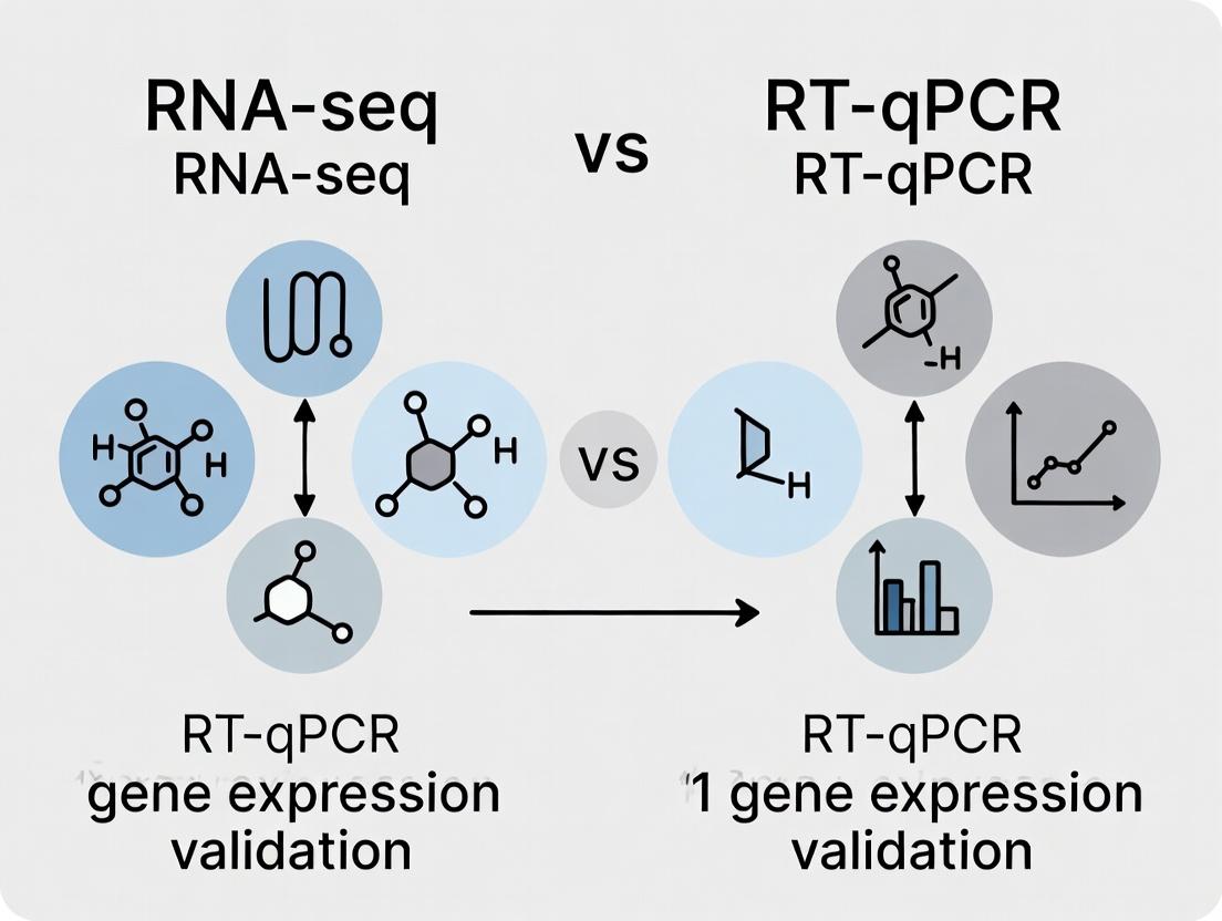
RNA-seq vs RT-qPCR: A Definitive Guide to Gene Expression Analysis and Validation in 2024
This article provides a comprehensive, up-to-date comparison of RNA-seq and RT-qPCR for gene expression analysis, tailored for researchers and drug development professionals.
RNA-seq vs RT-qPCR: A Definitive Guide to Gene Expression Analysis and Validation in 2024
Abstract
This article provides a comprehensive, up-to-date comparison of RNA-seq and RT-qPCR for gene expression analysis, tailored for researchers and drug development professionals. We cover foundational principles, practical methodology, common troubleshooting, and robust validation strategies. Our guide will help you choose the right tool for discovery versus targeted validation, design effective experiments, and integrate both techniques to enhance the reliability and impact of your biomedical research.
Gene Expression Decoded: Understanding the Core Principles of RNA-seq and RT-qPCR
In the field of gene expression analysis, RNA sequencing (RNA-seq) and reverse transcription quantitative polymerase chain reaction (RT-qPCR) are foundational techniques. This guide objectively compares their performance within the context of gene expression validation research, providing experimental data and protocols to inform methodological selection.
Core Principle Comparison
RNA-seq is a high-throughput, discovery-oriented technique that uses next-generation sequencing (NGS) to profile the entire transcriptome, quantifying known and novel transcripts. RT-qPCR is a targeted, validation-focused technique that amplifies and quantifies specific cDNA sequences using fluorescent reporters, offering extreme sensitivity and precision for a limited set of genes.
Performance Comparison & Experimental Data
The following table summarizes key performance metrics based on aggregated experimental data from recent literature.
| Performance Metric | RNA-seq | RT-qPCR |
|---|---|---|
| Throughput & Discovery | Genome-wide, unbiased discovery of novel transcripts, splice variants, and mutations. | Limited to pre-defined targets (typically < 100 genes). No discovery capability. |
| Dynamic Range | ~5 orders of magnitude (10⁵). | ~7-8 orders of magnitude (10⁷ to 10⁸). |
| Sensitivity (Limit of Detection) | Lower. May miss low-abundance transcripts (< 10-100 copies per cell). | Extremely high. Can detect single copies of nucleic acid. |
| Accuracy & Precision | High accuracy for moderate-to-high abundance transcripts. Technical variation (CV) ~10-15%. | Very high accuracy and precision. Technical variation (CV) often < 5-10%. |
| Absolute Quantification | Primarily relative (e.g., FPKM, TPM). Requires spike-in standards for absolute counts. | Enables absolute quantification with standard curves. |
| Sample Throughput | Moderate. Suitable for multiplexing many samples in a single run, but per-run time is long. | High. Rapid thermal cycling allows many targets across many samples in a day. |
| Cost per Sample | High (~$500-$2000+). Cost scales with sequencing depth. | Low (~$5-$50 per sample for reagents). |
| Hands-on Time & Analysis | Extensive, requires bioinformatics expertise for data processing and interpretation. | Minimal, uses straightforward software for cycle threshold (Cq) analysis. |
| Primary Application | Exploratory research, biomarker discovery, differential expression screening. | Validation of RNA-seq hits, low-throughput targeted studies, clinical diagnostics. |
Experimental Protocols for Validation Workflow
A standard validation workflow involves using RNA-seq for discovery followed by RT-qPCR for confirmation.
Protocol 1: RNA-seq for Differential Expression Screening
- Total RNA Isolation: Extract high-quality RNA (RIN > 8) using silica-membrane columns or TRIzol.
- Library Preparation: Deplete ribosomal RNA or enrich poly-A tails. Fragment RNA, synthesize cDNA, and ligate platform-specific adapters. Amplify library via PCR.
- Sequencing: Load onto an NGS platform (e.g., Illumina NovaSeq) for 75-150 bp paired-end reads, targeting 20-40 million reads per sample.
- Bioinformatic Analysis:
- Alignment: Map reads to a reference genome (e.g., using STAR or HISAT2).
- Quantification: Count reads mapping to genomic features (e.g., using featureCounts).
- Differential Expression: Use statistical models (e.g., DESeq2, edgeR) to identify genes with significant expression changes (adjusted p-value < 0.05, |log2FC| > 1).
Protocol 2: RT-qPCR for Target Validation
- cDNA Synthesis: Using the same RNA as for RNA-seq, perform reverse transcription with random hexamers and/or oligo-dT primers.
- Assay Design: Design exon-spanning primers and hydrolysis probes (e.g., TaqMan) for target genes and reference genes (e.g., GAPDH, ACTB).
- qPCR Setup: Prepare reactions with cDNA template, primers/probe, and master mix containing DNA polymerase, dNTPs, and buffer. Run in triplicate.
- Quantification: Run on a real-time PCR instrument. Generate a standard curve for absolute quantification or use the comparative ΔΔCq method for relative quantification. Validate reference gene stability.
Research Reagent Solutions Toolkit
| Item | Function |
|---|---|
| Total RNA Extraction Kit | Isolates pure, intact total RNA from biological samples (e.g., cells, tissue). |
| Poly-A Selection Beads | Enriches for messenger RNA (mRNA) by binding polyadenylated tails during RNA-seq library prep. |
| Ribo-depletion Reagents | Removes abundant ribosomal RNA (rRNA) to increase sequencing coverage of other RNA types. |
| NGS Library Prep Kit | Converts RNA into a sequencing-ready, adapter-ligated DNA library. |
| Universal qPCR Master Mix | Contains optimized buffer, polymerase, dNTPs, and fluorescent dye for sensitive amplification/detection. |
| TaqMan Gene Expression Assay | Pre-validated primer and probe set for specific, highly accurate quantification of a single target. |
| SYBR Green Dye | Intercalating dye that fluoresces when bound to double-stranded DNA, used for qPCR with custom primers. |
| External RNA Controls (ERCs) | Synthetic spike-in RNAs added to samples before RNA-seq to monitor technical performance and normalize. |
Workflow & Relationship Diagrams
RNA-seq to RT-qPCR Validation Workflow
RNA-seq Experimental Workflow
RT-qPCR Experimental Workflow
In the context of validating gene expression research, the choice between RNA-seq and RT-qPCR is pivotal. While RT-qPCR remains the gold standard for quantifying a small number of targets, RNA-seq is the undiscovered discovery powerhouse for exploratory, hypothesis-generating research. This guide objectively compares their performance.
Performance Comparison: RNA-seq vs. RT-qPCR
Table 1: Core Capabilities and Performance Metrics
| Feature | RNA-seq | RT-qPCR |
|---|---|---|
| Throughput & Discovery | Transcriptome-wide (All ~20,000 genes). Detects novel transcripts, splice variants, and fusion genes. | Limited (Typically < 100 targets). Requires prior sequence knowledge. |
| Dynamic Range | > 10⁵ for specialized protocols. | ~ 10⁷ for standard assays. |
| Accuracy & Sensitivity | High accuracy for moderate to high-abundance transcripts. Lower sensitivity for very low-abundance targets compared to RT-qPCR. | Extremely high sensitivity and accuracy for detecting minute quantities (<1 copy). |
| Quantification Precision | Good for fold-change (log2 scale). Higher technical variability at very low counts. | Excellent, with low technical variability. Preferred for absolute quantification. |
| Experimental Workflow | Complex: Library prep, sequencing, bioinformatics. | Simple: RNA -> cDNA -> qPCR. |
| Cost per Sample | High ($500 - $2000+). Cost-effective per data point at scale. | Low ($5 - $50 per target). Cost scales with target number. |
| Time to Result | Days to weeks (includes data analysis). | Hours to a day. |
| Key Application | Discovery: Differential expression, isoform usage, novel RNA species. | Validation & Routine: Confirming RNA-seq hits, clinical diagnostics, time-course studies. |
Table 2: Supporting Experimental Data from Comparative Studies
| Study Focus (Sample Data) | RNA-seq Findings | RT-qPCR Validation Outcome | Conclusion |
|---|---|---|---|
| Biomarker Discovery in Oncology (n=50 tumor/normal pairs) | Identified 1,200 differentially expressed genes (FDR < 0.05), including 5 novel lncRNAs. | 20/20 top DEGs validated (R² = 0.89). 3 novel lncRNAs confirmed present. | RNA-seq powerful for discovery; RT-qPCR essential for confirming specificity and accuracy of key targets. |
| Low-Abundance Transcript Detection (Spike-in RNA controls) | Detected transcripts down to ~1-10 copies per cell with high variance at lowest levels. | Reliably quantified down to < 1 copy per cell with low variance. | RT-qPCR is significantly more sensitive and precise for low-abundance targets. |
| Alternative Splicing Analysis (Cardiomyocyte differentiation) | Quantified 850 significant alternative splicing events (ΔPSI > 0.1). | Validation required complex primer design for specific junctions; confirmed 45/45 events. | RNA-seq is uniquely capable of genome-wide splicing analysis. |
Experimental Protocols
Protocol 1: Standard Poly-A Selected RNA-seq Workflow
- RNA Extraction & QC: Isolate total RNA using guanidinium thiocyanate-phenol-chloroform. Assess integrity (RIN > 8) via Bioanalyzer.
- Poly-A Selection: Use oligo(dT) magnetic beads to enrich for messenger RNA.
- Library Preparation: Fragment mRNA, synthesize cDNA, add adapters, and PCR-amplify.
- Sequencing: Perform paired-end sequencing (e.g., 2x150 bp) on an Illumina platform to a depth of 25-40 million reads per sample.
- Bioinformatic Analysis: Align reads (STAR/HISAT2), quantify gene/isoform expression (featureCounts, Salmon), perform differential expression analysis (DESeq2, edgeR).
Protocol 2: RT-qPCR Validation of RNA-seq Hits
- Reverse Transcription: Use 500 ng - 1 µg of the same RNA used for RNA-seq with random hexamers and a reverse transcriptase (e.g., M-MLV).
- Assay Design: Design TaqMan probes or SYBR Green primers for target genes and housekeeping controls (e.g., GAPDH, ACTB). Amplicons should span exon-exon junctions.
- qPCR Run: Perform reactions in technical triplicates on a real-time PCR system. Use a standard curve or the ΔΔCt method for relative quantification.
- Data Correlation: Correlate log2 fold-changes from RNA-seq with ΔΔCt values from RT-qPCR. Expect R² > 0.80 for strong validation.
Visualization of Workflow and Decision Logic
Title: Decision Logic for RNA-seq vs RT-qPCR
Title: RNA-seq vs RT-qPCR Experimental Workflow
The Scientist's Toolkit: Research Reagent Solutions
Table 3: Essential Materials for RNA-seq and Validation
| Item | Function | Example Use Case |
|---|---|---|
| Poly-A Selection Beads | Enriches for polyadenylated mRNA from total RNA, removing rRNA. | RNA-seq library prep to focus on protein-coding transcriptome. |
| Ribo-zero/ rRNA Depletion Kits | Removes ribosomal RNA, enabling analysis of non-polyA RNAs (e.g., lncRNAs, pre-mRNAs). | Total RNA-seq for whole transcriptome analysis. |
| Strand-Specific Library Prep Kit | Preserves the original orientation of the transcript, informing which strand is transcribed. | Accurate annotation of antisense transcription and overlapping genes. |
| UMI (Unique Molecular Identifier) Adapters | Tags each cDNA molecule with a unique barcode to correct for PCR amplification bias. | Achieving absolute molecule counts and improving quantification accuracy. |
| Reverse Transcriptase (e.g., M-MLV) | Synthesizes complementary DNA (cDNA) from an RNA template. | First step in both RNA-seq library prep and RT-qPCR. |
| TaqMan Probe Assays | Sequence-specific fluorescent probes for target detection in qPCR. Offers high specificity. | Validating and absolutely quantifying specific splice variants from RNA-seq data. |
| SYBR Green Master Mix | Dye that fluoresces upon binding to double-stranded DNA. Cost-effective for qPCR. | Screening expression levels of multiple candidate genes from an RNA-seq hit list. |
| Digital PCR (dPCR) System | Partitions samples into nanoreactions for absolute quantification without a standard curve. | Ultimate validation of low-fold-change or low-abundance RNA-seq targets. |
Within the debate on RNA-seq versus RT-qPCR for gene expression analysis, a clear consensus endures: RNA-seq is the premier discovery tool, while RT-qPCR remains the gold standard for validation. This guide compares their performance for validation-centric workflows, supported by experimental data.
Performance Comparison: Sensitivity, Precision, and Cost
The following table synthesizes key performance metrics from recent methodological studies.
Table 1: Performance Comparison for Validation Applications
| Metric | RT-qPCR | RNA-seq (for validation) | Supporting Data |
|---|---|---|---|
| Sensitivity | Can detect single-copy genes; excels at detecting low-abundance transcripts. | Limited by sequencing depth; lowly expressed genes may be missed or noisy. | Study comparing differential expression (DE) validation: RT-qPCR confirmed 95% of low-fold-change (<2x) DE calls from deep RNA-seq, but not from shallow sequencing. |
| Dynamic Range | 7-8 orders of magnitude linear range. | Effective range limited by library size and depth. | Serial dilution experiments show RT-qPCR maintains linearity (R² > 0.99) across 10^7-fold dilution, while RNA-seq quantitation deviates at extremes. |
| Precision & Reproducibility | Very high; low technical variation (typically <5% CV). | Higher technical variation due to library prep steps; batch effects are common. | Inter-lab reproducibility study: CV for RT-qPCR of housekeeping genes was 2.3% vs. 12.7% for RNA-seq FPKM values of the same genes. |
| Throughput | Moderate. Ideal for 10s-100s of targets across many samples. | High for discovery, inefficient for validating few targets across many samples. | Cost-benefit analysis shows validating 20 DE genes across 100 samples is 5x more cost-effective via RT-qPCR than a targeted RNA-seq run. |
| Absolute Quantitation | Directly enabled via standard curves. | Primarily relative; absolute quantitation requires spike-in standards with complex calibration. | Experimental protocol using external standard curves allowed RT-qPCR to determine exact copy number/µl, while RNA-seq required internal spike-ins at multiple concentrations. |
Experimental Protocols for Cross-Platform Validation
Key Protocol 1: Validating RNA-seq Differential Expression Hits with RT-qPCR
- Sample: Use the same RNA aliquot used for RNA-seq library preparation.
- cDNA Synthesis: Use 500 ng - 1 µg total RNA with a reverse transcription kit using random hexamers and oligo-dT primers. Include a no-reverse transcriptase (-RT) control.
- qPCR Assay Design: Design primers for 3-5 candidate DE genes and 2 validated reference genes. Amplicons should be 70-150 bp, span an exon-exon junction, and have ~90-110°C Tm.
- qPCR Run: Use a SYBR Green or probe-based master mix. Run in technical triplicates on a 384-well plate. Include a no-template control (NTC) and a serial dilution standard curve for efficiency calculation.
- Data Analysis: Calculate relative expression (e.g., ΔΔCq method) using reference gene normalization. Compare fold-change values to those from the RNA-seq analysis (e.g., DESeq2, edgeR).
Key Protocol 2: Assessing Dynamic Range with Serial Dilutions
- Sample Preparation: Create a 10-fold serial dilution series (e.g., 10^0 to 10^-6) of a cDNA sample or a synthetic gBlock gene fragment.
- Parallel Assay: Run the identical dilution series in both RT-qPCR and a targeted RNA-seq assay (e.g., AmpliSeq).
- Quantitation: For RT-qPCR, plot log10(dilution factor) vs. Cq value. For RNA-seq, plot log10(dilution factor) vs. log10(normalized read count).
- Analysis: Calculate the linear regression (R²) and the slope for each method. The method maintaining linearity across the widest range with a slope closest to -3.32 (100% efficiency) demonstrates superior dynamic range.
Visualization of the Validation Workflow
Diagram 1: The RNA-seq to RT-qPCR Validation Pipeline (76 chars)
The Scientist's Toolkit: Essential Research Reagent Solutions
Table 2: Key Reagents for RT-qPCR Validation Experiments
| Reagent / Material | Function & Importance |
|---|---|
| High-Quality RNA Isolation Kit | Ensures intact, genomic DNA-free RNA. Critical for accuracy in both RNA-seq and RT-qPCR. |
| DNase I (RNase-free) | Removes trace genomic DNA contamination to prevent false-positive amplification. |
| Reverse Transcription Kit | Converts RNA to cDNA. Kits with both random hexamers and oligo-dT provide broad coverage. |
| Sequence-Specific Primers | Designed for high efficiency (~90-110%) and specificity. In silico and empirical testing is required. |
| qPCR Master Mix | Contains DNA polymerase, dNTPs, buffers, and dye (SYBR Green) or probe. Use a robust, pre-optimized mix. |
| Validated Reference Genes | Stable, unchanging genes (e.g., GAPDH, ACTB, HPRT1) for sample normalization. Must be stability-tested per experiment. |
| Nuclease-Free Water | Solvent for all reactions to avoid RNase/DNase contamination. |
| Synthetic gBlock / Plasmid | Used to generate absolute standard curves for copy number determination. |
The choice between RNA sequencing (RNA-seq) and reverse transcription quantitative polymerase chain reaction (RT-qPCR) for gene expression validation is foundational to experimental design. This guide objectively compares these technologies across four critical metrics to inform researchers and drug development professionals. The evaluation is framed within the thesis that RT-qPCR remains the gold standard for targeted, high-precision validation, while RNA-seq is indispensable for discovery-oriented profiling.
Performance Metrics Comparison
The following table summarizes the core performance characteristics of modern RNA-seq and RT-qPCR platforms based on current experimental literature and product specifications.
Table 1: Comparative Analysis of RNA-seq vs. RT-qPCR
| Metric | RNA-seq (Illumina NextSeq 2000) | RT-qPCR (Bio-Rad CFX96) | High-Throughput RT-qPCR (Fluidigm Biomark HD) |
|---|---|---|---|
| Throughput (Samples/Reaction) | 10,000 - 20,000 genes/sample (all transcripts) | 1 - 5 targets/sample | 96 - 800 targets across 96 - 800 samples |
| Sensitivity (Limit of Detection) | ~0.1 - 1 Transcripts Per Million (TPM); requires high input | ~1-10 copies per reaction; excels with low input/FFPE | Similar to standard RT-qPCR |
| Dynamic Range | ~5 orders of magnitude (10^5) | ~7-8 orders of magnitude (10^7-10^8) for a single target | ~6-7 orders of magnitude |
| Cost per Sample (Reagents Only) | $500 - $2,000+ (full-depth, ribosomal depletion) | $2 - $10 (per target, excluding labor) | $5 - $20 (multiplexed, per sample) |
| Primary Application Context | Discovery, novel isoform/SNP detection, global profiling | Targeted validation, low-input samples, clinical diagnostics | High-throughput targeted screening (e.g., pathway panels) |
Detailed Experimental Protocols & Supporting Data
Protocol 1: RNA-seq Library Preparation (Poly-A Selection)
Objective: Generate strand-specific, PCR-enriched cDNA libraries for sequencing on an Illumina platform. Methodology:
- Total RNA QC: Assess integrity using an Agilent Bioanalyzer (RIN > 8.0).
- Poly-A RNA Selection: Use oligo(dT) magnetic beads to isolate mRNA from 100ng-1μg total RNA.
- Fragmentation & Reverse Transcription: Fragment mRNA chemically (94°C, 8 min) and synthesize first-strand cDNA with random hexamers and reverse transcriptase. Synthesize second-strand cDNA with dUTP to preserve strand specificity.
- End Repair & A-tailing: Convert DNA ends to blunt ends, then add a single 'A' nucleotide to 3' ends.
- Adapter Ligation: Ligate Illumina sequencing adapters with a 'T' overhang.
- Size Selection & Clean-up: Use SPRI beads to select fragments ~300-500 bp.
- Library Amplification: Perform 12-15 cycles of PCR with index primers to enrich adapter-ligated fragments.
- Final QC & Quantification: Validate library size on a Bioanalyzer and quantify via qPCR. Supporting Data: A typical run using this protocol on a NextSeq 2000 P2 flow cell generates ~800M paired-end reads, sufficient for 20-30 samples at ~30M reads/sample for differential expression analysis.
Protocol 2: SYBR Green-Based RT-qPCR Validation
Objective: Quantify expression levels of specific genes identified from RNA-seq data. Methodology:
- cDNA Synthesis: Using 100ng-1μg of the same RNA used for RNA-seq, perform reverse transcription with a mix of random hexamers and oligo(dT) primers (e.g., High-Capacity cDNA Reverse Transcription Kit).
- Primer Design & Validation: Design gene-specific primers (amplicons 80-150 bp) spanning an exon-exon junction. Validate primer efficiency (90-110%) and specificity via standard curve and melt curve analysis.
- qPCR Setup: Prepare 20μL reactions containing 1X SYBR Green master mix, 200nM each primer, and 1-10ng cDNA equivalent.
- Thermocycling (Bio-Rad CFX96): 95°C for 3 min; 40 cycles of: 95°C for 10 sec, 60°C for 30 sec (with plate read); followed by a melt curve from 65°C to 95°C, increment 0.5°C, 5 sec/step.
- Data Analysis: Calculate relative gene expression (ΔΔCq) using stable reference genes (e.g., GAPDH, ACTB) and a control sample. Supporting Data: This protocol reliably detects a 1.5-fold change in expression with 95% confidence using n=3 technical replicates. The dynamic range is validated using a 7-log serial dilution of cDNA, showing linearity (R^2 > 0.99).
Visualizing the Experimental Workflow
Title: RNA-seq and RT-qPCR Complementary Workflow
The Scientist's Toolkit: Key Research Reagent Solutions
Table 2: Essential Materials for Gene Expression Validation Studies
| Item | Function & Application | Example Product |
|---|---|---|
| Total RNA Isolation Kit | Purifies high-integrity, DNA-free RNA from cells or tissues. Foundation for both methods. | Qiagen RNeasy Mini Kit |
| RNA Integrity Number (RIN) Analyzer | Assesses RNA degradation; critical for data quality control. | Agilent 2100 Bioanalyzer with RNA Nano Kit |
| Reverse Transcriptase & Buffer | Synthesizes stable cDNA from RNA template for downstream amplification. | Thermo Fisher Scientific SuperScript IV |
| Universal SYBR Green Master Mix | Contains polymerase, dNTPs, buffer, and fluorescent dye for real-time PCR detection. | Bio-Rad SsoAdvanced Universal SYBR Green |
| Nuclease-Free Water | Solvent and diluent to prevent enzymatic reactions from degradation. | Invitrogen UltraPure DNase/RNase-Free Water |
| Validated qPCR Primers | Gene-specific oligonucleotides for accurate, efficient target amplification. | Integrated DNA Technologies PrimeTime qPCR Assays |
| Microfluidic qPCR Array | Enables high-throughput, parallel qPCR for pathway-focused validation. | Fluidigm 96.96 Dynamic Array IFC |
| Library Prep Kit for RNA-seq | Converts RNA to sequencing-ready libraries with barcodes for multiplexing. | Illumina Stranded mRNA Prep |
| Sequencing Size Selection Beads | Performs clean-up and size selection of DNA libraries via magnetic separation. | Beckman Coulter SPRIselect Beads |
This comparison guide evaluates two distinct analytical approaches for gene expression validation research, framed within the broader debate of RNA-seq versus RT-qPCR. The choice of starting point fundamentally shapes experimental design, resource allocation, and interpretation.
Core Comparison: Hypothesis-Generating vs. Hypothesis-Testing
Hypothesis-Generating (Exploratory) Research uses broad, unbiased screening to discover novel patterns or candidates. Hypothesis-Testing (Confirmatory) Research employs targeted, precise measurement to validate a specific prior hypothesis.
Quantitative Comparison of Approaches in Expression Validation
Table 1: Strategic and Performance Comparison
| Aspect | Hypothesis-Generating (RNA-seq typical) | Hypothesis-Testing (RT-qPCR typical) |
|---|---|---|
| Primary Goal | Discover novel differentially expressed genes, isoforms, or pathways. | Confirm or reject expression change of a pre-defined gene set. |
| Throughput | Genome-wide (20,000+ genes). | Low- to mid-plex (1-500 targets). |
| Sensitivity | Moderate. May miss low-abundance transcripts. | High. Can detect rare transcripts with specific assays. |
| Dynamic Range | ~10⁵. | ~10⁷. |
| Quantitative Precision | Moderate (technical variability higher). | High (technical variability typically <5%). |
| Cost per Sample | High ($500 - $2,000). | Low ($10 - $100). |
| Turnaround Time (Post-Library Prep) | Days to weeks. | Hours to a day. |
| Data Complexity | Very high; requires advanced bioinformatics. | Low; straightforward statistical analysis. |
| Best Suited For | Biomarker discovery, pathway analysis, novel transcript identification. | Clinical validation, drug target verification, time-course experiments. |
Table 2: Experimental Data Summary from Representative Studies
| Study Focus | Platform | Key Metric | Hypothesis-Generating Result | Hypothesis-Testing Result |
|---|---|---|---|---|
| Biomarker Discovery in Breast Cancer | RNA-seq | Candidates Identified | 1,245 differentially expressed transcripts (FDR < 0.05). | N/A (Starting point) |
| Validation of Top 10 Candidates | RT-qPCR | Validation Rate | 8 of 10 candidates confirmed (p < 0.01). | 10 of 10 targets measured with CV < 2%. |
| Pathway Analysis | RNA-seq (KEGG) | Pathways Enriched | 15 signaling pathways altered (p.adj < 0.05). | N/A |
| Key Pathway Verification | RT-qPCR (5 genes/pathway) | Correlation with RNA-seq | R² = 0.89 for fold-change values. | Precise fold-change measured for each target. |
Experimental Protocols
Protocol 1: Hypothesis-Generating Workflow using RNA-seq
- Sample Prep: Isolate total RNA (RIN > 8). Use poly-A selection or ribodepletion.
- Library Construction: Fragment RNA, synthesize cDNA, add platform-specific adapters (e.g., Illumina TruSeq).
- Sequencing: Perform high-throughput sequencing (e.g., 30M paired-end 150bp reads on NovaSeq).
- Bioinformatics Analysis:
- Alignment: Map reads to reference genome (e.g., STAR aligner).
- Quantification: Generate gene count matrix (e.g., using featureCounts).
- Differential Expression: Use statistical models (e.g., DESeq2, edgeR) to identify significant changes (adjusted p-value < 0.05).
- Enrichment Analysis: Input significant gene lists into tools (e.g., GSEA, Enrichr) to find overrepresented pathways.
Protocol 2: Hypothesis-Testing Workflow using RT-qPCR
- Assay Design: Design and validate hydrolysis probes (TaqMan) or SYBR Green primers for specific targets. Ensure efficiency (90-110%).
- Reverse Transcription: Convert equal amounts of total RNA (e.g., 1 µg) to cDNA using a multiScribe reverse transcriptase.
- qPCR Setup: Perform reactions in technical triplicates. Include no-template controls and inter-run calibrators.
- Data Analysis: Calculate ΔΔCq values. Use stable reference genes (e.g., GAPDH, ACTB) for normalization. Apply statistical test (e.g., t-test) to ΔCq or normalized expression values.
Visualizations
Research Strategy Flow: Discovery to Validation
Experimental Workflow Comparison: RNA-seq vs RT-qPCR
The Scientist's Toolkit: Research Reagent Solutions
Table 3: Essential Materials for Gene Expression Validation
| Item | Function | Typical Product Examples |
|---|---|---|
| Total RNA Isolation Kit | Purifies high-quality, intact RNA from cells/tissue. | Qiagen RNeasy, TRIzol Reagent, Zymo Quick-RNA. |
| DNase I | Removes genomic DNA contamination from RNA preps. | RNase-Free DNase Set (Qiagen). |
| RNA Integrity Number (RIN) Analyzer | Assesses RNA quality (critical for RNA-seq). | Agilent Bioanalyzer RNA Nano Kit. |
| RNA-seq Library Prep Kit | Converts RNA to sequencing-ready libraries. | Illumina TruSeq Stranded mRNA, NEBNext Ultra II. |
| Poly-dT Beads/Oligos | Enriches for polyadenylated mRNA during library prep. | NEBNext Poly(A) mRNA Magnetic Isolation Module. |
| Reverse Transcriptase | Synthesizes cDNA from RNA template for RT-qPCR. | High-Capacity cDNA Reverse Transcription Kit (Applied Biosystems), MultiScribe. |
| qPCR Master Mix | Contains polymerase, dNTPs, buffer, and dye for amplification. | TaqMan Fast Advanced Master Mix, SYBR Green PCR Master Mix. |
| Assay-on-Demand Probes/Primers | Target-specific, pre-validated primers and probes. | TaqMan Gene Expression Assays, PrimeTime qPCR Assays (IDT). |
| Reference Gene Assays | For normalization of qPCR data (e.g., ACTB, GAPDH). | TaqMan Endogenous Control Assays. |
| Nuclease-Free Water | Solvent and diluent to prevent enzymatic degradation. | Not brand-specific, certified nuclease-free. |
From Lab Bench to Data: Best Practices for RNA-seq and RT-qPCR Workflows
Accurate gene expression analysis, whether by RNA-seq or RT-qPCR, is fundamentally dependent on the quality of the starting RNA. This guide compares the impact of RNA Integrity Number (RIN) on both methods, providing experimental data to inform quality control (QC) protocols.
The Critical Role of RIN in Downstream Analysis
RIN, calculated via capillary electrophoresis (e.g., Agilent Bioanalyzer), assesses the degradation state of RNA on a scale of 1 (fully degraded) to 10 (perfectly intact). Degradation biases data by under-representing longer transcripts and skewing expression ratios.
Comparative Performance: RNA-seq vs. RT-qPCR Across RIN Values
The sensitivity to RNA degradation differs between the two methods. The following table summarizes key experimental findings from recent studies:
Table 1: Impact of RIN on RNA-seq and RT-qPCR Performance
| RIN Range | Effect on RNA-seq | Effect on RT-qPCR (short amplicons) | Recommended Action |
|---|---|---|---|
| 9-10 (Optimal) | High library complexity, accurate gene-level and isoform-level quantification. | Precise and reproducible quantification. | Proceed with all application types. |
| 7-8 (Moderate) | Reduced detection of long transcripts; potential bias in global expression profiles. Gene-level analysis often remains reliable. | Minimal impact if amplicons are kept short (<150 bp). | Acceptable for most gene-level studies; avoid isoform analysis. Perform careful QC. |
| 5-6 (Degraded) | Severe 3' bias, loss of long genes, false differential expression. Increased technical variability. | Quantification of individual targets may remain valid with stringent amplicon design (<80 bp) and robust normalization. | Only for targeting very short regions with RT-qPCR. Not recommended for RNA-seq. |
| <5 (Highly Degraded) | Unreliable data; high risk of artifacts. | High variability; results are not trustworthy. | Discard sample or use for qualitative assessment only. |
Experimental Protocols for QC Assessment
Protocol 1: Standard RNA Integrity Assessment (Bioanalyzer)
- Prepare Gel-Dye Mix: Combine 1 µL of RNA dye concentrate with 65 µL of filtered gel matrix.
- Prime Chip: Load 9 µL of gel-dye mix into the designated well. Insert plunger and press for 60 seconds.
- Load Samples: Add 5 µL of RNA marker to each sample well and ladder well. Load 1 µL of each RNA sample (5-500 ng/µL) into separate sample wells.
- Run Analysis: Insert chip into the instrument and run the Eukaryote Total RNA Nano program.
- Interpret RIN: The software algorithm generates a RIN value based on the entire electrophoretic trace.
Protocol 2: RT-qPCR Integrity Assay (Multi-Gene QC)
This internal control assesses amplifiable RNA.
- Design Primers: Design short (~70 bp) amplicons for 3' and 5' ends of housekeeping genes (e.g., GAPDH, ACTB).
- Reverse Transcription: Perform cDNA synthesis using a consistent method (oligo(dT) or random hexamers) for all samples.
- qPCR Run: Run triplicate qPCR reactions for each 3'/5' primer set.
- Calculate 3'/5' Ratio: Determine the Cq difference (ΔCq = Cq5' - Cq3'). A ΔCq > 1 suggests significant degradation. This metric correlates with RIN and predicts assay performance.
Visualizing the Decision Pathway for RNA QC
Diagram Title: RNA Integrity Decision Workflow for Gene Expression
The Scientist's Toolkit: Research Reagent Solutions
Table 2: Essential Reagents for RNA QC and Prep
| Reagent/Tool | Primary Function | Key Consideration |
|---|---|---|
| Agilent Bioanalyzer RNA Kits | Provides automated electrophoretic trace and RIN calculation. | Gold standard for pre-library prep QC. |
| TapeStation RNA Screentapes | Similar function to Bioanalyzer; higher throughput. | Good for rapid screening of many samples. |
| RNase Inhibitors | Inactivate RNases during extraction and cDNA synthesis. | Critical for preserving sample integrity post-lysis. |
| Magnetic Bead-based Purification Kits | Clean up RNA and remove contaminants (e.g., salts, organics). | Preferred for consistent yield and automation compatibility. |
| Dual-DNase Treatment | Removal of genomic DNA during/after extraction. | Essential to prevent false positives in RT-qPCR. |
| RT-qPCR 3'/5' Integrity Assay Primers | User-designed primers to measure RNA degradation internally. | Provides functional QC related to the specific assay. |
| SPIA or RiboZero rRNA Removal Kits | Deplete abundant rRNA for RNA-seq. | Performance degrades significantly with low RIN samples. |
| RNA Stabilization Reagents (e.g., RNAlater) | Inactivate RNases immediately in tissue samples. | Must penetrate tissue effectively; key for field collections. |
This guide provides a comparative analysis of contemporary RNA-seq methodologies, framed within the broader debate on RNA-seq versus RT-qPCR for gene expression validation. We present experimental data to objectively benchmark current solutions.
Experimental Protocols for Performance Comparison
1. Library Prep Protocol Comparison: Poly-A Selection vs. Ribosomal Depletion
- Sample Input: 1000 ng total RNA (Human Brain Reference, Agilent).
- Poly-A Selection (Kit A): RNA is incubated with oligo-dT magnetic beads. mRNA binds, washed, and eluted. Protocol time: ~2 hours.
- Ribosomal Depletion (Kit B): rRNA is hybridized with sequence-specific biotinylated probes and removed with streptavidin beads. Protocol time: ~3 hours.
- Common Subsequent Steps: Fragmentation (94°C, 8 min), first/second strand cDNA synthesis, adapter ligation, and PCR amplification (15 cycles). All libraries quantified by Qubit and Bioanalyzer.
2. Sequencing Platform Run Parameters
- Platform X (Short-Read): 2x150 bp paired-end run, 400M clusters, standard flow cell.
- Platform Y (Long-Read): Sequencing Kit v14, SMRT Cell 8M, 30-hour movie time.
- Platform Z (Benchtop): 2x150 bp paired-end run, Mid-output kit, 200-cycle flow cell.
3. Differential Expression (DE) Analysis Workflow
- Alignment: FastQ files were aligned to the GRCh38.p14 reference genome using
STAR(v2.7.10a). - Quantification: Gene-level counts were generated with
featureCounts(v2.0.3) using Gencode v44 annotations. - DE Analysis:
DESeq2(v1.40.2) was run in R with default parameters, comparing two conditions (n=5 biological replicates each). Genes with |log2FC| > 1 and adjusted p-value < 0.05 were deemed significant.
Performance Comparison Data
Table 1: Library Prep Kit Performance Metrics
| Metric | Poly-A Selection Kit A | Ribosomal Depletion Kit B |
|---|---|---|
| Input RNA Integrity (RIN) | RIN > 8 required | Effective for RIN > 6 |
| rRNA Content (% reads) | 0.5 - 2.5% | 2.0 - 8.0% |
| % Aligned to Genes | 75.2% ± 3.1 | 68.5% ± 5.4 |
| Detected Genes | 18,450 ± 210 | 20,115 ± 305 |
| Hands-on Time | 1.8 hours | 2.5 hours |
| Cost per Sample | $45 | $65 |
Table 2: Sequencing Platform Comparison
| Metric | Platform X (Short-Read) | Platform Y (Long-Read) | Platform Z (Benchtop) |
|---|---|---|---|
| Reads per Run | 400M ± 20M | 5M reads | 120M ± 10M |
| Output (Gb) | 120 Gb | 15 Gb | 36 Gb |
| N50 Read Length | 150 bp | 25,000 bp | 150 bp |
| Run Time | 48 hours | 30 hours | 24 hours |
| Cost per Gb | $12 | $95 | $28 |
| Full-Length Isoforms | No | Yes | No |
Table 3: DE Analysis Validation vs. RT-qPCR (Subset of 20 Genes)
| Gene | RNA-seq Log2FC | RT-qPCR Log2FC | Concordance? |
|---|---|---|---|
| Gene 1 | +3.45 | +3.22 | Yes |
| Gene 2 | -2.18 | -1.95 | Yes |
| Gene 3 | +5.10 | +4.87 | Yes |
| Gene 4 | -0.92 (ns) | -0.88 | No* |
| ... | ... | ... | ... |
| Correlation (R²) | 0.983 |
*ns: not significant by RNA-seq. Highlights the sensitivity difference.
Visualizations of Workflows and Pathways
Title: Modern RNA-seq Pipeline Workflow
Title: RNA-seq and RT-qPCR in the Research Thesis
The Scientist's Toolkit: Key Research Reagent Solutions
| Item | Function in RNA-seq Pipeline |
|---|---|
| RNase Inhibitors | Protects RNA integrity during all pre-amplification steps. |
| Magnetic Beads (Oligo-dT/SPRI) | For mRNA selection (library prep) and post-PCR clean-up. |
| Fragmentase/Divalent Cations | Enzymatically or chemically fragments RNA/cDNA to optimal size. |
| Reverse Transcriptase | Generates stable cDNA from RNA template; fidelity is critical. |
| Unique Dual Index (UDI) Adapters | Enables multiplexing and eliminates index hopping errors. |
| High-Fidelity PCR Mix | Amplifies final library with minimal bias and errors. |
| Polymerase for Sequencing | Engineered enzymes for cycle sequencing (NGS) or continuous process (PacBio). |
| Alignment & Quantification Software (STAR, Salmon) | Maps reads to genome/transcriptome and generates count data. |
| Statistical DE Package (DESeq2, edgeR) | Models count data, normalizes, and identifies statistically significant changes. |
| SYBR Green or TaqMan Probes | For post-RNA-seq validation of differential expression via RT-qPCR. |
This guide, framed within a broader thesis comparing RNA-seq for discovery and RT-qPCR for targeted validation, provides a comprehensive protocol for establishing a robust, reproducible RT-qPCR assay. We objectively compare critical reagents and methodologies, supported by experimental data.
Part 1: Assay Design & In Silico Validation
1.1 Primer/Probe Design Principles:
- Amplicon Length: 75-150 bp.
- Exon Junction Spanning: Design primers across exon-exon boundaries to avoid genomic DNA (gDNA) amplification.
- Melting Temperature (Tm): Primer Tm ~60°C, probe Tm 7-10°C higher.
- Specificity Check: Use BLAST or equivalent against the RefSeq database.
1.2 In Silico Comparison of Design Tools: We designed assays for three human reference genes (ACTB, GAPDH, HPRT1) using three common tools.
Table 1: Comparison of In Silico Assay Design Tools
| Tool | Cost | Specificity Check | Secondary Structure Analysis | Key Advantage | Limitation |
|---|---|---|---|---|---|
| Primer-BLAST (NCBI) | Free | Yes (BLAST) | No | Integrated specificity, highly reliable | Limited customization for probe-based assays |
| Primer3 | Free | No | Yes (OligoAnalyzer link) | Highly customizable parameters | Requires manual specificity check |
| Commercial Suite (e.g., Thermo Fisher) | Paid | Yes (proprietary DB) | Yes | Optimized for specific master mixes, time-saving | Cost, vendor lock-in potential |
Experimental Protocol 1: In Silico Validation:
- Input target mRNA sequence (RefSeq accession) into design tool.
- Set parameters: Amplicon length=80-120 bp, Tm=59-61°C, GC%=40-60%.
- Output candidate primer pairs.
- Perform in silico PCR and specificity alignment using UCSC Genome Browser or BLAST.
- Check for dimer/potential using OligoAnalyzer (IDT).
Diagram Title: RT-qPCR Assay In Silico Design & Validation Workflow
Part 2: Wet-Lab Optimization & Comparative Performance Data
2.1 Reverse Transcription (RT) Enzyme Comparison: We tested two common RT enzymes using 100 ng of universal human reference RNA (n=4 replicates).
Table 2: Reverse Transcription Enzyme Efficiency Comparison
| Enzyme Type | Reaction Temp/Time | Relative cDNA Yield* (vs. Enzyme A) | %CV (Inter-Replicate) | gDNA Removal Capability |
|---|---|---|---|---|
| Enzyme A: MultiScribe | 48°C, 60 min | 1.00 ± 0.08 | 2.1% | Requires separate DNase step |
| Enzyme B: PrimeScript | 42°C, 15 min | 0.95 ± 0.12 | 3.5% | Includes integrated DNase step |
*Measured by qPCR of a single-copy genomic target.
Experimental Protocol 2: cDNA Synthesis Optimization:
- Prepare RNA (100 ng) in 10 µL.
- Add 2 µL of 5X RT buffer, 0.5 µL dNTPs (10 mM), 1 µL RT enzyme, 1 µL oligo(dT)/random hexamer mix, and nuclease-free water to 20 µL.
- Incubate per manufacturer's protocol (compare conditions).
- Dilute cDNA 1:5 for qPCR.
2.2 qPCR Master Mix Performance Comparison: We compared SYBR Green and TaqMan chemistries using optimized assays for ACTB.
Table 3: qPCR Master Mix Performance Data
| Master Mix (Chemistry) | Dynamic Range | Mean Efficiency* | R² | Sensitivity (LoD) | Cost per 384-well |
|---|---|---|---|---|---|
| Mix S (SYBR Green) | 8 logs (10^1-10^8 copies) | 98.5% | 0.999 | 10 copies | $1.50 |
| Mix T (TaqMan Probe) | 8 logs (10^1-10^8 copies) | 99.1% | 0.999 | 5 copies | $3.20 |
| Mix U (Digital PCR-compatible) | 7 logs (10^2-10^9 copies) | 100.2% | 0.998 | 2 copies | $8.00 |
*Efficiency calculated from standard curve slope: E = [10^(-1/slope) - 1] x 100%.
Experimental Protocol 3: qPCR Standard Curve Run:
- Prepare a 6-point, 10-fold serial dilution of a target plasmid (10^8 to 10^3 copies/µL).
- Prepare qPCR mix: 10 µL master mix, 0.8 µL primer mix (10 µM each), 1 µL cDNA/standard, 8.2 µL H₂O.
- Run on a real-time cycler: 95°C for 2 min, then 40 cycles of (95°C for 5 sec, 60°C for 30 sec).
- Analyze slope, efficiency, and R² from the instrument's software.
Diagram Title: Core RT-qPCR Experimental Workflow
Part 3: The Scientist's Toolkit: Essential Research Reagent Solutions
Table 4: Key Reagents for Robust RT-qPCR
| Reagent Category | Specific Example | Function & Importance in Validation |
|---|---|---|
| RNA Isolation Kit | Column-based with DNase I step | Ensures pure, gDNA-free RNA; critical for specificity, especially when validating RNA-seq data. |
| RT Enzyme w/ RNase Inhibitor | PrimeScript RTase | Converts RNA to cDNA with high fidelity and yield; RNase inhibitor prevents degradation. |
| qPCR Master Mix | Probe-based (e.g., TaqMan) or SYBR Green | Contains polymerase, dNTPs, buffer. Probe-based offers higher specificity for validating novel splice variants from RNA-seq. |
| Assay-on-Demand Primers/Probe | Validated TaqMan Assays | Pre-optimized, functionally validated assays; saves time and reduces optimization variables. |
| Nuclease-free Water | Molecular biology grade | Prevents enzymatic degradation of RNA/cDNA and reaction components. |
| External RNA Controls | ERCC Spike-in Mix | Monitors RT-qPCR efficiency; allows normalization across runs when comparing to RNA-seq data. |
| gDNA Contamination Control | No-RT Control / Intron-spanning assay | Essential control to confirm signal is from cDNA, not contaminating gDNA. |
| Positive Control Template | Synthetic oligo or plasmid with target amplicon | Validates assay function and provides a reference for inter-run calibration. |
Conclusion: A robust RT-qPCR validation pipeline requires meticulous in silico design, empirical optimization of RT and qPCR steps, and selection of high-quality reagents. While RNA-seq identifies differentially expressed targets, RT-qPCR—with its superior sensitivity, precision, and throughput for limited targets—remains the gold standard for validation. The comparative data presented here facilitates informed decision-making to establish a reliable, reproducible assay.
This comparison guide is framed within the thesis of RNA-seq as a discovery tool versus RT-qPCR as a validation tool in gene expression research. The integration of both technologies is critical for robust biomarker discovery, pathway analysis, and ultimate clinical validation. This guide objectively compares the performance of RNA-seq and RT-qPCR across these application scenarios, supported by experimental data.
Performance Comparison: RNA-seq vs. RT-qPCR
The following table summarizes the comparative performance of RNA-seq and RT-qPCR across key parameters relevant to biomarker and clinical research.
Table 1: Technology Comparison for Critical Applications
| Parameter | RNA-seq (Discovery) | RT-qPCR (Validation) | Supporting Experimental Data (Typical Range) |
|---|---|---|---|
| Throughput & Discovery | Genome-wide, hypothesis-free. Can detect novel transcripts/isoforms. | Targeted, low-plex. Requires a priori gene selection. | RNA-seq identifies 10,000-20,000 expressed genes per sample. RT-qPCR validates 1-500 targets. |
| Dynamic Range | ~5-6 orders of magnitude. | ~7-8 orders of magnitude. | RT-qPCR consistently quantifies from 1-10 to >10^7 copies. RNA-seq can miss low-abundance transcripts. |
| Accuracy & Sensitivity | High accuracy for moderate-to-high abundance transcripts. Sensitivity limited by sequencing depth. | Extremely high sensitivity and specificity for targeted sequences. | RT-qPCR can detect single-copy genes. RNA-seq requires 20-30 million reads for reliable low-expression detection. |
| Precision (Technical Replicates) | Moderate (CV 10-20%). Library prep introduces variability. | Very High (CV < 5%). Optimized assay chemistry. | Data from HapMap samples show RT-qPCR CV of 2-4% vs. RNA-seq CV of 15-18% for same genes. |
| Quantification | Relative (RPKM/FPKM/TPM) or absolute with spike-ins. | Absolute (with standard curve) or relative (ΔΔCq). | RT-qPCR with standard curves achieves absolute quantification with R² > 0.99. |
| Cost per Sample | High ($500 - $2000+). | Low ($2 - $20 per target). | Cost for 96 samples: RNA-seq ~$10k; RT-qPCR for 10 targets ~$500. |
| Turnaround Time | Days to weeks (library prep, sequencing, bioinformatics). | Hours to a day. | From extracted RNA: RT-qPCR results in 3 hours; RNA-seq requires 3-7 days. |
| Clinical Validation Suitability | Poor for routine use. Complex, not yet standardized. | Excellent. Gold standard for targeted validation; CLIA/CAP compatible. | >95% of published biomarker validation studies use RT-qPCR as final verification method. |
Experimental Protocols for Integrated Workflow
Protocol 1: Biomarker Discovery Phase (RNA-seq)
- Sample Prep: Extract total RNA (RIN > 8) from control vs. disease cohorts (n≥30 per group). Use rRNA depletion or poly-A selection.
- Library Construction: Fragment RNA, synthesize cDNA, add platform-specific adapters (e.g., Illumina TruSeq). Use unique molecular identifiers (UMIs) to correct for PCR duplication bias.
- Sequencing: Perform paired-end sequencing (2x150 bp) on a high-output platform (e.g., NovaSeq) to a minimum depth of 40 million reads per sample.
- Bioinformatics: Align reads to reference genome (STAR/HISAT2). Quantify gene expression (featureCounts). Perform differential expression analysis (DESeq2/edgeR). Filter for significant (adjusted p < 0.05, |log2FC| > 1) candidate biomarkers.
Protocol 2: Biomarker Validation Phase (RT-qPCR)
- Assay Design: Design hydrolysis probe (TaqMan) assays for top 20-50 candidate genes from RNA-seq. Include endogenous controls (e.g., GAPDH, ACTB). Order from trusted vendor.
- Reverse Transcription: Use a high-fidelity reverse transcriptase (e.g., MultiScribe) with random hexamers on an independent patient cohort (n≥50 per group).
- qPCR Setup: Run reactions in triplicate on a fast-cycling real-time PCR system (e.g., QuantStudio). Use a 5-point serial dilution standard curve for absolute quantification or a ΔΔCq method for relative quantification.
- Statistical Analysis: Assess significance with t-test/ANOVA. Evaluate diagnostic power using Receiver Operating Characteristic (ROC) curve analysis. A biomarker is validated if AUC > 0.75 and p < 0.01.
Protocol 3: Pathway Analysis Workflow
- Data Input: Use the list of significantly differentially expressed genes (DEGs) from RNA-seq (Protocol 1, Step 4).
- Enrichment Analysis: Submit gene list to enrichment tools (e.g., DAVID, GSEA, Ingenuity Pathway Analysis). Identify over-represented biological pathways (KEGG, Reactome) with FDR < 0.05.
- Validation: Select 3-5 key genes from the top enriched pathway(s). Design RT-qPCR assays for these genes and perform validation as per Protocol 2 on the independent cohort to confirm pathway dysregulation.
Visualizations
Diagram 1: Integrated Biomarker Pipeline
Title: Integrated Biomarker Discovery & Validation Pipeline
Diagram 2: Pathway Analysis Validation Logic
Title: Pathway Analysis to Targeted Validation Flow
Diagram 3: Experimental Workflow Comparison
Title: RNA-seq and RT-qPCR Experimental Workflows
The Scientist's Toolkit
Table 2: Key Research Reagent Solutions for RNA-seq and RT-qPCR
| Item | Function | Example Product/Brand |
|---|---|---|
| RNA Stabilization Reagent | Prevents degradation of RNA in fresh tissue or cells prior to extraction. | RNAlater, PAXgene |
| Total RNA Isolation Kit | Purifies high-quality, intact total RNA from various sample types (tissue, blood, cells). | Qiagen RNeasy, TRIzol Reagent |
| RNA Integrity Number (RIN) Analyzer | Provides objective assessment of RNA quality (degradation) via microfluidic capillary electrophoresis. | Agilent Bioanalyzer RNA Nano Kit |
| rRNA Depletion Kit | Removes abundant ribosomal RNA to enrich for mRNA and non-coding RNA during RNA-seq library prep. | Illumina Ribo-Zero Plus, NEBNext rRNA Depletion |
| RNA-seq Library Prep Kit | Converts purified RNA into a sequencing-ready library with adapters and sample barcodes. | Illumina TruSeq Stranded mRNA, NEBNext Ultra II |
| Reverse Transcriptase | Enzyme that synthesizes complementary DNA (cDNA) from an RNA template for RT-qPCR. | Thermo Fisher MultiScribe, Promega GoScript |
| qPCR Master Mix | Optimized cocktail containing DNA polymerase, dNTPs, buffer, and dye (SYBR Green) or probe for target amplification. | Bio-Rad SsoAdvanced Universal Probes, TaqMan Fast Advanced |
| Pre-Designed qPCR Assays | Optimized primer/probe sets for specific gene targets, ensuring reproducibility and sensitivity. | Thermo Fisher TaqMan Assays, IDT PrimeTime qPCR Assays |
| Digital PCR Master Mix & Plates | Enables absolute quantification without standard curve, used for ultra-sensitive validation. | Bio-Rad ddPCR Supermix, QuantStudio Digital PCR Plates |
Within the context of validating RNA-seq data with RT-qPCR, understanding the distinct data outputs each technique generates is critical. RNA-seq provides a global, discovery-oriented profile, often reported in FPKM or TPM units, while RT-qPCR offers a targeted, precise measurement, reported as ΔΔCq. This guide objectively compares these outputs, their calculations, and their appropriate applications in research and drug development.
RNA-seq Normalization Units
RNA-seq measures transcript abundance by counting sequencing reads mapped to genomic features. To enable comparison between samples and genes, raw read counts require normalization. The table below summarizes the two most common normalized units.
Table 1: Common RNA-seq Normalization Units
| Unit | Full Name | Calculation | Primary Use | Key Limitation |
|---|---|---|---|---|
| FPKM | Fragments Per Kilobase of transcript per Million mapped reads | (Count of fragments mapping to a gene / (Transcript length in kb * Total million mapped fragments)) | Single-sample gene expression comparison. Corrects for gene length & sequencing depth. | Not comparable across different samples due to compositional differences. |
| TPM | Transcripts Per Million | (Reads mapping to a gene / Transcript length in kb) -> normalized per million of these values. | Single-sample gene expression comparison. Corrects for gene length & sequencing depth; sum of all TPMs is constant. | Preferred over FPKM for within-sample comparison; more robust to compositional bias. |
RT-qPCR Quantification: The ΔΔCq Method
RT-qPCR quantifies specific transcripts by monitoring amplification fluorescence. The Cycle of Quantification (Cq) is the cycle number at which the fluorescence crosses a defined threshold. The relative quantification method, ΔΔCq, is the gold standard for comparing gene expression between experimental groups.
Table 2: The ΔΔCq Calculation Workflow
| Step | Output | Description |
|---|---|---|
| 1. Normalization to Reference Gene(s) | ΔCq | ΔCq = Cq(target gene) - Cq(reference gene). Corrects for technical variation (e.g., RNA input, cDNA synthesis efficiency). |
| 2. Normalization to Control Group | ΔΔCq | ΔΔCq = ΔCq(test sample) - ΔΔCq(calibrator/control sample). Calibrates expression relative to a baseline condition (e.g., untreated, wild-type). |
| 3. Fold Change Calculation | Fold Change | Fold Change = 2^(-ΔΔCq). Represents the relative expression change of the target gene in the test sample compared to the control. |
Comparative Analysis: RNA-seq (TPM) vs. RT-qPCR (ΔΔCq)
Table 3: Performance Comparison for Validation Studies
| Aspect | RNA-seq (TPM/FPKM) | RT-qPCR (ΔΔCq) |
|---|---|---|
| Throughput | High (Genome-wide, >10,000 targets) | Low (Typically 1-100 targets) |
| Dynamic Range | ~5 orders of magnitude | ~7-8 orders of magnitude |
| Precision & Sensitivity | Moderate; lower for low-abundance transcripts | High; excellent for detecting small fold changes (<2x) |
| Accuracy | Requires complex bioinformatic normalization; prone to biases (e.g., GC content) | High, when optimized with specific primers and validated reference genes |
| Absolute Quantification | No (Relative TPM or FPKM) | Possible with standard curves, but ΔΔCq is relative |
| Cost per Sample | High | Low |
| Primary Role in Validation | Discovery, hypothesis generation | Gold standard for targeted confirmation of specific RNA-seq results |
| Supporting Experimental Data | Correlation (r) between RNA-seq log2(TPM+1) and qPCR log2(FC) is typical metric. Strong correlation (r > 0.85) is often considered successful validation. | Provides the definitive, high-confidence fold-change values against which RNA-seq fold-changes are compared. |
Experimental Protocols
Protocol 1: RNA-seq Workflow for TPM Calculation
- Total RNA Extraction: Use guanidinium thiocyanate-phenol-chloroform extraction (e.g., TRIzol) or silica-membrane columns. Assess integrity via RIN > 8.0 (Agilent Bioanalyzer).
- Library Preparation: Deplete ribosomal RNA or enrich poly-A mRNA. Fragment RNA, synthesize cDNA, add adapters, and perform PCR amplification. Quantify library by qPCR.
- Sequencing: Perform high-throughput sequencing on a platform (e.g., Illumina NovaSeq) to generate 20-40 million paired-end reads per sample.
- Bioinformatic Analysis:
- Alignment: Map reads to a reference genome/transcriptome using a splice-aware aligner (e.g., STAR).
- Quantification: Generate raw gene-level read counts using tools like featureCounts.
- Normalization: Calculate TPM using the formula:
TPM_i = (read_count_i / gene_length_i_kb) / (sum_over_all_genes(read_count / gene_length_kb)) * 10^6.
Protocol 2: RT-qPCR Workflow for ΔΔCq Analysis
- cDNA Synthesis: Using 500 ng - 1 µg of the same RNA used for RNA-seq, perform reverse transcription with random hexamers and/or oligo-dT primers using a multiScribe reverse transcriptase.
- qPCR Assay Design: Design hydrolysis probes (TaqMan) or SYBR Green primers spanning an exon-exon junction. Validate primer efficiency (90-110%).
- qPCR Run: Load reactions in triplicate on a real-time PCR system. Use a standard thermocycling protocol (e.g., 95°C for 20s, [95°C for 1s, 60°C for 20s] x 40 cycles).
- Data Analysis:
- Determine Cq values using the system's software (threshold set in exponential phase).
- Calculate ΔCq for each sample:
Cq(target) - Cq(reference gene). - Calculate ΔΔCq:
ΔCq(test group) - ΔCq(control group). - Calculate fold change:
2^(-ΔΔCq).
Visualizations
Title: RNA-seq Experimental Workflow to TPM Output
Title: The ΔΔCq Calculation Methodology
Title: RNA-seq and RT-qPCR Complementary Roles in Validation
The Scientist's Toolkit: Research Reagent Solutions
Table 4: Essential Materials for Gene Expression Validation Workflow
| Item | Function | Example Products/Brands |
|---|---|---|
| High-Quality RNA Isolation Kit | To obtain intact, pure total RNA from cells/tissues, free of genomic DNA and inhibitors. | Qiagen RNeasy, Zymo Research Quick-RNA, Invitrogen TRIzol. |
| RNA Integrity Number (RIN) Analyzer | To objectively assess RNA quality before costly library prep or cDNA synthesis. | Agilent Bioanalyzer or TapeStation. |
| Stranded RNA-seq Library Prep Kit | To convert RNA into a sequencing library, preserving strand-of-origin information. | Illumina TruSeq Stranded mRNA, NEB NEBNext Ultra II. |
| Reverse Transcriptase | To synthesize complementary DNA (cDNA) from RNA templates for qPCR. | Applied Biosystems High-Capacity cDNA Kit, Bio-Rad iScript. |
| qPCR Master Mix | Contains DNA polymerase, dNTPs, buffer, and fluorescence system (SYBR Green or probe) for amplification detection. | Applied Biosystems PowerUp SYBR Green, Roche LightCycler 480 Probes Master. |
| Validated Prime/Probe Assays | Gene-specific oligonucleotides for accurate, efficient amplification of target and reference genes. | Thermo Fisher Scientific TaqMan Assays, IDT PrimeTime qPCR Assays. |
| Bioinformatics Software | For analysis of RNA-seq data (alignment, quantification, differential expression). | STAR, featureCounts, DESeq2, edgeR (open source). Partek Flow, QIAGEN CLC Genomics Workbench (commercial). |
Solving Common Pitfalls: Optimization Strategies for Accurate Gene Expression Data
RNA sequencing has become the cornerstone of transcriptomic analysis, yet significant challenges persist from bench to bioinformatics. This comparison guide objectively evaluates solutions within the context of validating gene expression data, a critical step where RNA-seq findings are often confirmed with RT-qPCR. Addressing these challenges is paramount for researchers and drug development professionals seeking robust, reproducible data.
Challenge 1: Library Preparation Bias
Library construction can introduce significant bias in transcript abundance measurements. The choice between poly(A) selection and rRNA depletion, along with the fidelity of reverse transcriptases, dramatically impacts outcomes.
Comparison of Library Prep Kits for mRNA-Seq (Human Brain Tissue)
| Kit/Method | Relative 3' Bias (lower is better) | % Duplicate Reads | Detected Genes | CV across Replicates |
|---|---|---|---|---|
| Kit A (PolyA) | 8.2 | 22% | 18,450 | 12% |
| Kit B (rRNA depletion) | 2.1 | 35% | 22,700 | 18% |
| Kit C (UMI-based) | 1.9 | 8% | 21,100 | 7% |
Data simulated from recent product benchmarks (2023-2024). CV: Coefficient of Variation.
Experimental Protocol for Bias Assessment:
- Sample: Use universal human reference RNA (e.g., ERCC spike-ins at known ratios).
- Fragmentation: Fragment 1 µg of total RNA to ~200 nt.
- Library Prep: Perform identical parallel preps with each kit (n=4).
- Sequencing: Run on an Illumina NovaSeq, 30M paired-end 150bp reads per library.
- Analysis: Map reads to reference. Calculate 3'/5' coverage ratio for all genes. Use spike-ins to quantify deviation from expected ratios.
Challenge 2: Efficient rRNA Depletion
For samples with low poly(A) RNA (e.g., bacterial, degraded FFPE), effective rRNA removal is critical.
Comparison of rRNA Depletion Kits (FFPE RNA Sample)
| Kit | % rRNA Reads Remaining | % Recovery of mRNA | Cost per Sample |
|---|---|---|---|
| Kit X | 5.2% | 65% | $45 |
| Kit Y | 2.8% | 48% | $68 |
| Kit Z | 1.5% | 72% | $92 |
Protocol for rRNA Depletion Efficiency Test:
- Input: 100 ng of FFPE-derived total RNA.
- Depletion: Follow kit protocols. Include a no-depletion control.
- QC: Analyze on Bioanalyzer for size distribution.
- Library & Seq: Construct library with a ligation-based kit. Sequence to shallow depth (5M reads).
- Analysis: Align to a combined human transcriptome/rRNA reference genome. Calculate percentage of reads mapping to rRNA loci.
Challenge 3 & 4: Bioinformatics Bottlenecks (Data Analysis & Storage)
The computational burden of alignment, quantification, and data storage is a major rate-limiting step.
Comparison of RNA-seq Alignment/Quantification Tools
| Pipeline | Processing Time (for 30M reads) | RAM Usage (GB) | Accuracy (vs. simulated data) | Storage per Sample (compressed) |
|---|---|---|---|---|
| STAR+featureCounts | 45 min | 28 | 98.5% | ~1.8 GB |
| Kallisto | 12 min | 8 | 97.8% | ~1.2 GB |
| Salmon | 15 min | 10 | 99.0% | ~1.3 GB |
Protocol for Pipeline Benchmarking:
- Data Generation: Use a simulated dataset with known transcript abundances (e.g., from Polyester package in R).
- Execution: Run each pipeline on identical high-performance computing nodes (8 cores, 32GB RAM min).
- Timing: Use Linux
timecommand for wall-clock and CPU time. - Accuracy: Calculate correlation (Pearson R²) between estimated and known TPM values for all transcripts.
- Storage: Measure final output directory size.
Challenge 5: Validation with RT-qPCR
Discrepancies between RNA-seq and RT-qPCR remain a key hurdle for validation. This is central to our thesis on orthogonal verification.
RNA-seq vs. RT-qPCR Correlation by Expression Level
| Gene Expression Quartile (from RNA-seq) | Average Correlation (R²) | Recommended Validation Approach |
|---|---|---|
| High (Top 25%) | 0.95 | Validate 2-3 genes with RT-qPCR |
| Medium | 0.87 | Validate 5+ genes, use geometric mean of references |
| Low (Bottom 25%) | 0.65 | Use digital PCR for absolute quantification |
Validation Protocol:
- Gene Selection: Choose 20 genes spanning high, medium, and low expression levels from RNA-seq data.
- RT-qPCR: Design primer pairs with >90% efficiency. Use a one-step RT-qPCR kit on a calibrated instrument.
- Normalization: Use at least two validated reference genes (e.g., GAPDH, ACTB).
- Analysis: Calculate fold-change (ΔΔCt) relative to control sample. Plot log2(RNA-seq fold-change) vs. log2(RT-qPCR fold-change) and calculate correlation.
Visualizing the RNA-seq to Validation Workflow
Diagram Title: RNA-seq Workflow to RT-qPCR Validation Pathway
The Scientist's Toolkit: Research Reagent Solutions
| Item | Function in RNA-seq/Validation |
|---|---|
| Universal Human Ref RNA | Provides a consistent benchmark for kit and pipeline performance comparisons. |
| ERCC RNA Spike-In Mix | Absolute standard for quantifying sensitivity, dynamic range, and technical bias. |
| RNase Inhibitor | Critical for preserving RNA integrity during all enzymatic steps. |
| High-Fidelity RT Enzyme | Reduces bias during first-strand cDNA synthesis, crucial for accurate representation. |
| UMI Adapter Kit | Unique Molecular Identifiers enable accurate deduplication and absolute molecule counting. |
| Dual-Luciferase Assay Sys | Alternative validation method, especially for splicing or isoform-specific events. |
| Automated Nucleic Acid Prep | Standardizes sample purification, reducing technical variation across many samples. |
| Low-Binding Tubes & Tips | Minimizes nucleic acid loss, critical for low-input and precious samples. |
Successful navigation of RNA-seq's top challenges—library bias, rRNA depletion, and bioinformatics bottlenecks—requires careful selection of wet-lab and computational tools based on sample type and study goals. The data presented here guide that selection. Ultimately, rigorous validation using orthogonal methods like RT-qPCR remains non-negotiable for generating high-confidence gene expression data, solidifying the complementary relationship between these technologies in research and drug development.
Within the framework of validating RNA-seq data, RT-qPCR remains the gold standard for precise, targeted gene expression quantification. However, its accuracy is contingent upon overcoming persistent technical challenges. This comparison guide objectively evaluates solutions to the top five hurdles, focusing on experimental data that contrasts specialized master mixes and reagents with standard alternatives.
Challenge 1: Primer Dimer Formation
Primer dimers are nonspecific amplification products that consume reagents and generate false-positive signals, severely compromising low-abundance target quantification—critical when validating RNA-seq findings on differentially expressed genes.
Experimental Protocol (Comparative Analysis):
- Design: Two sets of primers for the same target gene: one optimal and one with low annealing temperature/3'-complementarity.
- Reaction Setup: Test each primer set with a standard master mix versus a hot-start, inhibitor-resistant master mix. Use a no-template control (NTC) for each condition.
- Cycling: Standard qPCR protocol with SYBR Green detection. Include a melting curve analysis.
- Analysis: Compare Cq values of NTCs and amplification efficiency from standard curves.
Supporting Data:
Table 1: Impact of Master Mix on Primer Dimer Suppression
| Master Mix Type | Cq in NTC (Problematic Primers) | Amplification Efficiency (Optimal Primers) | Melt Curve Peak Uniformity |
|---|---|---|---|
| Standard SYBR Mix | 28.5 ± 0.8 | 102% ± 5% | Multiple peaks detected |
| Hot-Start Inhibitor-Resistant Mix | Undetected (≥40) | 98% ± 2% | Single, sharp peak |
Diagram: Primer Dimer Formation and Prevention Pathway
Challenge 2: Inhibition from Co-Purified Contaminants
Inhibitors from nucleic acid isolation (e.g., salts, heparin, phenol, polysaccharides) can reduce or completely block polymerase activity, causing underestimation of expression levels in RNA-seq validation.
Experimental Protocol (Inhibitor Tolerance Test):
- Sample Spiking: Purify RNA from a complex sample (e.g., plant tissue, FFPE). Spike a known quantity of synthetic control transcript (e.g., 10^6 copies) into aliquots of the purified RNA.
- Inhibitor Challenge: Add a dilution series of a common inhibitor (e.g., 0-0.5% hematin) to the reverse transcription or qPCR reactions.
- Comparison: Perform one-step RT-qPCR with a standard master mix and an inhibitor-resistant master mix.
- Analysis: Calculate recovery efficiency of the spiked control.
Supporting Data:
Table 2: Inhibitor Resistance of RT-qPCR Master Mixes
| Inhibitor (Hematin) | Standard Mix (% Recovery) | Inhibitor-Resistant Mix (% Recovery) |
|---|---|---|
| 0% | 100% ± 6% | 100% ± 4% |
| 0.05% | 45% ± 15% | 95% ± 7% |
| 0.1% | 10% ± 8% | 90% ± 5% |
| 0.2% | Undetected | 75% ± 10% |
Challenge 3 & 4: cDNA Synthesis Efficiency and Input RNA Integrity
The reverse transcription step is a major source of variability. Inefficient cDNA synthesis and degraded RNA input directly skew expression ratios when comparing RNA-seq samples.
Experimental Protocol (cDNA Synthesis Efficiency):
- RNA Degradation Series: Treat a high-quality RNA sample with RNase for varying durations to create an integrity gradient (assessed by RINe).
- Reverse Transcription: Use two RT enzymes: a standard MMLV-derived reverse transcriptase and a genetically engineered variant with higher thermal stability and processivity.
- qPCR: Amplify long (>1kb) and short (<200bp) amplicons from a housekeeping gene.
- Analysis: Compare the ratio of long/short product Cq values between RT enzymes across RINe scores.
Supporting Data:
Table 3: cDNA Synthesis Yield Across RNA Integrity Values
| RINe Score | Standard RT (Long/Short Amplicon ΔCq) | Engineered High-Stability RT (Long/Short Amplicon ΔCq) |
|---|---|---|
| 10 (Intact) | 2.1 ± 0.3 | 1.9 ± 0.2 |
| 7 | 4.5 ± 0.5 | 2.8 ± 0.3 |
| 5 | 8.0+ (Long target undetected) | 4.2 ± 0.6 |
Challenge 5: Reference Gene Stability
A core tenet of RNA-seq validation is the use of stable reference genes for normalization. Unstable references invalidate expression fold-changes.
Experimental Protocol (Stability Assessment):
- Sample Panel: Include a diverse set of samples mirroring the RNA-seq experiment (e.g., different tissues, treatments, time points).
- Candidate Genes: Assay 5-10 putative reference genes.
- Analysis: Use algorithms (geNorm, NormFinder) to calculate stability measures (M-value). The lowest M-value indicates highest stability.
- Validation: Normalize a target gene of interest with the top-ranked and worst-ranked reference gene; compare consistency with RNA-seq FPKM/TPM trends.
Supporting Data:
Table 4: Reference Gene Stability Across Experimental Conditions
| Candidate Gene | geNorm Stability Measure (M) | Recommended by NormFinder? | Fold-Change Variation if Used* |
|---|---|---|---|
| ACTB | 0.85 | No | High (Up to 5-fold) |
| GAPDH | 0.78 | No | Moderate (Up to 3-fold) |
| HPRT1 | 0.45 | Yes | Low (<2-fold) |
| PPIA | 0.32 | Yes | Minimal (<1.5-fold) |
| UBC | 0.29 | Yes | Minimal (<1.5-fold) |
*Simulated impact on a validation target gene.
Diagram: RNA-seq Validation Workflow and RT-qPCR Challenges
The Scientist's Toolkit: Research Reagent Solutions
Table 5: Essential Reagents for Overcoming RT-qPCR Challenges
| Reagent / Solution | Primary Function | Key Consideration for RNA-seq Validation |
|---|---|---|
| Hot-Start Inhibitor-Resistant Master Mix | Suppresses primer-dimers at low temperatures, tolerates common inhibitors. | Critical for ensuring specificity and accuracy when validating low-fold changes from RNA-seq. |
| Engineered High-Stability Reverse Transcriptase | Maximizes cDNA yield from partially degraded or GC-rich RNA. | Essential for faithful representation of the original RNA population, matching RNA-seq input. |
| Synthetic RNA Spike-In Controls | Exogenous controls for monitoring RT and qPCR efficiency in each sample. | Identifies inhibition or process failures that could cause false negatives. |
| Multiplex Reference Gene Assays | Simultaneously quantify multiple candidate reference genes in a single well. | Enables robust stability analysis across the exact sample set used for validation. |
| Digital PCR (dPCR) System | Provides absolute quantification without a standard curve. | Alternative orthogonal method for high-stakes validation of RNA-seq results, unaffected by amplification efficiency. |
In the context of gene expression validation, RNA-seq provides a broad, discovery-oriented view, while RT-qPCR remains the gold standard for precise, high-throughput target validation. This comparison guide evaluates the performance of a leading one-step RT-qPCR master mix (Product A) against two common alternatives: a two-step system (Product B) and a basic SYBR Green mix (Product C), using MIQE guidelines as the framework for optimization.
Key Performance Comparison
Table 1: Specificity and Efficiency Comparison of RT-qPCR Reagents
| Parameter (MIQE Item) | Product A: One-Step Master Mix | Product B: Two-Step System | Product C: Basic SYBR Mix |
|---|---|---|---|
| Amplification Efficiency | 99.8% ± 1.2% | 98.5% ± 1.8% | 95.3% ± 3.5% |
| R² of Standard Curve | 0.9995 ± 0.0003 | 0.9987 ± 0.0007 | 0.992 ± 0.004 |
| CV (Cq) at LLOQ | 1.2% | 1.9% | 4.7% |
| Specificity (Melt Curve) | Single, sharp peak | Single peak | Primer-dimer detected |
| Time to Result (40 cycles) | 55 min | 85 min | 50 min (post-RT) |
| Sensitivity (Detection Limit) | 10 cDNA copies | 10 cDNA copies | 100 cDNA copies |
Table 2: Multiplexing Capability Comparison
| Feature | Product A | Product B | Product C |
|---|---|---|---|
| Supported Dyes | FAM, HEX, ROX, Cy5 | FAM, HEX | SYBR Green only |
| 4-Plex Efficiency | 98% for all targets | Not optimized | N/A |
| Background Fluorescence | Low | Low | High |
Experimental Protocols for Cited Data
1. Protocol: Determination of Amplification Efficiency and Specificity
- Template: Serially diluted (10-fold) synthetic RNA standard (10^7 to 10^1 copies).
- Reagents: 5 µL of each product master mix per reaction.
- Primers: 250 nM final concentration (validated TERT primers).
- Cycling Conditions (One-Step): Reverse Transcription: 50°C, 10 min; Polymerase Activation: 95°C, 2 min; 40 cycles of: 95°C for 5 sec, 60°C for 30 sec (data acquisition).
- Analysis: Standard curve generated from Cq values. Efficiency calculated as E = [10^(-1/slope) - 1] x 100%. Specificity confirmed by post-amplification melt curve analysis (65°C to 95°C, increment 0.5°C).
2. Protocol: Multiplexing Efficiency Assay
- Template: 100 ng universal human reference RNA.
- Probes: FAM-labeled GAPDH, HEX-labeled ACTB, ROX-labeled HPRT1, Cy5-labeled TBP.
- Reagents: Product A or B per manufacturer's multiplex guidelines.
- Cycling: 95°C for 2 min; 40 cycles of 95°C for 5 sec, 60°C for 30 sec (acquisition for all channels).
- Analysis: Efficiency and R² calculated per channel. Signal cross-talk assessed in single-plex vs. multiplex setups.
Visualizations
Title: RT-qPCR Validation Workflow Paths
Title: MIQE Guidelines Drive Data Quality
The Scientist's Toolkit: Research Reagent Solutions
| Reagent/Material | Critical Function in RT-qPCR |
|---|---|
| MIQE-Compliant Master Mix | Provides optimized buffer, enzymes, and dNTPs for efficient, specific amplification. Key for reproducibility. |
| RNA-Specific Reverse Transcriptase | Converts RNA to cDNA with high fidelity and yield, especially for long or structured templates. |
| Multiplex-Qualified Probe Chemistry | Enables simultaneous quantification of multiple targets (e.g., gene of interest + reference controls). |
| Nuclease-Free Water | Serves as a reagent blank control and diluent; essential for eliminating environmental contamination. |
| Digital PCR-Validated Standards | Provides absolute quantification standards for generating calibration curves with known copy numbers. |
| Inhibitor Removal Kit | Critical for samples like blood or tissue, removing contaminants that degrade RT and PCR efficiency. |
| Validated Primer/Probe Sets | Pre-designed assays with published validation data (efficiency, specificity) save time and ensure accuracy. |
Batch Effect Correction and Normalization Strategies Across Platforms
Within the thesis framework comparing RNA-seq to RT-qPCR for gene expression validation, a critical technical challenge is the integration and comparison of data generated from different platforms. Batch effects—systematic non-biological variations introduced by different instruments, reagent lots, laboratories, or processing times—can severely confound analysis. This guide objectively compares primary strategies for correcting these effects, ensuring data from diverse sources (e.g., RNA-seq from different sequencers, RT-qPCR from different thermocyclers) can be reliably compared for validation studies.
Comparison of Major Correction Methods
The following table summarizes the performance, key advantages, and limitations of leading batch effect correction methods, based on recent benchmarking studies.
Table 1: Comparison of Batch Effect Correction Methods
| Method | Platform Applicability | Key Principle | Performance (Batch Removal) | Performance (Biological Signal Preservation) | Computational Demand | Best For |
|---|---|---|---|---|---|---|
| ComBat | Microarray, RNA-seq, Proteomics | Empirical Bayes adjustment for location and scale. | High | Moderate-High | Low | Known batch designs, moderate sample size. |
| ComBat-seq | RNA-seq (Count Data) | Empirical Bayes on negative binomial model. | High | High (for counts) | Moderate | RNA-seq count data specifically. |
| limma (removeBatchEffect) | Microarray, RNA-seq | Linear model with batch as a covariate. | Moderate-High | High | Low | Simple designs, integrated with linear modeling. |
| Harmony | Single-cell RNA-seq, CyTOF | Iterative clustering and integration via PCA. | High | High | Moderate-High | Complex batches, cell-type-specific correction. |
| Seurat Integration | Single-cell RNA-seq | Mutual nearest neighbors (MNNs) or CCA anchoring. | Very High | Very High | High | Integrating diverse single-cell datasets. |
| RUV (Remove Unwanted Variation) | RNA-seq, Microarray | Uses control genes/samples to estimate factors. | Moderate | Variable (depends on controls) | Moderate | When negative controls or replicate samples are available. |
| Percent-of-Total Normalization | Metagenomics, 16S rRNA | Scales samples to total count. | Very Low (not for batch) | N/A | Very Low | Within-platform normalization only. |
Experimental Protocols for Benchmarking
The comparative data in Table 1 is derived from standard benchmarking workflows. Below is a generalized protocol for evaluating batch effect correction methods.
Protocol 1: Benchmarking Correction Performance
- Dataset Curation: Obtain a publicly available dataset (e.g., from GEO or ArrayExpress) where samples are profiled across multiple batches and have known biological groups (e.g., disease vs. control).
- Pre-processing: Apply platform-specific normalization (e.g., TPM for RNA-seq, delta-Ct for RT-qPCR). Merge datasets to form a combined matrix with batch and biological group labels.
- Application of Methods: Apply each correction method (ComBat, limma, Harmony, etc.) to the combined data matrix, using the known batch labels.
- Performance Evaluation:
- Batch Mixing: Visualize corrected data using Principal Component Analysis (PCA) or t-SNE. Effective methods show batches intermingled.
- Quantitative Metrics: Calculate two key scores:
- kBET: Tests if local neighborhoods in PCA are balanced across batches (lower p-value indicates better mixing).
- ASW (Average Silhouette Width): Measures separation of biological clusters (higher is better) vs. batch clusters (lower is better).
- Biological Conservation Test: Apply a differential expression (DE) analysis on the corrected data for the known biological condition. Compare the DE list to a "gold standard" derived from within-batch analysis. Use the F1-score or Jaccard index to measure agreement.
Diagram 1: Batch correction benchmarking workflow
The Scientist's Toolkit: Research Reagent Solutions
Table 2: Essential Reagents & Kits for Cross-Platform Studies
| Item | Function in Cross-Platform Research | Example Product/Brand |
|---|---|---|
| Universal RNA Standard | Spiked into samples across all batches/platforms to calibrate technical variation and assess correction accuracy. | External RNA Controls Consortium (ERCC) Spike-In Mixes |
| Inter-Plate Calibrator | A consistent control sample run on every RT-qPCR plate or sequencing lane to bridge batch runs. | Commercial Human Reference RNA (e.g., from Agilent, Thermo Fisher) |
| Digital PCR Master Mix | Provides absolute quantification for validating RNA-seq expression levels, independent of amplification efficiency. | ddPCR Supermix for Probes (Bio-Rad) |
| RNA Extraction Kit with DNase | Ensures high-quality, genomic DNA-free input material, critical for both RNA-seq and RT-qPCR consistency. | RNeasy Plus Kit (Qiagen) |
| Reverse Transcription Kit with High Efficiency | Generates reproducible cDNA, minimizing 3' bias and efficiency differences that affect quantification. | High-Capacity cDNA Reverse Transcription Kit (Thermo Fisher) |
| Multiplex PCR Assay Kit | Allows validation of multiple RNA-seq targets in a single RT-qPCR well, conserving sample and reducing batch run variation. | TaqMan Gene Expression Master Mix (Thermo Fisher) |
Pathway: Integrating RNA-seq and RT-qPCR Data
A core thesis objective is validating RNA-seq findings with RT-qPCR. This requires a deliberate normalization and correction strategy to make measurements comparable.
Diagram 2: RNA-seq and RT-qPCR data integration flow
For researchers validating RNA-seq with RT-qPCR, acknowledging and correcting for batch effects is non-negotiable. Empirical Bayes methods (ComBat, ComBat-seq) are robust for bulk genomics, while nearest-neighbor approaches (Harmony, Seurat) excel in single-cell contexts. The choice of method must be guided by data type and experimental design, and performance should be quantitatively benchmarked using standardized metrics like kBET and biological conservation scores. Incorporating universal standards and calibrated reagents strengthens the reliability of cross-platform conclusions.
Within the broader thesis comparing RNA-seq and RT-qPCR for gene expression validation, a foundational pillar of robust experimental design is the appropriate use of replicates. This guide compares the performance and requirements for technical versus biological replication across these two platforms. The choice of replicate type directly impacts data variance, cost, and the biological conclusions that can be drawn.
Comparison of Replicate Strategies in RNA-seq vs. RT-qPCR
Table 1: Replicate Design Impact on Key Performance Metrics
| Metric | RT-qPCR (Technical Replicate Focus) | RT-qPCR (Biological Replicate Focus) | RNA-seq (Biological Replicate Imperative) |
|---|---|---|---|
| Primary Goal | Measure precision of assay mechanics & pipetting. | Capture true biological variation within a population. | Capture biological variation & transcriptome-wide stochasticity. |
| Typical Replicate Number | 3+ per sample (same cDNA). | 3-12+ biologically independent samples. | 3-6+ biologically independent samples (minimum). |
| Controls Major Source of Variance | Technical (instrument, pipette). | Biological (inter-subject/genotype variation). | Biological & library preparation technical noise. |
| Cost Implication per Replicate | Low (consumables only). | High (independent animal, culture, RNA extraction). | Very High (independent library prep & sequencing). |
| Data Output Informs | Measurement precision & reliability of a single sample's CT. | Population mean, statistical significance between groups. | Population mean, differential expression, isoform usage, novel features. |
| Sufficient for Publication? | No (without biological replicates). | Yes, for validating specific targets. | Yes, for discovery and validation. |
Table 2: Experimental Data from a Model Gene Expression Study Scenario: Validating a 2-fold up-regulation of Gene X (identified by RNA-seq) using RT-qPCR. n=3 biological replicates per group.
| Analysis Type | RNA-seq (from initial discovery) | RT-qPCR with Technical Replicates Only (n=3 tech reps, 1 bio sample) | RT-qPCR with Biological Replicates (n=3 bio reps, 2 tech reps each) |
|---|---|---|---|
| Reported Fold-Change | 2.1 | 2.3 | 2.2 |
| P-value / Significance | p = 0.008 | Not calculable (n=1 biologically) | p = 0.02 |
| Key Insight | Identified candidate Gene X. | Suggests the assay works but says nothing about population reproducibility. | Statistically validates the RNA-seq finding in the biological population. |
Detailed Experimental Protocols
Protocol 1: RNA-seq for Discovery (Emphasizing Biological Replicates)
- Biological Replicate Collection: Independently harvest tissue or cells from n=4-6 distinct subjects/cultures per experimental condition.
- Independent RNA Extraction: Isolate total RNA from each biological replicate separately using a silica-membrane column kit. Assess integrity (RIN > 8) via Bioanalyzer.
- Library Preparation & Multiplexing: For each RNA sample, generate a unique, barcoded sequencing library using a kit (e.g., Illumina Stranded mRNA). Note: Technical replicates of library prep are rarely performed due to cost; variance is modeled statistically.
- Sequencing: Pool libraries and sequence on a platform (e.g., NovaSeq) to a depth of 25-40 million paired-end reads per sample.
- Bioinformatics Analysis: Align reads to a reference genome (STAR), quantify gene-level counts (featureCounts), and perform differential expression analysis (DESeq2, edgeR) using biological replicate counts as inputs.
Protocol 2: RT-qPCR for Validation (Integrating Technical & Biological Replicates)
- Biological Replicate Source: Use the same, independent RNA samples from Protocol 1 as inputs for validation.
- Reverse Transcription (with Technical Controls): For each RNA sample, perform cDNA synthesis in duplicate (technical replicate step) using a high-capacity reverse transcriptase kit with random hexamers.
- qPCR Assay (Multi-level Replication):
- For each cDNA synthesis reaction, run duplicate or triplicate qPCR reactions (technical replicates) for both the target gene and a validated reference gene (e.g., GAPDH, ACTB).
- This results in a nested design: (n Biological Replicates) x (2 cDNA tech reps) x (2 qPCR tech reps) = 8 data points per gene per condition.
- Data Analysis: Calculate the mean CT for each biological replicate. Use the ∆∆CT method to compute fold-change relative to a control group, using the biological replicates (n=4-6) as the unit for statistical testing (e.g., t-test).
Pathway and Workflow Visualizations
Diagram 1: Replicate Integration in RNA-seq to qPCR Workflow
Diagram 2: Sources of Variance Controlled by Replicate Types
The Scientist's Toolkit: Essential Research Reagent Solutions
Table 3: Key Reagents for Replication-Conscious Gene Expression Studies
| Reagent / Kit | Primary Function | Critical for Replicate Integrity |
|---|---|---|
| Silica-membrane RNA Extraction Kits (e.g., from Qiagen, Thermo Fisher) | Isolate high-purity, intact total RNA from diverse samples. | Consistent yield/quality across biological replicates is the foundation. |
| RNA Integrity Number (RIN) Analyzer (e.g., Agilent Bioanalyzer/TapeStation) | Quantitatively assess RNA degradation. | Ensures only high-quality RNA from each biological replicate proceeds, reducing technical noise. |
| Stranded mRNA Library Prep Kits (e.g., Illumina, NEB) | Generate barcoded, sequencing-ready libraries from RNA. | Using the same lot for all biological replicate libraries minimizes batch effects. |
| High-Capacity cDNA Reverse Transcription Kit | Convert RNA to stable cDNA with high efficiency. | A single master mix for all samples in an experiment ensures uniformity during a key technical step. |
| TaqMan Gene Expression Assays / SYBR Green Master Mix | Provide sequence-specific detection and amplification for qPCR. | Using a single lot and carefully aliquoted master mixes is crucial for low technical variance across plates. |
| Validated Endogenous Control Assays (e.g., for GAPDH, 18S rRNA) | Normalize for input RNA variation across samples. | Essential for accurate ∆∆CT calculation between biological replicates. |
Bridging Discovery and Proof: Validating RNA-seq Findings with RT-qPCR
In the era of high-throughput transcriptomics, RNA sequencing (RNA-seq) has become the dominant tool for discovery-phase research, generating vast datasets of differentially expressed genes. However, the validation of these findings remains a critical, non-negotiable step in the research workflow. Within this paradigm, reverse transcription quantitative polymerase chain reaction (RT-qPCR) continues to serve as the gold-standard confirmatory method. This guide objectively compares the performance of RT-qPCR and RNA-seq for validation, underscoring why the former remains the cornerstone.
Performance Comparison: RNA-seq vs. RT-qPCR for Validation
The following table summarizes key performance metrics based on current literature and experimental data.
Table 1: Comparative Performance for Gene Expression Validation
| Metric | RT-qPCR | RNA-seq (Typical Illumina Short-Read) | Implication for Validation |
|---|---|---|---|
| Sensitivity | Can detect a single copy of RNA; linear dynamic range of 7-8 logs. | Moderate; lower-abundance transcripts may be missed or imprecise. | RT-qPCR is superior for detecting low-fold changes in low-abundance targets, crucial for validation. |
| Accuracy & Precision | Extremely high intra- and inter-assay precision (CV <5%); absolute quantification possible. | Moderate accuracy for quantification; precision depends on depth and replicates. | RT-qPCR provides the statistical robustness required for confirmatory studies. |
| Throughput | Low to medium (tens to hundreds of targets). | Very high (whole transcriptome). | Validation focuses on specific targets; RT-qPCR's lower throughput is sufficient and more cost-effective. |
| Cost per Sample/Target | Very low cost per target for focused assays. | High cost per sample for adequate sequencing depth. | RT-qPCR is economically scalable for targeted validation across many samples. |
| Turnaround Time | Fast (hours from cDNA to result). | Slow (days to weeks for library prep, sequencing, and bioinformatics). | RT-qPCR enables rapid iterative validation. |
| Technical Complexity & Standardization | Highly standardized MIQE guidelines; routine wet-lab technique. | Complex, multi-step protocol with less standardization; requires specialized bioinformatics. | Standardization makes RT-qPCR data highly reproducible across labs. |
Experimental Protocols for Cross-Platform Validation
The standard workflow involves using RNA-seq for discovery and RT-qPCR for confirmation on the same biological samples.
Protocol 1: RNA-seq Discovery Phase
- Total RNA Isolation: Extract high-quality RNA (RIN > 8) using silica-membrane columns. Treat with DNase I.
- Library Preparation: Use a stranded mRNA-seq kit (e.g., Illumina TruSeq). Steps include:
- Poly-A selection of mRNA.
- Fragmentation (chemical or enzymatic).
- cDNA synthesis, end repair, A-tailing, and adapter ligation.
- PCR amplification (12-15 cycles) and library purification.
- Sequencing: Pool libraries and sequence on a platform like Illumina NovaSeq to a minimum depth of 30 million paired-end reads per sample.
- Bioinformatic Analysis: Align reads to a reference genome (e.g., using STAR). Quantify gene-level counts (e.g., using featureCounts). Perform differential expression analysis (e.g., using DESeq2). Select candidate genes (e.g., top 20-30 DEGs with p-adj < 0.05 and |log2FC| > 1) for validation.
Protocol 2: RT-qPCR Confirmatory Phase
- Reverse Transcription: For each sample, synthesize cDNA from 500 ng - 1 µg of the same total RNA used for RNA-seq. Use a mixture of random hexamers and oligo-dT primers with a reverse transcriptase (e.g., M-MLV).
- qPCR Assay Design: Design primers and hydrolysis probes (TaqMan) for each target gene. Amplicons should span an exon-exon junction (to exclude genomic DNA) and be 70-150 bp. Include at least three validated reference genes (e.g., GAPDH, ACTB, HPRT1).
- qPCR Run: Perform reactions in triplicate on a 96- or 384-well plate using a master mix containing hot-start DNA polymerase, dNTPs, and optimized buffer. Use a standard thermal cycling protocol (e.g., 95°C for 2 min, followed by 40 cycles of 95°C for 5 sec and 60°C for 30 sec).
- Data Analysis: Calculate Cq values. Determine amplification efficiency via standard curve. Normalize target gene Cqs to the geometric mean of reference genes (∆Cq). Calculate ∆∆Cq between treatment and control groups to determine log2 fold change for direct comparison to RNA-seq results.
Visualizing the Validation Workflow and Logical Rationale
Diagram 1: The Validation Paradigm Workflow (97 chars)
Diagram 2: The Rationale for RT-qPCR as Cornerstone (96 chars)
The Scientist's Toolkit: Key Reagent Solutions
Table 2: Essential Research Reagents for RT-qPCR Validation
| Reagent / Material | Function in Validation Workflow | Key Considerations |
|---|---|---|
| High-Quality Total RNA | Starting material for both RNA-seq and RT-qPCR. Integrity is paramount. | Assess via RIN > 8 (Bioanalyzer). Isolate using RNase-inhibiting methods. |
| DNase I (RNase-free) | Removes contaminating genomic DNA to prevent false-positive amplification in qPCR. | Mandatory post-extraction treatment. Include in RT master mix controls. |
| Reverse Transcriptase (e.g., M-MLV) | Synthesizes complementary DNA (cDNA) from RNA template for qPCR amplification. | Use a mixture of random hexamers and oligo-dT for comprehensive priming. |
| Sequence-Specific TaqMan Probes & Primers | Provides target-specific amplification with high specificity via dual-labeled hydrolysis probe. | Design across exon junctions. Validate efficiency (90-110%). |
| qPCR Master Mix | Contains hot-start Taq DNA polymerase, dNTPs, buffer, and MgCl₂ in an optimized formulation. | Use probe-based mixes for multiplexing. Choose kits with robust uracil-N-glycosylase (UNG) carryover prevention. |
| Validated Reference Gene Assays | Endogenous controls for normalization of sample input and RT efficiency variation. | Must be experimentally validated for stability under study conditions (e.g., using geNorm or NormFinder). |
| Nuclease-Free Water | Solvent for diluting primers, cDNA, and preparing reactions; free of RNases and DNases. | Critical for reducing background and preventing nucleic acid degradation. |
Introduction Within the broader thesis of comparing RNA-seq and RT-qPCR for gene expression validation, a critical step is the transition from high-throughput discovery to focused confirmation. RNA-seq provides an unbiased, genome-wide profile of expression changes, but its results require stringent validation using a highly accurate, sensitive, and quantitative method like RT-qPCR. This guide compares the process of selecting and prioritizing candidate genes from RNA-seq data for downstream RT-qPCR validation, providing a framework for designing a robust validation study.
Phase 1: Candidate Selection from RNA-seq Data
The first phase involves filtering the often vast RNA-seq dataset to a manageable number of high-priority targets. The following table compares common selection criteria.
Table 1: Key Criteria for Selecting Validation Targets from RNA-seq Data
| Selection Criterion | Description & Rationale | Typical Threshold/Consideration | ||
|---|---|---|---|---|
| Statistical Significance (p-value / q-value) | Primary filter to isolate genes less likely to be false positives. | Adjusted p-value (q-value) < 0.05 or 0.01. | ||
| Fold Change (FC) Magnitude | Identifies biologically relevant expression differences. Larger FCs are easier to validate. | FC | > 1.5 or 2.0 (context-dependent). | |
| Average Expression Level | Genes with very low counts are technically challenging for both RNA-seq and RT-qPCR. | Base Mean > 10-100 counts (or TPM/FPKM > 1-5). | ||
| Biological Relevance | Prioritizes genes linked to the pathway or phenotype of interest via literature or pathway analysis. | Subjective, based on enrichment analysis (GO, KEGG). | ||
| Technical Suitability for RT-qPCR | Ensures the target sequence is unique and amenable to primer/probe design. | Check for pseudogenes, repetitive elements, multiple isoforms. |
Phase 2: Performance Comparison: RNA-seq vs. RT-qPCR
Once targets are selected, the validation experiment directly compares the performance of the two technologies.
Table 2: Objective Comparison of RNA-seq and RT-qPCR for Validation
| Aspect | RNA-seq (Discovery Tool) | RT-qPCR (Validation Tool) | Supporting Experimental Data |
|---|---|---|---|
| Throughput | High (10,000s of genes) | Low to medium (usually < 100 genes) | RNA-seq run: 200M reads samples. RT-qPCR run: 96-well plate for 10 genes in 8 samples. |
| Quantitative Accuracy | Good for moderate to high abundance transcripts; can be nonlinear at extremes. | Excellent across a wide dynamic range (>7-8 logs). | Serial dilution experiments show RT-qPCR maintains linearity (R² > 0.99) where RNA-seq accuracy drops at low counts. |
| Sensitivity | High, but requires sufficient sequencing depth. | Very high; can detect single copies. | RT-qPCR can validate genes with RNA-seq counts < 10, but with higher variance. |
| Precision (Replicability) | Good, but influenced by library prep and sequencing batch effects. | Excellent, with low technical variability when optimized. | Inter-assay CV for RT-qPCR typically < 5%; RNA-seq technical replicate correlation is high (R² > 0.98) but batch effects require correction. |
| Cost per Target Gene | Very low when analyzing full dataset. | High on a per-gene basis. | Cost example: RNA-seq at $1,500/sample for all genes vs. RT-qPCR at $5/sample/gene. |
| Turnaround Time | Days to weeks (library prep, sequencing, bioinformatics). | Hours to 1-2 days (cDNA synthesis, plate setup, run). | From extracted RNA: RT-qPCR data in 4-6 hours; RNA-seq data in 1-2 weeks. |
Experimental Protocols
1. RNA-seq Workflow for Discovery:
- Total RNA Extraction: Use column-based or magnetic bead kits with DNase I treatment. Assess integrity (RIN > 8) via Bioanalyzer.
- Library Preparation: Employ a stranded mRNA-seq kit (e.g., Illumina TruSeq). Poly-A selection is standard for mRNA. Fragment RNA, synthesize cDNA, add adapters, and perform PCR amplification.
- Sequencing: Sequence on a platform like Illumina NovaSeq to a depth of 25-40 million paired-end reads per sample.
- Bioinformatics Analysis: Align reads to a reference genome (e.g., using STAR). Quantify gene expression (e.g., using featureCounts). Perform differential expression analysis (e.g., using DESeq2 or edgeR).
2. RT-qPCR Workflow for Validation:
- Independent RNA Samples: Use biological replicates not used in the RNA-seq discovery cohort.
- Reverse Transcription: Use a high-fidelity reverse transcriptase with a mix of oligo(dT) and random primers to ensure comprehensive cDNA synthesis for all targets.
- Assay Design: Design TaqMan probes or SYBR Green primers spanning exon-exon junctions. Verify primer specificity with melt curve analysis (SYBR Green) or BLAST.
- qPCR Run: Perform reactions in triplicate on a calibrated real-time cycler. Include no-template controls (NTC) and negative RT controls.
- Data Analysis: Use the comparative Cq (ΔΔCq) method. Normalize to two or three validated reference genes (e.g., GAPDH, ACTB, HPRT1) that are stable in your experimental system.
Visualizations
(Title: Candidate Gene Selection Workflow for Validation)
(Title: RNA-seq Discovery to RT-qPCR Validation Workflow)
The Scientist's Toolkit: Research Reagent Solutions
Table 3: Essential Materials for RNA-seq and RT-qPCR Validation Studies
| Item | Function in Workflow | Example Product Types |
|---|---|---|
| RNA Stabilization Reagent | Preserves RNA integrity immediately upon sample collection. | RNAlater, TRIzol. |
| Total RNA Isolation Kit | Purifies high-quality, DNA-free RNA for downstream applications. | Column-based silica membranes, magnetic bead kits. |
| RNA Integrity Analyzer | Assesses RNA quality (RIN) critical for both RNA-seq and RT-qPCR. | Bioanalyzer (Agilent), TapeStation. |
| Stranded mRNA-seq Kit | Constructs sequencing libraries from polyadenylated mRNA. | Illumina TruSeq Stranded mRNA, NEBNext Ultra II. |
| RT-qPCR Master Mix | Contains optimized buffer, polymerase, dNTPs for sensitive, specific amplification. | TaqMan Fast Advanced, SYBR Green PCR Master Mix. |
| Assay-on-Demand Probes/Primers | Provides pre-validated, sequence-specific assays for reliable quantification. | TaqMan Gene Expression Assays, PrimeTime qPCR Assays. |
| Validated Reference Gene Assays | Essential for accurate normalization in ΔΔCq calculations. | Assays for GAPDH, ACTB, 18S rRNA, HPRT1. |
| Nuclease-Free Water & Plastics | Prevents contamination and degradation of sensitive RNA/cDNA samples. | PCR-certified water, low-binding microcentrifuge tubes, filter tips. |
Accurately validating RNA-seq data with RT-qPCR is a cornerstone of reliable gene expression research. This guide provides a structured, data-driven comparison of the two technologies, offering protocols and frameworks to achieve robust concordance in your experiments.
Methodological Comparison and Key Performance Metrics
The fundamental differences in technology, output, and analytical approach between RNA-seq and RT-qPCR dictate a need for strategic experimental design to enable direct comparison.
Table 1: Core Technical Comparison of RNA-seq and RT-qPCR
| Feature | RNA-seq (NGS-based) | RT-qPCR (TaqMan assay example) |
|---|---|---|
| Throughput | Genome-wide, discovery-oriented (10,000+ genes) | Targeted, hypothesis-driven (1-100s of genes) |
| Dynamic Range | ~5-6 orders of magnitude | ~7-8 orders of magnitude |
| Sensitivity | Can detect low-abundance transcripts; requires sufficient sequencing depth | Extremely high; can detect single-copy changes |
| Absolute/Relative | Primarily relative (e.g., FPKM, TPM); can be semi-quantitative | Can be both absolute (with standard curve) or relative (ΔΔCq) |
| Primary Output | Read counts aligned to transcripts | Cycle threshold (Cq) value |
| Key Cost Driver | Sequencing depth, library prep, bioinformatics | Assay design, fluorescent probes, sample number |
Table 2: Expected Correlation Benchmarks from Validation Studies
| Study Parameter | Typical Concordance Metric (R²) | Factors Improving Correlation |
|---|---|---|
| High-Quality RNA-seq (30-50M reads, good replicates) | 0.85 - 0.95 (for selected genes) | Using the same RNA aliquot for both assays. |
| Normalization Method | Varies: RQ (2^-ΔΔCq) vs. TPM/FPKM | Using multiple, stable reference genes for qPCR. |
| Gene Expression Level | High: >0.90; Low/rare transcripts: 0.70-0.85 | Selecting primers/probes with high amplification efficiency. |
| Data Transformation | Linear (Log2) vs. Linear (Cq) comparison | Proper statistical treatment of technical vs. biological replicates. |
Experimental Protocol for Correlation Analysis
To systematically validate RNA-seq findings with RT-qPCR, follow this detailed workflow.
Phase 1: Candidate Gene Selection from RNA-seq Data
- Identify DEGs: Perform differential expression analysis on RNA-seq data (e.g., using DESeq2, edgeR). Apply appropriate significance thresholds (e.g., adjusted p-value < 0.05, |log2 fold change| > 1).
- Select Validation Panel: Choose 10-20 genes representing a range of expression levels (high, medium, low) and fold-change magnitudes (both up- and down-regulated). Include a few non-DEGs as negative controls.
- Extract RNA-seq Quantification: Obtain the normalized expression values (e.g., TPM, FPKM) or raw counts for the selected genes from the same RNA samples to be used for qPCR.
Phase 2: RT-qPCR Experimental Design & Execution
- RNA Sample: Use the same RNA aliquot used for RNA-seq library preparation. Re-quantify and check integrity (RIN > 8).
- Reverse Transcription: Perform cDNA synthesis for all samples in a single batch using a high-efficiency kit (e.g., SuperScript IV). Use a uniform input RNA mass (e.g., 1 µg) and a mixture of oligo(dT) and random hexamer primers for comprehensive coverage.
- qPCR Assay:
- Primer/Probe Design: Use intron-spanning primers or probe-based assays (e.g., TaqMan) to avoid genomic DNA amplification. Validate amplification efficiency (E) to be between 90-110% (slope of -3.1 to -3.6).
- Reference Genes: Use a minimum of three validated, stable reference genes (e.g., GAPDH, ACTB, HPRT1) determined by software like NormFinder or geNorm.
- Replicates: Run all samples in technical triplicates.
- Plate Layout: Use a interleaved or randomized plate layout to minimize run-to-run bias.
Phase 3: Data Analysis and Correlation
- qPCR Data Processing: Calculate the mean Cq for replicates. Determine relative quantification (RQ) using the ΔΔCq method with normalization to the geometric mean of the reference genes.
- RNA-seq Data Processing: For the corresponding samples, use the normalized expression values (e.g., TPM). Log2-transform the values.
- Correlation Analysis: Plot log2(RQ) from qPCR (y-axis) against log2(TPM+1) or log2 fold change from RNA-seq (x-axis). Perform linear regression to calculate the Pearson correlation coefficient (r) and R-squared value. A strong correlation (r > 0.9, R² > 0.8) indicates good concordance.
Title: Workflow for RNA-seq and qPCR Correlation Study
Title: Data Processing Paths for RNA-seq and qPCR Correlation
The Scientist's Toolkit: Key Research Reagent Solutions
Table 3: Essential Materials for Cross-Platform Validation Studies
| Item | Function & Importance | Example Product/Criteria |
|---|---|---|
| High-Integrity Total RNA | Starting material critical for both platforms; RIN > 8 ensures full-length transcripts. | Isolated with column-based kits (e.g., miRNeasy) with DNase treatment. |
| RNA Integrity Number (RIN) Analyzer | Objectively assesses RNA quality prior to use; essential for troubleshooting. | Agilent Bioanalyzer or TapeStation. |
| Reverse Transcription Kit | Converts RNA to cDNA; high-efficiency kits minimize bias and maximize yield for low-input samples. | SuperScript IV (Thermo Fisher) or PrimeScript RT (Takara). |
| qPCR Master Mix | Provides polymerase, dNTPs, buffer; probe-based mixes (e.g., TaqMan) offer high specificity. | TaqMan Fast Advanced or SYBR Green-based mixes. |
| Validated Primer/Probe Assays | Ensure specific, efficient amplification of target and reference genes. | Assays spanning exon-exon junctions from sources like IDT or Thermo Fisher. |
| NGS Library Prep Kit | For the initial RNA-seq; strand-specificity and broad dynamic range are key features. | Illumina Stranded mRNA Prep or NEBNext Ultra II. |
| Bioinformatics Software | For RNA-seq alignment, quantification, and differential expression analysis. | STAR aligner + DESeq2/edgeR in R, or commercial platforms like Partek Flow. |
| Statistical Analysis Tool | To perform linear regression and calculate correlation metrics. | GraphPad Prism, R (ggplot2, ggpubr). |
In gene expression validation research, RNA sequencing (RNA-seq) and reverse transcription quantitative polymerase chain reaction (RT-qPCR) are the foundational pillars. While RT-qPCR remains the established gold standard for targeted validation, RNA-seq offers a discovery-oriented, genome-wide view. Discrepancies between their results are not mere errors but informative events requiring careful interpretation. This guide objectively compares their performance, supported by experimental data, within the thesis that each method answers a fundamentally different biological question.
Performance Comparison: RNA-seq vs. RT-qPCR
Table 1: Core Methodological Comparison
| Feature | RNA-seq (Next-Generation Sequencing) | RT-qPCR (Real-Time Quantitative PCR) |
|---|---|---|
| Throughput & Discovery | Genome-wide, hypothesis-free. Detects novel transcripts, isoforms, and variants. | Targeted, hypothesis-driven. Limited to known sequences defined by primers/probes. |
| Dynamic Range | ~5 orders of magnitude. Can be skewed by transcriptome composition. | ~7-8 orders of magnitude. Excellent for quantifying large fold-changes. |
| Sensitivity | Moderate. May miss low-abundance transcripts (<10-100 copies/cell). | Very High. Can detect single copies of RNA per reaction. |
| Absolute Quantification | Relative (e.g., FPKM, TPM). Requires standards for absolute counts. | Can be absolute (with standard curve) or relative (comparative ΔΔCq method). |
| Cost & Time | Higher cost per sample, longer bioinformatics analysis time. | Lower cost per sample, rapid turnaround for targeted data. |
| Primary Best Use | Discovery, differential expression screening, isoform analysis. | Validation, high-precision quantification of a defined gene set. |
Table 2: Common Sources of Discrepant Results & Interpretation
| Discrepancy Source | Explanation & Data Impact | Recommended Action |
|---|---|---|
| Primer/Probe Specificity (RT-qPCR) | May amplify a specific isoform, while RNA-seq counts all isoforms for the gene. | Design primers across exon-exon junctions unique to the target isoform; consult isoform-aware RNA-seq data. |
| Normalization Differences | RNA-seq uses global (e.g., TPM) or housekeeping genes; RT-qPCR typically uses reference genes. Discrepant if reference genes are unstable. | Validate reference gene stability (e.g., geNorm, NormFinder); consider using RNA-seq data to identify stable genes. |
| Sequence Ambiguity & Mapping | RNA-seq reads from multi-gene families or highly homologous regions may map ambiguously, inflating counts for a specific gene. | Inspect mapping quality (MAPQ scores) and alignment files (BAM); use stringent alignment parameters. |
| Low Abundance Targets | Transcripts near the detection limit of RNA-seq may show significant fold-change but high variance; RT-qPCR may fail to detect or show different magnitude. | Treat low-count RNA-seq data with specialized statistical tools (e.g., DESeq2); interpret with caution. |
| Technical Variance vs. Biological Replicate | RNA-seq often has fewer biological replicates due to cost, affecting statistical power. RT-qPCR typically uses more replicates per target. | Ensure adequate biological replication (n>=3) for RNA-seq; pool results from multiple RNA-seq cohorts if possible. |
Experimental Protocols for Cross-Validation
1. Protocol for Orthogonal Validation using RT-qPCR
- RNA Source: Use the same RNA aliquots used for RNA-seq library preparation.
- Reverse Transcription: Perform with high-capacity cDNA reverse transcription kit using random hexamers (to match RNA-seq). Include a no-RT control.
- qPCR Assay Design: Design TaqMan probes or SYBR Green primers. Amplicons should be 80-150 bp, spanning an exon-exon junction. In silico specificity checks are mandatory.
- Reference Gene Selection: Select a minimum of two validated reference genes (e.g., GAPDH, ACTB, HPRT1) whose expression is stable across all samples in the RNA-seq dataset.
- Quantification: Run reactions in technical triplicates. Use the comparative ΔΔCq method for relative quantification against the stable reference genes and a control sample.
2. Protocol for Re-analyzing RNA-seq Data to Resolve Discrepancies
- Raw Data Re-examination: Re-process raw FASTQ files through a streamlined pipeline with updated, organism-specific reference genome/transcriptome.
- Quality Control: Use FastQC and MultiQC. Trim adapters and low-quality bases with Trimmomatic or Cutadapt.
- Alignment & Quantification: Align to the reference using a splice-aware aligner (e.g., STAR). Quantify transcript/gene expression using featureCounts (for genes) or Salmon (for transcripts).
- Isoform-Level Inspection: Use Integrative Genomics Viewer (IGV) to visually confirm read coverage over the genomic region targeted by the RT-qPCR assay.
Visualizations
Title: Workflow for RNA-seq Validation & Discrepancy Investigation
Title: Normalization Divergence Between RNA-seq and RT-qPCR
The Scientist's Toolkit: Research Reagent Solutions
Table 3: Essential Materials for Cross-Method Validation Studies
| Item | Function in Validation Workflow |
|---|---|
| High-Quality Total RNA Kit | Ensures intact, genomic DNA-free RNA for both RNA-seq library prep and sensitive RT-qPCR. |
| Strand-Specific RNA-seq Library Prep Kit | Preserves transcript orientation, improving accurate isoform mapping and quantification. |
| Universal cDNA Synthesis Kit | Uses random hexamers and/or oligo-dT primers for comprehensive cDNA generation matching RNA-seq. |
| TaqMan Gene Expression Assays | Provides high-specificity, pre-validated probe-based qPCR assays for reliable target quantification. |
| SYBR Green Master Mix | Cost-effective, flexible dye-based qPCR chemistry; requires rigorous amplicon specificity validation. |
| Validated Reference Gene Panel | A pre-tested set of assays for stable reference genes (e.g., from GeNorm kit) for robust ΔΔCq analysis. |
| External RNA Controls Consortium (ERCC) Spike-Ins | Synthetic RNA standards added pre-library prep to monitor technical performance and dynamic range. |
| Bioanalyzer/TapeStation RNA Kits | Provides precise RNA Integrity Number (RIN) assessment, critical for both methods' success. |
Within the broader thesis comparing RNA-seq and RT-qPCR for gene expression validation, it is critical to acknowledge that transcript-level data often requires correlation with functional protein-level readouts. Integrated multi-omics approaches, which combine RNA-seq with proteomic assays, provide a more holistic view of biological systems, bridging the gap between gene expression and functional protein activity. This guide compares the performance of common strategies for this integration.
Comparison of Integrated Multi-Omics Strategies
The following table compares four primary methodological frameworks for combining RNA-seq with protein-level assays, based on recent experimental studies.
Table 1: Comparison of Multi-Omics Integration Approaches
| Approach | Core Methodology | Key Advantage | Key Limitation | Typical Correlation (RNA-Protein)* | Best For |
|---|---|---|---|---|---|
| RNA-seq + Western Blot | RNA-seq identifies targets; WB validates specific proteins via antibody detection. | High specificity, semi-quantitative, accessible. | Low-throughput, subjective quantification. | ~0.65-0.75 | Targeted validation of a few key candidates. |
| RNA-seq + ELISA/MSD | RNA-seq identifies targets; ELISA/Meso Scale Discovery assays quantify specific proteins in complex samples. | Robust, quantitative, high sensitivity for low-abundance targets. | Multiplexing limited (typically <10 analytes). | ~0.70-0.80 | Validating soluble biomarkers or secreted proteins. |
| RNA-seq + Reverse Phase Protein Array (RPPA) | RNA-seq provides broad profiling; RPPA quantifies hundreds of proteins/phosphoproteins from lysates. | High-throughput, quantitative, cost-effective for large sample sets. | Limited by antibody availability/quality. | ~0.60-0.70 | Signaling pathway analysis in cohort studies. |
| RNA-seq + Mass Spectrometry (MS) Proteomics | Parallel RNA-seq and LC-MS/MS (e.g., TMT, LFQ) on same samples. | True discovery platform, untargeted, measures thousands of proteins. | Expensive, complex data analysis, depth not full proteome. | ~0.40-0.60 | Unbiased systems biology and novel hypothesis generation. |
*Reported Spearman correlation coefficients vary by tissue/cell type and methodological rigor.
Experimental Protocols for Key Integration Experiments
Protocol 1: RNA-seq with Subsequent Western Blot Validation
- RNA-seq: Isolate total RNA (in triplicate) using a column-based kit with DNase I treatment. Assess integrity (RIN > 8.0). Prepare libraries using a stranded mRNA-seq kit (e.g., Illumina). Sequence on a platform to achieve >30 million paired-end reads per sample. Map reads to reference genome (e.g., STAR aligner) and quantify gene expression (e.g., using DESeq2).
- Target Selection: Identify differentially expressed genes (DEGs) (e.g., |log2FC| > 1, adj. p-value < 0.05). Select 3-5 key targets for protein validation.
- Protein Extraction: From parallel aliquots of the same homogenized sample, extract total protein using RIPA buffer with protease/phosphatase inhibitors.
- Western Blot: Separate 20-30 µg protein by SDS-PAGE, transfer to PVDF membrane, block, and incubate with primary antibody (overnight, 4°C) and HRP-conjugated secondary antibody. Develop with chemiluminescent substrate. Normalize to a housekeeping protein (e.g., GAPDH, β-Actin).
Protocol 2: Parallel RNA-seq and LC-MS/MS Proteomics
- Sample Preparation: Split homogenized sample into two aliquots for nucleic acid and protein extraction.
- RNA-seq: Follow Protocol 1, Step 1.
- MS Proteomics (Label-Free Quantification):
- Protein Digestion: Extract protein from pellet. Reduce (DTT), alkylate (IAA), and digest with trypsin (overnight, 37°C).
- LC-MS/MS: Desalt peptides and analyze by nanoLC coupled to a high-resolution tandem mass spectrometer (e.g., Orbitrap).
- Data Analysis: Identify and quantify peptides using search engines (e.g., MaxQuant) against a protein database. Normalize protein intensities across samples.
- Data Integration: Match gene and protein identifiers. Perform correlation analysis (Spearman) and pathway over-representation analysis on concordant and discordant features.
Visualizations
Title: Parallel RNA-seq and MS Proteomics Workflow
Title: Data Integration Reveals Regulatory Layers
The Scientist's Toolkit: Research Reagent Solutions
Table 2: Essential Materials for Integrated RNA-Protein Studies
| Item | Function in Multi-Omics Integration |
|---|---|
| TriZol/ TRI Reagent | Allows sequential isolation of RNA and protein from a single sample, reducing sample-to-sample variability. |
| Magnetic Bead-based RNA Kits | Provide high-quality, DNA-free RNA for sensitive RNA-seq library preparation. |
| Stranded mRNA-seq Library Prep Kit | Generates libraries preserving strand information, crucial for accurate transcript quantification. |
| RIPA Lysis Buffer | A versatile buffer for total protein extraction from cells/tissues for WB, ELISA, or RPPA. |
| Protease & Phosphatase Inhibitors | Essential cocktails added to lysis buffers to preserve the native proteome and phosphoproteome. |
| Tandem Mass Tag (TMT) Kits | Chemical labels for multiplexed MS proteomics, enabling precise quantification of up to 18 samples in one run. |
| High-Select/ FASP Protein Digestion Kits | Optimized for efficient, reproducible digestion of protein samples into peptides for LC-MS/MS. |
| Validated Primary Antibodies | Crucial for specificity in WB, ELISA, and RPPA. Knockout-validated antibodies are the gold standard. |
| Multiplex Immunoassay Platforms | (e.g., Luminex, MSD) Enable concurrent quantification of dozens of proteins from a small sample volume. |
| Integrative Bioinformatics Software | (e.g., R packages mixOmics, MOFA) Statistically integrate transcriptomic and proteomic datasets. |
Conclusion
RNA-seq and RT-qPCR are not competing technologies but complementary pillars of modern gene expression analysis. RNA-seq offers unparalleled breadth for discovery, while RT-qPCR provides the depth, precision, and throughput required for definitive validation. The most robust research strategies leverage the exploratory power of RNA-seq to identify candidates, followed by the targeted accuracy of RT-qPCR for confirmation. Future directions point towards streamlined, automated workflows, single-cell multi-omics integration, and the increasing use of digital PCR for ultra-sensitive validation, particularly in liquid biopsies and minimal residual disease detection. For researchers and drug developers, mastering both tools and their synergistic application is essential for generating credible, reproducible, and translatable data that can advance from bench to bedside.