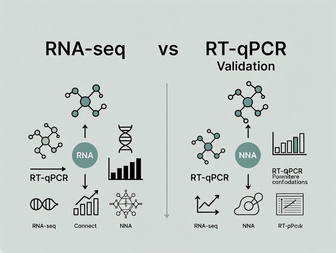
RNA-seq vs RT-qPCR: A Modern Guide to Validation, Best Practices, and Advanced Applications in Biomedical Research
This comprehensive guide provides researchers and drug development professionals with a detailed framework for validating RNA-seq findings with RT-qPCR.
RNA-seq vs RT-qPCR: A Modern Guide to Validation, Best Practices, and Advanced Applications in Biomedical Research
Abstract
This comprehensive guide provides researchers and drug development professionals with a detailed framework for validating RNA-seq findings with RT-qPCR. We explore the foundational principles of both technologies, contrasting their methodological workflows, sensitivity, and throughput. The article addresses common troubleshooting and optimization strategies for robust experimental design and data interpretation. Finally, we deliver a critical comparative analysis to establish when and how RT-qPCR validation is essential, necessary, or optional, empowering scientists to implement gold-standard validation protocols that enhance the credibility and translational impact of their gene expression studies.
Understanding the Core Technologies: RNA-seq Discovery and RT-qPCR Precision
This comparison guide is framed within a broader research thesis investigating the complementary roles of RNA sequencing (RNA-seq) and RT-qPCR. While RT-qPCR remains the gold standard for targeted, high-sensitivity validation of a limited number of transcripts, RNA-seq provides an unbiased, genome-wide discovery platform. The revolution lies in RNA-seq's ability to profile the entire transcriptome without prior sequence knowledge, enabling novel hypothesis generation regarding differential expression, alternative splicing, novel transcripts, and gene fusions.
Performance Comparison: RNA-seq vs. Microarrays and RT-qPCR Panels
The following table compares key performance metrics of modern RNA-seq with legacy microarray technology and targeted RT-qPCR panels. Data is synthesized from recent benchmark studies (2023-2024).
Table 1: Technology Performance Comparison for Transcriptome Analysis
| Feature | RNA-seq (Illumina NovaSeq X) | Microarray (Affymetrix Clariom S) | RT-qPCR Panel (Fluidigm) |
|---|---|---|---|
| Throughput | Entire transcriptome (20,000+ genes) | Pre-defined probeset (20,000 genes) | Pre-defined panel (50-500 targets) |
| Dynamic Range | >10⁵ (Wide) | 10³-10⁴ (Limited) | >10⁷ (Very Wide) |
| Sensitivity | High (Can detect low-abundance transcripts) | Moderate (Background noise limits) | Very High (Optimal for rare transcripts) |
| Discovery Power | Unbiased; detects novel transcripts, isoforms, fusions | Biased to known, annotated sequences | Biased to pre-selected targets |
| Sample Input | 10-1000 ng total RNA (varies by protocol) | 50-500 ng total RNA | 1-100 ng total RNA (cDNA) |
| Quantitative Accuracy | High (Linear correlation with RT-qPCR R²=0.85-0.95) | Moderate (Saturation at high expression) | Very High (Gold standard) |
| Typical Cost per Sample | $200 - $800 | $150 - $400 | $20 - $100 (for panel targets) |
| Best Application | Discovery, hypothesis generation, global profiling | Targeted profiling of known genes | Validation, high-precision targeted quantitation |
Experimental Data from Comparative Studies
Table 2: Representative Data from a Benchmarking Study (Cancer Cell Lines)
| Measurement | RNA-seq Result | Microarray Result | RT-qPCR Validation |
|---|---|---|---|
| Differentially Expressed Genes (FDR<0.05) | 1,245 | 987 | 50/50 randomly selected confirmed |
| Novel Alternative Splicing Events Detected | 42 | 0 (platform limitation) | 5/5 novel junctions confirmed |
| Gene Fusion Detected (Known oncogene) | 1 | Not detected | Confirmed by ddPCR |
| Correlation with RT-qPCR (for 50 genes) | Pearson R = 0.92 | Pearson R = 0.78 | N/A (Reference) |
| Time from library prep to data | ~3-5 days | ~2 days | ~1 day (for 50 targets) |
Detailed Methodologies for Key Experiments
Protocol 1: Standard Poly-A Selected RNA-seq Workflow
1. RNA Quality Control: Assess RNA integrity using Agilent Bioanalyzer (RIN > 8.0 required). 2. Library Preparation: Use stranded poly-A enrichment (e.g., Illumina Stranded mRNA Prep). Fragment purified mRNA, synthesize cDNA with random priming, add adapters, and amplify with index primers (12-16 PCR cycles). 3. Sequencing: Pool libraries and sequence on an Illumina NovaSeq X platform targeting 30-50 million 150bp paired-end reads per sample. 4. Bioinformatic Analysis: * Alignment: Use STAR aligner to map reads to the reference genome (e.g., GRCh38). * Quantification: Generate gene-level counts with featureCounts. * Differential Expression: Analyze with DESeq2 or edgeR, applying normalization (e.g., median-of-ratios) and statistical testing (Wald test).
Protocol 2: RT-qPCR Validation of RNA-seq Hits
1. cDNA Synthesis: Using the same RNA as RNA-seq, perform reverse transcription with random hexamers and a high-fidelity reverse transcriptase (e.g., SuperScript IV). 2. Assay Design: Design TaqMan hydrolysis probes or SYBR Green primers for targets of interest and housekeeping genes (e.g., GAPDH, ACTB). Ensure amplicons span exon-exon junctions. 3. qPCR Run: Perform reactions in technical triplicates on a qPCR instrument (e.g., QuantStudio 7 Pro). Use a standard curve or ΔΔCt method for absolute or relative quantification. 4. Statistical Correlation: Calculate Pearson correlation between RNA-seq normalized counts (e.g., TPM or log2 fold-change) and RT-qPCR ΔCt or log2 fold-change values.
Visualizations
Diagram 1: Standard RNA-seq Workflow
Diagram 2: RNA-seq to RT-qPCR Thesis Framework
The Scientist's Toolkit: Research Reagent Solutions
Table 3: Essential Reagents and Materials for RNA-seq & Validation
| Item | Function | Example Product |
|---|---|---|
| RNA Stabilization Reagent | Preserves RNA integrity immediately upon sample collection, inhibiting RNases. | RNAlater Stabilization Solution |
| High-Sensitivity RNA QC Kit | Assesses RNA Integrity Number (RIN) and concentration; critical for input quality. | Agilent RNA 6000 Nano Kit |
| Stranded mRNA Library Prep Kit | Converts RNA into sequencer-compatible DNA libraries with strand information. | Illumina Stranded mRNA Prep |
| Nuclease-Free Water | Solvent free of RNases and DNases for all molecular biology steps. | Ambion Nuclease-Free Water |
| High-Fidelity Reverse Transcriptase | Synthesizes cDNA from RNA template with high efficiency and accuracy for both RNA-seq and qPCR. | SuperScript IV Reverse Transcriptase |
| Universal ProbeLibrary (UPL) Probes | Pre-designed, hydrolysis-based probes for flexible and highly specific RT-qPCR assay design. | Roche Universal ProbeLibrary |
| Multiplex qPCR Master Mix | Enables simultaneous amplification and detection of multiple targets in a single well. | TaqMan Fast Advanced Master Mix |
| Digital PCR Assay | Provides absolute quantification for ultra-sensitive validation of gene fusions or low-abundance targets. | ddPCR Mutation Assay |
| Bioanalyzer DNA High Sensitivity Kit | Validates final library fragment size distribution and quantity before sequencing. | Agilent High Sensitivity DNA Kit |
Within the ongoing research discourse comparing RNA-seq and RT-qPCR for gene expression validation, RT-qPCR maintains its position as the indispensable gold standard. This status is predicated on its superior performance in three critical analytical parameters: sensitivity, dynamic range, and specificity. This guide objectively compares RT-qPCR's performance in these areas against common alternatives, supported by experimental data.
Comparison of Analytical Performance
The following table summarizes key performance characteristics of RT-qPCR versus alternative methods, based on consolidated data from recent publications and technical literature.
Table 1: Performance Comparison of Gene Expression Quantification Methods
| Parameter | RT-qPCR (SYBR Green) | RT-qPCR (TaqMan Probe) | Microarray | RNA-seq (Standard Depth) | Digital PCR (dPCR) |
|---|---|---|---|---|---|
| Sensitivity | 1-10 copies | 1-5 copies | Medium-High | Low-Medium | <1 copy |
| Dynamic Range | 7-8 logs | 7-8 logs | 3-4 logs | >5 logs | 4-5 logs |
| Specificity | High (Primer-dependent) | Very High (Probe-based) | Medium (Cross-hybridization) | High (Mapping-dependent) | Very High (Endpoint) |
| Absolute Quantification | Yes (with standard curve) | Yes (with standard curve) | No | No (relative) | Yes (absolute, no standard curve) |
| Multiplexing Capacity | Low-Moderate | Moderate-High | Very High | Extremely High | Low |
| Throughput | High | High | Very High | High | Low-Moderate |
Experimental Protocols for Cited Performance Data
Protocol 1: Determining RT-qPCR Sensitivity & Dynamic Range
- Objective: To establish the limit of detection (LoD) and linear dynamic range.
- Method:
- Standard Curve Preparation: Serially dilute a known quantity of in vitro transcribed RNA or gDNA template across 8 orders of magnitude (e.g., from 10^7 to 10^0 copies).
- RT-qPCR Setup: Perform reverse transcription followed by qPCR for each dilution in triplicate. Use a hydrolysis probe (TaqMan) or intercalating dye (SYBR Green).
- Data Analysis: Plot the log10 of the starting template quantity against the quantification cycle (Cq). The linear range is defined where the coefficient of determination (R^2) > 0.99 and amplification efficiency is 90-110%. The LoD is the lowest concentration where ≥95% of replicates are detected.
Protocol 2: Assessing Specificity via Melt Curve or Probe Validation
- Objective: To confirm amplification of a single, intended product.
- SYBR Green Method:
- After amplification, perform a melt curve analysis from 65°C to 95°C.
- A single, sharp peak in the derivative melt curve indicates specific amplification. Multiple peaks suggest primer-dimer or non-specific products.
- TaqMan Probe Method:
- Specificity is inherent to the probe binding sequence.
- Validate by testing assays on genomic DNA and non-template controls (NTCs). Signal in NTCs indicates probe degradation or contamination.
Visualizations
RT-qPCR Workflow from RNA to Quantification
RNA-seq Discovery to RT-qPCR Validation Workflow
The Scientist's Toolkit: Key Research Reagent Solutions
Table 2: Essential Materials for RT-qPCR Validation Experiments
| Item | Function & Importance |
|---|---|
| High-Quality RNA Isolation Kit | Removes genomic DNA, RNases, and inhibitors. Purity (A260/A280 ratio) is critical for reverse transcription efficiency. |
| Reverse Transcriptase with RNase Inhibitor | Converts RNA to stable cDNA. Enzyme fidelity and processivity impact representation and downstream quantification. |
| qPCR Master Mix | Contains DNA polymerase, dNTPs, and optimized buffer. Probe-based mixes include fluorescein for normalization. Stabilizes reaction chemistry. |
| Sequence-Specific Primers & Probes | Primers define amplification region. Hydrolysis probes (e.g., TaqMan) provide unmatched specificity via an additional oligonucleotide. |
| Nuclease-Free Water | Reaction diluent. Prevents RNase/DNase contamination that degrades samples and reagents. |
| Validated Reference Gene Assays | For relative quantification (ΔΔCq). Essential for normalizing biological and technical variation (e.g., GAPDH, ACTB, HPRT1). |
| Calibrator Sample or Standard Curve Material | Provides a known quantity benchmark for relative (ΔΔCq) or absolute (standard curve) quantification across runs. |
| Optical Plates & Seals | Ensure consistent thermal conductivity and prevent evaporation and contamination during cycling. |
In the validation of genomic and transcriptomic findings, researchers must navigate a critical choice between high-throughput, discovery-focused technologies like RNA-seq and targeted, accuracy-driven methods like RT-qPCR. This comparison guide objectively evaluates these platforms within the context of validation research, providing current data and experimental frameworks to inform decision-making for scientists and drug development professionals.
Core Metric Comparison
Table 1: Platform Comparison for Validation Studies
| Metric | RNA-seq (NGS) | RT-qPCR (TaqMan) | Best for Validation Phase |
|---|---|---|---|
| Throughput (Samples/Run) | 16-96+ (multiplexed) | 1-384 (multiplex limited to 4-6) | RT-qPCR (for <100 targets) |
| Cost per Sample (Reagents) | ~$50 - $200 | ~$2 - $10 | RT-qPCR |
| Discovery Power | High (Whole transcriptome) | None (Targeted only) | RNA-seq (Discovery) |
| Targeted Accuracy | Moderate (Dynamic range issues) | Very High (Wide dynamic range) | RT-qPCR |
| Experimental Turnaround | 3-7 days | 1-2 days | RT-qPCR |
| Primary Validation Role | Hypothesis Generation, Isoform Discovery | Gold-Standard Confirmatory Quantification | N/A |
Table 2: Experimental Performance Data from Comparative Studies
| Study Focus | RNA-seq Correlation (vs. qPCR) | Key Limitation Cited | Recommended Use |
|---|---|---|---|
| Differential Expression Validation (10 genes) | R² = 0.72 - 0.85 | Lower concordance for low-abundance transcripts | Use qPCR for final validation of key targets. |
| Absolute Quantification | Poor (Relative) | RNA-seq lacks internal standard for copy number | qPCR is required for absolute copy number. |
| Detection of Rare Transcripts | Moderate (High depth costly) | High false-negative rate at low expression | qPCR offers sensitive, specific detection. |
Experimental Protocols for Cross-Platform Validation
Protocol 1: RNA-seq Workflow for Initial Discovery
- Total RNA Isolation: Use column-based kits with DNase I treatment. Assess integrity (RIN > 8) via Bioanalyzer.
- Library Preparation: Employ stranded, poly-A-selection kits (e.g., Illumina). Input: 100 ng - 1 µg total RNA.
- Sequencing: Perform on platform (e.g., NovaSeq) for minimum 30 million paired-end 150bp reads per sample.
- Bioinformatic Analysis: Align to reference genome (STAR), quantify gene counts (featureCounts), perform differential expression (DESeq2).
Protocol 2: RT-qPCR Workflow for Targeted Validation
- cDNA Synthesis: Use same RNA as RNA-seq. Perform reverse transcription with random hexamers and oligo-dT primers.
- Assay Design: Use pre-validated, exon-spanning TaqMan assays or design SYBR Green primers with melt curve analysis.
- qPCR Run: Perform in technical triplicates on 384-well block cycler. Include no-template controls and serial dilutions for standard curve.
- Data Analysis: Calculate relative quantification (ΔΔCq) using 2-3 validated reference genes (e.g., GAPDH, ACTB).
Visualizations
Title: Sequential Use of RNA-seq and RT-qPCR in Validation
Title: Technology Positioning by Cost and Target Throughput
The Scientist's Toolkit: Research Reagent Solutions
| Item | Function in Validation Workflow |
|---|---|
| High-Capacity cDNA Reverse Transcription Kit | Converts RNA to stable cDNA for downstream qPCR; includes RNase inhibitor. |
| TaqMan Gene Expression Assays | Pre-optimized, target-specific primers & FAM-labeled probe for highest accuracy. |
| SYBR Green Master Mix | Cost-effective dye for qPCR; requires post-run melt curve for specificity check. |
| Universal RNA-seq Library Prep Kit | Allows for strand-specific, whole-transcriptome library construction for NGS. |
| Digital PCR Master Mix | Provides absolute quantification without standard curve; used for ultra-sensitive validation. |
| Bioanalyzer RNA Nano Kit | Assesses RNA Integrity Number (RIN) to ensure input quality for both platforms. |
Within the broader thesis of RNA-seq discovery versus RT-qPCR validation, selecting the appropriate technology at each pipeline stage is critical for efficiency and accuracy. RNA-seq excels in unbiased, genome-wide transcriptome profiling, while RT-qPCR remains the gold standard for targeted, high-precision validation of a limited number of transcripts. This guide compares their performance based on current experimental data to inform deployment strategies.
Performance Comparison: RNA-seq vs. RT-qPCR
Table 1: Core Performance Metrics Comparison
| Parameter | RNA-seq (Illumina NGS) | RT-qPCR (TaqMan Probe-Based) | Experimental Support |
|---|---|---|---|
| Dynamic Range | >10⁵ (linear) | >10⁷ (log-linear) | Zhao et al., 2021 BMC Genomics |
| Accuracy (vs. known spikes) | High (R² >0.98) | Very High (R² >0.99) | SEQC/MAQC-III Consortium, 2019 Nat Commun |
| Precision (Replicate CV) | 5-15% (library prep dominant) | 1-5% (assay dependent) | Everaert et al., 2019 Sci Rep |
| Sample Throughput | High (multiplexed, 100s/s run) | Medium (96-384 well plates) | Platform-dependent |
| Gene Throughput | Whole transcriptome (10,000s) | Targeted (1-500 genes/run) | Fundamental design |
| Input RNA Requirement | 10 ng - 1 µg (standard) | 1 pg - 100 ng (high sensitivity) | Comparison studies |
| Cost per Sample | $$-$$$ (decreasing with plex) | $-$$ (increasing with plex) | Lab benchmarking 2023 |
| Turnaround Time | 3-7 days (library to data) | 1-2 days (cDNA to data) | Standard workflows |
| Primary Use Case | Discovery, differential expression, isoform detection, novel transcript ID | Validation, low-abundance targets, absolute quantification, high-throughput screening | Consensus application |
Detailed Experimental Protocols
Protocol 1: RNA-seq for Differential Expression Discovery
- RNA QC: Assess integrity (RIN >8 via Bioanalyzer) and quantity (Qubit).
- Library Prep: Use poly-A selection or rRNA depletion (Illumina Stranded TruSeq). Fragment RNA, synthesize cDNA, add adapters, and perform PCR amplification (12-15 cycles).
- Sequencing: Pool libraries and sequence on Illumina NovaSeq (PE 150 bp) to a depth of 25-40 million reads per sample.
- Bioinformatics: Align reads to reference genome (STAR aligner). Quantify gene counts (featureCounts). Perform differential expression analysis (DESeq2 R package, FDR-adjusted p-value <0.05, |log2FC|>1).
Protocol 2: RT-qPCR for Target Validation
- Reverse Transcription: Use high-capacity cDNA reverse transcription kit with random hexamers on 500 ng total RNA per sample (no RT control essential).
- Assay Design: Use pre-validated, exon-spanning TaqMan probe/primer sets. Include three technical replicates.
- qPCR Run: Use 384-well format. Prepare reaction mix with cDNA, primers, probe, and TaqMan Fast Advanced Master Mix. Run on QuantStudio 7 Pro: 50°C (2 min), 95°C (20 sec), then 40 cycles of 95°C (1 sec) and 60°C (20 sec).
- Analysis: Use comparative ΔΔCt method. Normalize to two validated reference genes (e.g., GAPDH, ACTB). Include no-template controls (NTC).
Workflow Diagrams
Decision Logic for Technology Deployment
RNA-seq Experimental Workflow
RT-qPCR Validation Workflow
The Scientist's Toolkit: Research Reagent Solutions
Table 2: Essential Materials for Featured Experiments
| Item | Function | Example Product (Non-exhaustive) |
|---|---|---|
| RNA Integrity Number (RIN) Analyzer | Assesses RNA quality pre-library prep; critical for both technologies. | Agilent Bioanalyzer / TapeStation |
| High-Sensitivity DNA/RNA Quantitation | Accurately measures low-concentration nucleic acids for normalization. | Thermo Fisher Qubit Fluorometer |
| Stranded mRNA Library Prep Kit | For RNA-seq: captures poly-A mRNA, preserves strand information. | Illumina Stranded TruSeq |
| rRNA Depletion Kit | For RNA-seq of degraded or non-polyA samples (e.g., FFPE). | Illumina Ribo-Zero Plus |
| Universal cDNA Synthesis Kit | For RT-qPCR: generates high-efficiency, reproducible cDNA. | Thermo Fisher High-Capacity Kit |
| TaqMan Gene Expression Assays | For RT-qPCR: pre-validated, highly specific primer/probe sets. | Thermo Fisher TaqMan Assays |
| Fast Advanced Master Mix | For RT-qPCR: contains polymerase, dNTPs, optimized buffer. | Thermo Fisher TaqMan Fast Advanced |
| Nuclease-Free Water | Critical reagent for all molecular biology steps to avoid degradation. | Various certified suppliers |
| Digital PCR System (Emerging) | Provides absolute quantification for orthogonal validation. | Bio-Rad QX200 / Thermo Fisher QuantStudio 3D |
Designing a Robust Validation Workflow: From RNA-seq Data to RT-qPCR Confirmation
Within the broader thesis of RNA-seq vs. RT-qPCR validation research, the initial selection of optimal candidates from RNA-seq data is the most critical determinant of validation success. This guide compares common selection strategies and their outcomes based on published experimental data.
Comparison of Candidate Selection Strategies
The table below summarizes the performance of four primary selection criteria when subsequent RT-qPCR validation is performed.
Table 1: Validation Success Rates by Selection Strategy
| Selection Criterion | Avg. Validation Success Rate (RT-qPCR) | Key Advantages | Key Limitations | Typical Use Case |
|---|---|---|---|---|
| Fold Change (FC) Only (e.g., |log2FC| > 2) | 60-75% | Simple, identifies large effect sizes. | High false positive rate from low-abundance transcripts. | Preliminary screens where sensitivity is prioritized. |
| Statistical Significance Only (e.g., adj. p-value < 0.05) | 65-80% | Controls false discoveries. | May miss biologically relevant, low-count transcripts. | Hypothesis-driven, focused studies. |
| FC + Statistical Significance | 80-92% | Robust balance; industry standard. | Can exclude important transcripts with moderate FC. | Most differential expression studies. |
| FC + Significance + Abundance (e.g., Base Mean > 50) | 90-98% | Highest validation success; ensures reliable detection. | May filter out key low-expressed regulators. | High-stakes validation for drug targets or biomarkers. |
Experimental Protocols for Key Studies
Protocol 1: Benchmarking Selection Criteria (Reference: Conesa et al., 2016)
- RNA-seq Analysis: Process three public datasets (human, mouse, rat) through a standardized pipeline (Hisat2 → StringTie → DESeq2).
- Candidate Lists: Generate four gene lists per dataset using the criteria in Table 1.
- RT-qPCR Validation: Design primers for 50 random genes from each list. Use 500 ng total RNA for cDNA synthesis (SuperScript IV). Perform qPCR in triplicate (SYBR Green) on a QuantStudio 6.
- Success Calculation: A gene is "validated" if its direction of change and statistical significance (p<0.05, t-test) by RT-qPCR match the RNA-seq prediction.
Protocol 2: Impact of Expression Level on Validation (Reference: Everaert et al., 2017)
- Stratification: From a human cancer RNA-seq dataset, categorize differentially expressed genes (adj. p<0.01, \|log2FC\|>1) into quartiles based on their base mean expression.
- Validation: Subject 20 genes from each quartile to RT-qPCR using TaqMan assays (more specific than SYBR).
- Correlation Analysis: Calculate the Pearson correlation between RNA-seq log2FC and RT-qPCR log2FC for each quartile.
Table 2: Validation Correlation by Expression Quartile
| Expression Quartile (Base Mean) | Avg. Pearson Correlation (RNA-seq vs. RT-qPCR) |
|---|---|
| Q1 (Lowest) | 0.45 |
| Q2 | 0.78 |
| Q3 | 0.92 |
| Q4 (Highest) | 0.97 |
Visualizing the Optimal Selection Workflow
Title: Workflow for Selecting High-Confidence Validation Candidates
The Scientist's Toolkit: Key Research Reagent Solutions
Table 3: Essential Reagents for RNA-seq to qPCR Validation Pipeline
| Item | Function & Rationale | Example Product |
|---|---|---|
| RNA Isolation Kit | Ensures high-integrity, DNA-free RNA for both sequencing and sensitive qPCR. | Qiagen RNeasy Mini Kit with DNase I step. |
| High-Sensitivity cDNA Synthesis Kit | Critical for faithful reverse transcription of low-abundance candidates. | Thermo Fisher SuperScript IV VILO. |
| qPCR Master Mix | Provides consistent amplification efficiency for accurate fold-change calculation. | Bio-Rad SsoAdvanced Universal SYBR Green. |
| Specific Assays | For low-expressed or homologous targets, specificity is paramount. | Thermo Fisher TaqMan Gene Expression Assays. |
| RNA-seq Library Prep Kit | Stranded, ribosomal RNA-depleted kits provide accurate directional transcriptome data. | Illumina Stranded Total RNA Prep. |
| Digital Pipettes | Essential for precise, reproducible liquid handling in low-volume qPCR setups. | Eppendorf Research Plus. |
Within the context of validating RNA-seq data, RT-qPCR remains the gold standard for quantifying specific transcripts. The accuracy of this validation hinges critically on the specificity of the primer and probe design. This guide compares key design strategies and their impact on assay performance, providing experimental data to inform best practices for researchers and drug development professionals.
Design Parameter Comparison and Experimental Data
The following table summarizes experimental outcomes from assays designed with different stringency parameters, measuring specificity via melt curve analysis and efficiency.
Table 1: Impact of Primer Design Parameters on RT-qPCR Specificity
| Design Parameter | Target Tm (°C) | Amplicon Length (bp) | Specificity (Melt Curve Peak) | Avg. Efficiency (%) | Key Advantage |
|---|---|---|---|---|---|
| Standard Design (Primer3) | 58-60 | 80-150 | Single, broad peak | 95 ± 5 | Simplicity, robust yield |
| Stringent Design (NCBI Primer-BLAST) | 60-62 | 65-90 | Single, sharp peak | 98 ± 2 | High specificity, minimal primer-dimer |
| Exon-Exon Junction Spanning | 59-61 | 70-120 | Single, defined peak | 96 ± 3 | Excludes genomic DNA amplification |
| Locked Nucleic Acid (LNA) Probes | Probe Tm +5-10 | 70-100 | N/A (Probe-based) | 99 ± 1 | Enhanced allele discrimination, high stability |
Data derived from validation experiments on a panel of 10 human cytokine genes. Specificity assessed by SYBR Green melt curve analysis or TaqMan probe fluorescence.
Detailed Experimental Protocol for Specificity Validation
Protocol: Comparative Specificity Testing of Primer Sets
- In Silico Design: Design three primer sets per target using (a) basic Primer3 defaults, (b) stringent Primer-BLAST with cross-species check, and (c) primers spanning a known exon-exon junction.
- Template Preparation: Dilute cDNA (reverse-transcribed from total RNA) to a working concentration series (e.g., 5-log dilution series).
- qPCR Setup: Perform reactions in triplicate using a SYBR Green master mix. Include a no-template control (NTC) for each primer set.
- Run Conditions: Use a standard two-step cycling protocol (95°C for 10 min, followed by 40 cycles of 95°C for 15 sec and 60°C for 1 min) with a subsequent melt curve analysis (65°C to 95°C, increment 0.5°C).
- Data Analysis: Calculate amplification efficiency from the standard curve. Analyze melt curves for single, sharp peaks indicative of specific product. Confirm product size by gel electrophoresis.
Visualizing the RT-qPCR Validation Workflow
Diagram 1: RNA-seq to RT-qPCR validation workflow.
The Scientist's Toolkit: Essential Reagents for RT-qPCR Validation
Table 2: Key Research Reagent Solutions
| Reagent / Material | Function in Experiment | Key Consideration |
|---|---|---|
| High-Fidelity Reverse Transcriptase | Converts RNA to cDNA, minimizing enzyme-induced bias. | Essential for accurate representation of transcript abundance. |
| Hot-Start DNA Polymerase | Reduces non-specific amplification and primer-dimer formation during reaction setup. | Critical for high-specificity assays, especially with SYBR Green. |
| Sequence-Specific TaqMan Probes | Provides fluorescence signal upon cleavage during amplification, adding a layer of specificity. | Ideal for multiplexing or detecting single nucleotide polymorphisms (SNPs). |
| Nuclease-Free Water | Serves as the reaction diluent. | Must be certified nuclease-free to prevent degradation of primers, probes, and template. |
| qPCR Plates with Optical Seals | Holds reactions and allows for fluorescence detection by the instrument. | Ensure seal is compatible with the thermocycler block to prevent evaporation and well-to-well contamination. |
The integrity of RNA and the efficiency of its reverse transcription (RT) are foundational steps that critically influence the accuracy and reproducibility of both RNA-seq and RT-qPCR data. Within the broader thesis of comparing these technologies for validation research, this step represents a major point of convergence where methodological rigor is non-negotiable. The quality of cDNA synthesized during RT directly dictates the dynamic range and reliability of downstream quantification.
Impact of RNA Integrity on Downstream Assays
RNA Integrity Number (RIN) is a standard metric. Degraded RNA (low RIN) leads to biased representation, particularly affecting the 3' ends of transcripts.
| RIN Value | Effect on RNA-seq | Effect on RT-qPCR | Recommended Action |
|---|---|---|---|
| ≥ 9.0 | Optimal coverage, even across transcript length. | High efficiency, accurate Cq values across all amplicons. | Proceed with standard protocols. |
| 7.0 - 8.9 | Moderate 3' bias detectable; usable for most analyses. | Amplicons >300 bp may show variable efficiency; prefer shorter targets. | Proceed with caution; note potential bias. |
| 6.0 - 6.9 | Significant 3' bias; gene-level analysis possible but avoid isoform detection. | Only short amplicons (<150 bp) are reliable; requires careful assay design. | Use for targeted assays only; not recommended for full RNA-seq. |
| < 6.0 | Severe bias and high technical noise; data largely unreliable. | Extreme variability; results are not quantifiable. | Do not proceed; re-extract RNA. |
Reverse Transcription Enzyme and Protocol Comparison
The choice of reverse transcriptase and priming method introduces systematic variation.
| Kit/Enzyme Type | Processivity | Thermal Stability | Recommended for RNA-seq | Recommended for RT-qPCR |
|---|---|---|---|---|
| Moloney Murine Leukemia Virus (MMLV) | Moderate | Low (42°C max) | Not optimal for complex or GC-rich RNA. | Standard for routine assays with high-quality RNA. |
| M-MLV RNase H⁻ | High | Moderate (50°C) | Good for standard libraries, reduces secondary structure. | Excellent for most applications, reduces primer-dimer artifacts. |
| ArrayScript (Thermostable) | Very High | High (55-60°C) | Optimal. Superior for GC-rich templates and full-length cDNA. | Optimal for demanding targets (long amplicons, high GC). |
| Template-Switching (SMART) | N/A | Varies | Required for specific protocols (e.g., single-cell, low-input). | Less common; used for specific whole-transcript amplification. |
Priming Strategy:
- Oligo(dT) Priming: Enriches for polyadenylated mRNA. Introduces strong 3' bias in RNA-seq. Ideal for RT-qPCR when targeting the 3' end.
- Random Hexamer Priming: Provides uniform coverage across transcriptome, including non-coding RNA. Can generate primer artifacts. Standard for RNA-seq and whole-transcriptome RT-qPCR.
- Gene-Specific Priming: Used exclusively in RT-qPCR for maximum sensitivity and specificity for a single target. Not applicable for RNA-seq.
Experimental Protocols for Key Validation Experiments
Protocol 1: Assessing RT Efficiency for RT-qPCR
- Serially Diluted RNA Input: Perform identical RT reactions on a dilution series of a high-quality RNA sample (e.g., 1 µg, 100 ng, 10 ng, 1 ng).
- qPCR Amplification: Perform qPCR on a stable, moderately expressed reference gene from each cDNA dilution.
- Efficiency Calculation: Plot log10(RNA input) vs. Cq value. The slope of the line determines efficiency: Efficiency % = [10^(-1/slope) - 1] x 100. Ideal efficiency is 90-110%, with an R² > 0.99.
- Interpretation: A decline in efficiency with lower input indicates RT inhibition or suboptimal enzyme performance.
Protocol 2: Evaluating 3' Bias for RNA-seq
- Library Preparation & Sequencing: Prepare RNA-seq libraries from a sample using standard protocols (e.g., poly-A selection, random priming).
- Bioinformatic Analysis: Map reads to the reference genome. Using tools like
PicardorRSeQC, calculate the gene body coverage metric. - Visualization: Plot the normalized read coverage from the 5' end to the 3' end of genes (aggregated across many genes).
- Interpretation: A flat coverage profile indicates no bias. A descending slope from 5' to 3' indicates degradation (low RIN). A 3'-enriched profile indicates severe degradation or poly-A priming bias.
Visualizations
Diagram Title: Workflow Divergence Post-Reverse Transcription
Diagram Title: RT Priming Method Determines Coverage Bias
The Scientist's Toolkit: Research Reagent Solutions
| Reagent / Material | Function & Importance |
|---|---|
| Agilent Bioanalyzer / TapeStation | Microfluidics-based system for precisely calculating RNA Integrity Number (RIN) or DV200. Essential for objective QC before costly library prep or RT. |
| RNase Inhibitors (e.g., Recombinant RNasin) | Protects RNA templates from degradation during RT reaction setup, crucial for long transcripts and low-input samples. |
| High-Efficiency RT Kits (e.g., SuperScript IV, PrimeScript RT) | Engineered reverse transcriptases with high thermal stability and processivity, minimizing bias and maximizing cDNA yield from challenging samples. |
| dNTP Mix | Deoxynucleotide triphosphates (dATP, dCTP, dGTP, dTTP) are the building blocks for cDNA synthesis. Balanced, high-purity mixes are critical. |
| Anchored Oligo(dT) Primers | Primers with a short anchor sequence (e.g., VN) ensure priming from the beginning of the poly-A tail, improving consistency over simple dT primers. |
| RNA Spike-in Controls (e.g., ERCC ExFold RNA Spike-in Mix) | Known, exogenous RNA molecules added to the sample before RT. Allow absolute normalization and detection of technical biases in both RNA-seq and RT-qPCR. |
| qPCR Master Mix with Hot-Start Taq | Contains optimized buffer, dNTPs, polymerase, and fluorescence dye/intercalator. Hot-start technology prevents non-specific amplification during reaction setup. |
In the validation phase of RNA-seq vs. RT-qPCR research, confirming differential expression (DE) findings through independent replication is paramount. This guide compares the performance of these platforms in validation studies, focusing on experimental design and statistical rigor.
Performance Comparison: RNA-seq vs. RT-qPCR in Validation Table 1: Platform Comparison for Validation Studies
| Metric | RT-qPCR (Validation Standard) | RNA-seq (Discovery & Validation) | Key Implication for Validation |
|---|---|---|---|
| Throughput | Low to medium (tens of targets) | High (whole transcriptome) | RT-qPCR is efficient for ≤100 targets; RNA-seq validates entire DE lists. |
| Dynamic Range | ~9 logs (Excellent) | ~5 logs (Very Good) | Both suitable; RT-qPCR superior for extreme fold-changes. |
| Sensitivity | Can detect single copies | Requires moderate expression level | RT-qPCR is preferred for low-abundance transcripts. |
| Precision | Very High (low technical variance) | High (higher technical variance) | RT-qPCR often yields tighter confidence intervals. |
| Absolute Quantification | Yes (with standard curves) | Indirect (relative, normalized counts) | RT-qPCR provides copy numbers; RNA-seq provides relative abundance. |
| Cost per Sample | Low (for few targets) | High | Budget scales with RT-qPCR target count. |
| Statistical Power Consideration | High power per target cost-effective. | Requires careful sample size calc for whole transcriptome. | Underpowered RNA-seq validation fails to confirm true DE. |
Table 2: Example Validation Outcomes from a Hypothetical DE Study
| Gene ID | RNA-seq Discovery (Log2FC) | RT-qPCR Validation (Log2FC) | p-value (qPCR) | Validated? |
|---|---|---|---|---|
| Gene A | +3.5 | +3.1 | 0.003 | Yes |
| Gene B | -2.1 | -1.9 | 0.021 | Yes |
| Gene C | +1.8 | +0.7 | 0.185 | No (Lack of power/artifact) |
| Gene D | -4.0 | -3.8 | 0.001 | Yes |
*Synthetic data for illustrative purposes.*
Experimental Protocol: Technical Replication for RT-qPCR Validation
- Sample: Use independent biological replicates (n≥3) not used in the discovery RNA-seq.
- RNA Isolation: Repeat extraction using same method (e.g., column-based with DNase I treatment).
- Reverse Transcription: Use 1µg total RNA, random hexamers, and a high-fidelity reverse transcriptase. Include a no-RT control.
- qPCR Assay: Design exon-spanning primers (amplicon 80-150bp). Perform reactions in triplicate (technical replicates) using a SYBR Green or probe-based master mix on a calibrated instrument.
- Data Analysis: Calculate average Cq values. Use stable reference genes (e.g., GAPDH, ACTB) for ΔΔCq analysis. Determine fold-change and statistical significance (e.g., Student's t-test) relative to control.
Statistical Power Analysis Protocol for Validation
- Define Parameters:
- Effect Size: Minimum fold-change (e.g., 1.5x) deemed biologically relevant.
- Significance Level (α): Typically 0.05.
- Desired Power (1-β): 80% or higher.
- Variance Estimate: Use standard deviation from pilot qPCR data or published studies for similar genes.
- Calculate Sample Size: Use power analysis software (e.g., GPower, R
pwrpackage). For a two-group comparison with a t-test, input the above parameters to determine the required number of *biological replicates per group. - Apply to Study Design: If RNA-seq discovery suggests 50 DE genes, and power analysis dictates n=8 per group for qPCR, then 8 independent samples per condition must be processed for validation.
Workflow for qPCR Validation of RNA-seq Data
Key Inputs for Statistical Power Calculation
The Scientist's Toolkit: Research Reagent Solutions Table 3: Essential Reagents for RNA-seq/qPCR Validation Workflow
| Item | Function in Validation | Example Solutions |
|---|---|---|
| DNase I, RNase-free | Removes genomic DNA contamination from RNA preps, critical for accurate qPCR. | Thermo Fisher RapidOut, Qiagen RNase-Free DNase Set. |
| High-Capacity cDNA Reverse Transcription Kit | Converts purified RNA to stable cDNA with high efficiency and consistency. | Applied Biosystems High-Capacity cDNA Reverse Transcription Kit. |
| SYBR Green or TaqMan Master Mix | Provides enzymes, dNTPs, and fluorescence chemistry for quantitative PCR. | Bio-Rad SsoAdvanced SYBR, Thermo Fisher TaqMan Universal MM. |
| Validated PrimeTime qPCR Assays | Pre-designed, optimized primer-probe sets for specific, reproducible target amplification. | Integrated DNA Technologies (IDT) PrimeTime assays. |
| RNA Spike-in Controls | Added to samples pre-extraction to monitor technical variability across entire workflow. | External RNA Controls Consortium (ERCC) spikes. |
| Statistical Power Analysis Software | Calculates necessary sample size to avoid underpowered, inconclusive validation experiments. | G*Power, R/Bioconductor pwr package. |
Accurate normalization is critical in RT-qPCR, the gold standard for validating RNA-seq data. The choice of reference genes (RGs) is the most significant source of error, necessitating a move beyond assumed "housekeepers" like GAPDH or ACTB to empirically validated, stable targets. This guide compares traditional housekeepers with modern stability validation approaches.
The Problem with Traditional Housekeepers
Traditional housekeeping genes are involved in basic cellular maintenance and were historically assumed to be constitutively expressed. However, extensive research shows their expression can vary significantly across different experimental conditions, tissues, and disease states, leading to normalization errors and inaccurate gene expression quantification.
Comparison of Validation Methods and Gene Performance
The following table summarizes key metrics from recent studies comparing traditional RGs with those selected via stability validation algorithms.
Table 1: Performance Comparison of Reference Gene Selection Methods
| Method / Gene | Principle | Key Metric (M Value) | Recommended Use Case | Limitation |
|---|---|---|---|---|
| Traditional GAPDH | Assumed constitutive expression | M = 1.2 (High variability in hypoxia studies) | Preliminary, single-condition studies | Highly unstable under metabolic stress, tumor samples. |
| Traditional ACTB | Assumed constitutive expression | M = 1.0 (Variable in proliferating cells) | Cell lines with stable cytoskeleton. | Unstable during cell differentiation, migration. |
| geNorm Algorithm | Pairwise variation; calculates M (stability) | M < 0.5 is preferred | Determines the optimal number of RGs. | Requires at least two RGs; can't rank the best single gene. |
| NormFinder Algorithm | Model-based; estimates intra/inter-group variation | Stability Value < 0.2 is preferred | Experiments with defined sample subgroups. | Less robust with small sample sizes (n < 8). |
| BestKeeper | Uses raw Cq values and CV% | CV% < 10% is stable | Quick assessment of candidate genes. | Sensitive to outliers; less effective with high sample diversity. |
| Combined Refs (e.g., RPLP0, PPIA) | Selected via geNorm/NormFinder | M < 0.15, CV% < 5% | High-precision studies (e.g., drug response, biomarker validation). | Requires upfront validation effort. |
Experimental Protocol for Reference Gene Validation
Objective: To empirically identify the most stable reference genes for normalizing RT-qPCR data in a specific experimental system (e.g., liver tissue from drug-treated vs. control mice).
- Candidate Gene Selection: Select 8-12 candidate RGs from literature. Include traditional genes (GAPDH, ACTB, 18S rRNA) and newer candidates (e.g., RPLP0, PPIA, HPRT1, TBP, YWHAZ, UBC).
- RNA Extraction & QC: Extract total RNA using a silica-membrane column kit. Assess purity (A260/A280 ~2.0) and integrity (RIN > 8.0 via Bioanalyzer).
- cDNA Synthesis: Use a high-capacity reverse transcription kit with random hexamers and uniform input RNA mass (e.g., 1 µg) across all samples.
- qPCR Amplification: Perform qPCR in triplicate for each candidate gene using SYBR Green or TaqMan assays. Use a standardized thermal cycling protocol.
- Data Analysis & Stability Ranking:
- Export Cq values.
- Input data into dedicated algorithms (geNorm, NormFinder, BestKeeper).
- geNorm: Generates an M value (average pairwise variation); lower M = greater stability. Also determines the pairwise variation (Vn/Vn+1) to identify the optimal number of RGs (V < 0.15).
- NormFinder: Provides a stability value considering intra- and inter-group variation; lower value = greater stability.
- BestKeeper: Calculates the coefficient of variance (CV) and standard deviation (SD) of raw Cq values.
- Consensus Selection: Create a final ranking based on the composite results from all algorithms. Select the top 2-3 most stable genes for normalization.
The Scientist's Toolkit: Key Reagents & Materials
Table 2: Essential Research Reagent Solutions for RG Validation
| Item | Function | Example/Note |
|---|---|---|
| Total RNA Isolation Kit | Purifies high-integrity, DNA-free RNA. | Silica-membrane columns (e.g., Qiagen RNeasy, Zymo Research). |
| DNAse I Enzyme | Removes genomic DNA contamination post-extraction. | Essential for accurate Cq values. |
| Reverse Transcription Kit | Synthesizes cDNA from RNA template. | Use kits with random hexamers for comprehensive coverage. |
| qPCR Master Mix | Contains polymerase, dNTPs, buffer, and fluorescent dye. | SYBR Green for cost-efficiency; probe-based for specificity. |
| Validated Primer/Probe Sets | Gene-specific assays for amplification. | PrimeTime qPCR Assays (IDT) or TaqMan Gene Expression Assays. |
| Stability Analysis Software | Computes stability rankings from Cq data. | RefFinder (web tool), qbase+ (Biogazelle), or standalone algorithms. |
The Critical Role in RNA-seq Validation
Within the thesis framework of RNA-seq vs. RT-qPCR validation, RG selection is the linchpin of the qPCR arm. RNA-seq data, normalized by global methods (e.g., TPM, DESeq2), identifies differentially expressed genes. To validate these hits with RT-qPCR, the normalization must be equally robust. Using an unstable RG can invalidate confirmation, creating false discordance between the two platforms. Therefore, Step 5 is not optional; it is a prerequisite for credible translational research.
Workflow and Pathway Diagrams
Title: Reference Gene Validation Workflow
Title: Impact of RG Choice on RNA-seq Validation
Solving Common Pitfalls: Optimizing Both Techniques for Reproducible Results
Within the broader thesis of RNA-seq versus RT-qPCR validation research, a critical and frequently encountered challenge is the low concordance between high-throughput sequencing results and quantitative polymerase chain reaction (qPCR) data. This discrepancy can stem from both technical variance inherent to each platform and true biological variance. This guide objectively compares the performance characteristics of RNA-seq and qPCR, dissecting sources of variance to provide a framework for troubleshooting.
Performance Comparison: RNA-seq vs. RT-qPCR
Table 1: Platform Characteristics and Sources of Variance
| Feature | RNA-seq (NGS Platform) | RT-qPCR | Primary Impact on Concordance |
|---|---|---|---|
| Dynamic Range | ~5-6 orders of magnitude | ~7-8 orders of magnitude | qPCR may better quantify very high/low expression genes. |
| Accuracy & Specificity | Prone to mapping errors, isoform ambiguity. | High, determined by primer/probe specificity. | qPCR often considered the "gold standard" for defined targets. |
| Throughput | High (genome-wide) | Low (targeted, <100 genes/run) | RNA-seq captures global noise; qPCR focuses on pre-selected targets. |
| Normalization | Relies on global methods (e.g., TPM, DESeq2). | Uses carefully selected reference gene(s). | Improper normalization is a major source of technical discordance. |
| Technical Replicates | Often low (2-3) due to cost. | Routinely high (3+) . | RNA-seq may undersample technical variance. |
| Input Requirement | High (ng-μg of total RNA) | Low (pg-ng of total RNA) | RNA-seq requires amplification, introducing bias. |
Table 2: Common Experimental Findings from Validation Studies
| Observed Discrepancy | Likely Primary Source | Supporting Data (Typical Range) |
|---|---|---|
| Systematic fold-change differences | Normalization Error | RNA-seq fold-change ±1.5-2x vs qPCR after re-normalization. |
| High variance for low-expression genes | Technical (RNA-seq) | Concordance (R²) drops from >0.9 to <0.6 for genes with <10 TPM/FPKM. |
| Disagreement for specific gene families | Technical (qPCR) | e.g., Pseudogenes or highly homologous isoforms; specificity verified with sequencing. |
| Inconsistent results across sample types | Biological Variance | e.g., Differential isoform usage not captured by qPCR assay; supported by IGV visualization. |
| Poor inter-lab reproducibility | Technical (Both) | Standardized protocols (e.g., MIQE, SEQC) improve R² from 0.7 to >0.9. |
Experimental Protocols for Diagnosis
Protocol 1: Diagnosing Normalization Errors
Objective: To determine if discordance stems from inappropriate normalization.
- Select 6-12 candidate reference genes using tools like geNorm or NormFinder.
- Perform RT-qPCR for these genes across all RNA-seq samples.
- Calculate a normalization factor from the stable reference genes.
- Re-normalize the RNA-seq data using a single matching housekeeping gene (e.g., GAPDH) and compare fold-changes to qPCR.
- Re-calculate using global RNA-seq normalization (e.g., TPM). The method yielding the highest concordance indicates the appropriate approach.
Protocol 2: Assessing Technical Specificity
Objective: To verify if primer/probe or mapping specificity is the cause.
- For discrepant genes, design multiple, non-overlapping qPCR assays.
- Run assays and compare Cq values. Significant variation suggests off-target amplification.
- For RNA-seq, visualize read alignment in IGV. Check for mis-mapping to homologous regions (e.g., pseudogenes, gene families).
- Perform PCR on RNA-seq library and sequence the product. Compare to expected amplicon.
Protocol 3: Distinguishing Biological from Technical Variance
Objective: To determine if discrepancies reflect true biological complexity.
- For genes showing discordance, design qPCR assays targeting specific exons or junctions identified by RNA-seq.
- Perform both RNA-seq and qPCR on the same technical sample (aliquot of pooled RNA).
- Compare results. If concordance is high on the technical sample but low across biological replicates, the variance is likely biological (e.g., alternative splicing, allele-specific expression).
Visualizing the Troubleshooting Workflow
Diagram Title: Troubleshooting Low Concordance Decision Tree
The Scientist's Toolkit: Research Reagent Solutions
Table 3: Essential Reagents and Materials for Concordance Studies
| Item | Function in Troubleshooting |
|---|---|
| High-Fidelity Reverse Transcriptase | Minimizes bias during cDNA synthesis, critical for both RNA-seq library prep and qPCR input. |
| Dual-Probe or TaqMan qPCR Assays | Provides superior specificity over intercalating dyes, reducing false positives from primer-dimer or mis-priming. |
| ERCC (External RNA Controls Consortium) Spike-Ins | Artificial RNA transcripts added pre-extraction to diagnose technical variance and normalize across platforms. |
| RNase H2-dependent PCR Assays | For qPCR; enables allele-specific discrimination, useful for verifying mapping errors in RNA-seq. |
| Ribosomal RNA Depletion Kits | Compared to poly-A selection, can improve coverage of non-polyadenylated transcripts and reduce 3' bias. |
| Unique Molecular Identifiers (UMIs) | Barcodes added to each RNA molecule before amplification to correct for PCR duplicate bias in RNA-seq. |
| Digital PCR (dPCR) System | Provides absolute quantification without a standard curve, serving as a higher-standard validator for qPCR and RNA-seq. |
Addressing low concordance between RNA-seq and qPCR requires a systematic dissection of technical and biological variance. As evidenced by comparative data, technical artifacts from normalization, specificity, and protocol sensitivity are frequent culprits. However, true biological variance, such as unaccounted isoform diversity, can also explain discrepancies. Implementing the diagnostic protocols and utilizing the recommended toolkit reagents will enable researchers to pinpoint the source of disagreement, strengthening the validity of their gene expression data in both basic research and drug development contexts.
Within RNA-seq validation research, RT-qPCR remains the definitive standard for quantifying gene expression. However, its accuracy is critically dependent on reaction efficiency and purity. This guide compares core reagent systems for preventing enzymatic inhibition and optimizing amplification fidelity, providing experimental data to inform reagent selection.
Comparative Analysis of Reverse Transcriptase Performance Under Inhibitory Conditions
Common inhibitors from RNA isolation, such as heparin, salts, or organics, can severely reduce cDNA yield and bias downstream qPCR. We evaluated the robustness of several commercially available reverse transcriptases (RTs) in the presence of added heparin.
Experimental Protocol: 500 ng of a standardized human total RNA (HEK-293) was spiked with heparin sodium salt at a final concentration of 0.1 U/µL. Reverse transcription was performed using each enzyme according to its standard protocol. cDNA was diluted 1:10, and qPCR was conducted in triplicate for two reference genes (GAPDH, ACTB) using a SYBR Green master mix. The Cq delay (ΔCq) relative to a no-inhibitor control was calculated.
Table 1: Reverse Transcriptase Inhibition Resistance
| Enzyme System | Proprietary Feature | ΔCq GAPDH (Mean ± SD) | ΔCq ACTB (Mean ± SD) | Relative cDNA Yield (%) |
|---|---|---|---|---|
| SuperScript IV | Thermostable mutant | 0.8 ± 0.2 | 1.1 ± 0.3 | 85 |
| PrimeScript RT | M-MLV variant | 1.9 ± 0.4 | 2.3 ± 0.5 | 62 |
| ImProm-II | Engineered stability | 1.5 ± 0.3 | 1.7 ± 0.4 | 70 |
| Standard M-MLV | Wild-type | 3.5 ± 0.6 | 4.0 ± 0.7 | 28 |
Evaluation of Hot-Start DNA Polymerases for Non-Specific Amplification Prevention
Primer-dimer and non-target amplification during qPCR setup lead to inefficient target amplification. Hot-start polymerases, activated by heat, mitigate this. We compared the specificity of three hot-start mechanisms using a low-template (10 pg) and a no-template control (NTC).
Experimental Protocol: A primer set with known dimerization propensity was used. Reactions were assembled on ice and subjected to a standard two-step qPCR protocol (95°C denaturation, 60°C annealing/extension). Fluorescence was monitored throughout 40 cycles. The Cq value for the target in the low-template sample and the maximum fluorescence in the NTC were recorded.
Table 2: Hot-Start qPCR Polymerase Specificity
| Polymerase | Activation Mechanism | Low-Template Cq | NTC Fluorescence (RFU) | Amplicon Melt Curve Peak Consistency |
|---|---|---|---|---|
| Antibody-Mediated | Monoclonal antibody | 28.5 | 120 | Single, sharp peak |
| Chemical Modification | Heat-labile blocker | 27.9 | 450 | Broad secondary peak |
| Affinity-Based | Inhibiting aptamer | 28.2 | 95 | Single, sharp peak |
| Non Hot-Start | N/A | 30.8 | 1850 | Multiple peaks |
The Scientist's Toolkit: Research Reagent Solutions
| Item | Function in Optimization |
|---|---|
| Inhibitor-Resistant RTase | Engineered reverse transcriptase that maintains high efficiency in the presence of common contaminants like heparin, salts, or phenol. |
| Hot-Start DNA Polymerase | Polymerase kept inactive until the initial denaturation step, preventing primer-dimer formation and non-specific amplification at low temperatures. |
| RNase Inhibitor | Recombinant protein that protects RNA templates and cDNA synthesis reactions from degradation by RNases. |
| dNTP Mix (Stabilized) | Deoxynucleotide solution with balanced concentrations and pH stabilizers to prevent hydrolysis and ensure consistent incorporation. |
| PCR Inhibitor Removal Kit | Silica-column or bead-based system designed to remove humic acids, polyphenols, heparin, and other amplification inhibitors from nucleic acid samples. |
| ROX Passive Reference Dye | Fluorescent dye used to normalize for non-PCR-related fluorescence fluctuations between wells in qPCR instruments requiring it. |
RT-qPCR Workflow Decision Points Leading to Success or Artifact
Mechanism of Hot-Start Polymerase Preventing Non-Specific Amplification
In the validation of RNA-seq data by RT-qPCR, a central methodological challenge is the fundamental disconnect between the normalized units reported by each technology. RNA-seq abundance estimates, such as Fragments Per Kilobase of transcript per Million mapped reads (FPKM) and Transcripts Per Million (TPM), are relative measures across a full transcriptome. In contrast, the RT-qPCR gold standard, the comparative Cq (ΔΔCq) method, yields a fold-change ratio relative to a control group and reference gene(s). This guide compares these normalization paradigms within the critical context of cross-platform validation research.
Core Conceptual Comparison
The table below summarizes the intrinsic differences that create the "normalization gap."
| Normalization Feature | RNA-seq (FPKM/TPM) | RT-qPCR (ΔΔCq) |
|---|---|---|
| Definition | Normalizes for sequencing depth and gene length. | Normalizes for input variation and reference gene stability. |
| Output Unit | Continuous abundance estimate (relative to total/featured reads). | Fold-change (relative to a calibrator sample, often control). |
| Scope | Global: Considers all expressed genes in the sample. | Targeted: Focuses only on genes of interest and reference genes. |
| Primary Control | Technical: Library size, sequencing depth. | Biological & Technical: Reference gene(s), sample input. |
| Assumption | Total output or feature counts are representative. | Reference genes are stably expressed across conditions. |
Experimental Validation Data
A typical validation experiment involves comparing log2 fold-changes from RNA-seq and RT-qPCR for a panel of differentially expressed genes. The following table illustrates common outcomes, highlighting the impact of normalization choices.
| Gene ID | RNA-seq Log2(TPM Fold-Change) | RT-qPCR Log2(ΔΔCq) | Absolute Difference | Key Discrepancy Factor |
|---|---|---|---|---|
| Gene A | 4.2 | 4.0 | 0.2 | High correlation; well-normalized. |
| Gene B | 3.5 | 2.1 | 1.4 | Low-abundance transcript; RNA-seq noise. |
| Gene C | -2.8 | -1.5 | 1.3 | Different transcript isoforms targeted. |
| Gene D (Ref) | 0.05 | 0.02 | 0.03 | Validates reference gene stability. |
Detailed Methodologies
1. RNA-seq Pipeline for FPKM/TPM Generation:
- Library Prep & Sequencing: Total RNA is extracted, rRNA-depleted or poly-A selected, and converted to a cDNA library for Illumina sequencing.
- Alignment & Quantification: Reads are aligned to a reference genome/transcriptome using aligners like STAR or HISAT2. Transcript-level counts are generated with tools like featureCounts or Salmon.
- Normalization: FPKM/TPM is calculated.
- FPKM = (Count of fragments mapping to gene / (Gene length in kb * Total million mapped fragments)).
- TPM = Similar but normalized per sample so the sum of all TPMs equals 1 million, allowing better cross-sample comparison.
2. RT-qPCR Validation via ΔΔCq Method:
- cDNA Synthesis: 1 µg of the same RNA used for RNA-seq is reverse-transcribed using random hexamers and/or oligo-dT primers.
- qPCR Assay: Gene-specific primers (designed to match the RNA-seq target region) are used with SYBR Green or TaqMan chemistry.
- Data Analysis:
- Calculate ΔCq = Cq(target gene) – Cq(reference gene) for each sample.
- Calculate ΔΔCq = ΔCq(test sample) – ΔCq(control calibrator sample).
- Fold-change = 2^(-ΔΔCq).
- Log2 fold-change is used for direct comparison with RNA-seq log2(TPM ratio).
Workflow Diagram: RNA-seq to qPCR Validation Pathway
The Scientist's Toolkit: Research Reagent Solutions
| Item | Function in Validation Workflow |
|---|---|
| High-Quality Total RNA Kit | Ensures intact, DNA-free RNA for both RNA-seq library prep and sensitive RT-qPCR. |
| rRNA Depletion Kit | For RNA-seq, enriches for mRNA and non-coding RNA, improving coverage vs. poly-A selection alone. |
| Stranded cDNA Library Prep Kit | Creates sequencing libraries that preserve transcript directionality, improving accuracy. |
| Reverse Transcriptase w/ Random Hexamers | Provides unbiased cDNA synthesis from all RNA species, critical for validating non-polyA transcripts. |
| SYBR Green qPCR Master Mix | For cost-effective, flexible qPCR assay development and validation of primer specificity. |
| TaqMan Gene Expression Assay | Provides superior specificity for challenging targets or paralog discrimination in qPCR. |
| Validated Reference Gene Assays | Pre-validated, stable reference genes (e.g., GAPDH, ACTB, HPRT1) are essential for reliable ΔΔCq. |
| Digital PCR System (ddPCR) | Offers absolute quantification without a standard curve, providing a third benchmark to resolve discrepancies. |
Normalization Decision Logic Diagram
Within the broader thesis context of RNA-seq vs. RT-qPCR validation research, a critical technical challenge is the accurate detection and quantification of low-abundance transcripts. This guide objectively compares the sensitivity limits of Next-Generation Sequencing (NGS)-based RNA-seq and Reverse Transcription Quantitative PCR (RT-qPCR) platforms, supported by experimental data.
Sensitivity Limits: A Quantitative Comparison
The limit of detection (LOD) is fundamentally different between the two technologies. RNA-seq sensitivity is driven by sequencing depth and library complexity, while RT-qPCR sensitivity is determined by amplification efficiency and template-specific probe/chemistry.
Table 1: Comparative Sensitivity Limits for Low-Abundance Transcripts
| Platform | Typical Limit of Detection (LOD) | Key Determinants of Sensitivity | Optimal Use Case for Low-Abundance Targets |
|---|---|---|---|
| RT-qPCR | 1-10 copies per reaction (≈0.1-1.0 attomolar) | Probe chemistry (TaqMan vs. SYBR), primer efficiency, reverse transcriptase fidelity, inhibitor absence. | Absolute quantification of specific, known rare transcripts; validation of RNA-seq hits. |
| Standard RNA-seq (Bulk) | 1-10 Transcripts Per Million (TPM)* (Highly depth-dependent) | Sequencing depth (million reads), library prep efficiency, ribosomal RNA depletion, transcript length. | Genome-wide discovery of rare transcripts; no a priori sequence requirement. |
| Ultra-Deep RNA-seq | <1 TPM (with >200M reads) | Extreme sequencing depth, unique molecular identifiers (UMIs), advanced depletion. | Discovery of extremely rare transcripts, splice variants, or fusion genes in heterogeneous samples. |
| Single-Cell RNA-seq | High per-cell dropout rate; aggregates sensitivity across population. | Capture efficiency, amplification bias, UMIs. | Identifying rare cell types by their transcriptomic signature, not quantifying single transcripts. |
Note: TPM values are not directly convertible to absolute copy number. Detection requires sufficient reads to map uniquely to the transcript.
Experimental Protocols for Sensitivity Assessment
Protocol 1: Establishing RT-qPCR Sensitivity (Standard Curve Method)
- Template Generation: Clone target cDNA sequence into a plasmid. Linearize and purify.
- Absolute Quantification: Use spectrophotometry (NanoDrop) and copy number calculation to determine stock concentration.
- Serial Dilution: Create a 10-fold dilution series in RNAse-free water, typically from 10^7 to 10^0 copies/µL.
- RT-qPCR Run: Perform reactions in triplicate for each dilution using optimized TaqMan probe/primers.
- Data Analysis: Plot Mean Cq (Quantification Cycle) vs. log10(copy number). The LOD is defined as the lowest dilution where amplification is consistently detected (Cq < 40) with 95% confidence, and efficiency (E = [10^(-1/slope)] - 1) is between 90-110%.
Protocol 2: Determining RNA-seq Sensitivity (Spike-In Control Experiment)
- Spike-In Selection: Use exogenous, non-cross-hosting RNA controls (e.g., ERCC Exfold RNA Spike-In Mix).
- Sample Preparation: Spike a known, graded concentration range (e.g., from high to single-digit copy numbers) into a constant mass of total RNA prior to library preparation.
- Library Prep & Sequencing: Proceed with standard RNA-seq protocol (poly-A selection/rRNA depletion, fragmentation, cDNA synthesis, adapter ligation) and sequence at varying depths (e.g., 30M, 100M, 200M reads).
- Bioinformatic Analysis: Map reads to a combined reference genome (host + spike-in). Calculate expression metrics (TPM, FPKM) for each spike-in transcript.
- Sensitivity Threshold: The LOD is identified as the lowest input spike-in concentration that yields a linear relationship between input and measured reads, above the technical noise floor.
Visualizing Sensitivity Determinants and Workflow
Diagram Title: Determinants of Sensitivity in qPCR vs. RNA-seq
The Scientist's Toolkit: Research Reagent Solutions
Table 2: Essential Materials for Low-Abundance Transcript Analysis
| Item | Function in Low-Abundance Work | Example Products/Technologies |
|---|---|---|
| High-Fidelity Reverse Transcriptase | Minimizes misincorporation and maximizes full-length cDNA yield from rare templates, critical for both platforms. | SuperScript IV, Maxima H Minus. |
| Unique Molecular Identifiers (UMIs) | Short random nucleotide sequences added during cDNA synthesis to tag each original molecule, enabling correction for PCR amplification bias in RNA-seq. | TruSeq UMI Adapters, Duplex-Specific Nuclease methods. |
| ERCC Spike-In Controls | Defined mix of exogenous RNA transcripts at known concentrations. Used to empirically determine sensitivity, accuracy, and dynamic range of an RNA-seq run. | Thermo Fisher ERCC Exfold RNA Spike-In Mixes. |
| Target-Specific Pre-Amplification | Limited-cycle PCR on cDNA to enrich specific low-abundance targets prior to qPCR, enhancing signal above detection threshold. | TaqMan PreAmp Master Mix. |
| Ribosomal RNA Depletion Kits | Removes abundant rRNA (>90% of total RNA), increasing the proportion of sequencing reads from rare mRNA and non-coding RNA. | Illumina Ribo-Zero Plus, QIAseq FastSelect. |
| Digital PCR (dPCR) Systems | Partitions sample into thousands of reactions for absolute quantification without a standard curve; often higher effective sensitivity than qPCR for very rare targets. | Bio-Rad ddPCR, Thermo Fisher QuantStudio Absolute Q. |
Quality Control Checkpoints for Each Stage of the Integrated Workflow
Within a broader thesis contrasting RNA-seq discovery with RT-qPCR validation, establishing rigorous, stage-specific quality control (QC) checkpoints is paramount for data integrity. This guide compares the performance of QC metrics and methodologies at critical workflow stages, supported by experimental data.
Stage 1: Nucleic Acid Isolation & QC
The initial QC checkpoint focuses on RNA integrity, which directly impacts downstream quantification accuracy.
Experimental Protocol: RNA Integrity Assessment
- Sample: Extract total RNA from a representative cell line (e.g., HEK293) or tissue using silica-membrane and organic solvent-based methods.
- Instrumentation: Analyze 1 µL of each RNA sample on an Agilent 2100 Bioanalyzer with an RNA 6000 Nano Kit.
- Procedure: Load samples according to manufacturer protocol. The system electrophoretically separates RNA and provides a digital electropherogram and RNA Integrity Number (RIN).
- Comparison Metric: Compare RIN values, 28S/18S ribosomal RNA ratios, and the presence of low-molecular-weight degradation products between extraction methods.
Performance Comparison Data: Table 1: RNA QC Metric Comparison for Downstream Applications
| QC Metric | Ideal Value (Bioanalyzer) | Acceptance Threshold for RNA-seq | Acceptance Threshold for RT-qPCR | Impact if Threshold Failed |
|---|---|---|---|---|
| RNA Integrity Number (RIN) | 10 | ≥ 8.0 | ≥ 7.0 | RNA-seq: 3' bias, false differential expression. RT-qPCR: Variable reverse transcription efficiency. |
| 28S/18S Ratio | ~2.0 (mammalian) | ≥ 1.8 | ≥ 1.5 | Not a standalone metric; used with RIN. |
| Concentration (Qubit) | N/A | > 50 ng/µL | > 10 ng/µL | Insufficient material for library prep or cDNA synthesis. |
| A260/A280 (Purity) | 2.0 | 1.8 - 2.2 | 1.8 - 2.2 | Protein/phenol contamination inhibits enzymatic steps. |
| DV200 (% >200nt) | N/A | ≥ 70% (for FFPE) | Not typically used | For degraded samples, predicts RNA-seq library yield. |
The Scientist's Toolkit: Research Reagent Solutions for RNA QC
- Agilent RNA 6000 Nano Kit: Microfluidics-based system for precise RIN and DV200 calculation.
- Qubit RNA HS Assay Kit: Fluorometric quantitation specific to RNA, unaffected by contaminants.
- RNase Inhibitors (e.g., Recombinant RNasin): Essential component in RT and pre-amplification steps to preserve sample integrity.
- SPRIselect Beads (Beckman Coulter): For accurate RNA and cDNA size selection and clean-up, critical for library prep.
Title: QC Checkpoint for RNA Isolation Stage
Stage 2: Library Preparation (RNA-seq) & cDNA Synthesis (RT-qPCR)
QC here ensures unbiased representation and efficient conversion.
Experimental Protocol: qPCR Assay for Library QC
- Sample: Use a standardized RNA sample (e.g., Universal Human Reference RNA) to generate RNA-seq libraries (using a kit like Illumina TruSeq Stranded mRNA) and cDNA (using random hexamers and oligo-dT primers).
- qPCR Assay: Perform SYBR Green qPCR on the final cDNA or diluted library using primers for housekeeping genes (e.g., GAPDH, ACTB) and potential low-abundance targets.
- Analysis: Compare Cq values and amplification efficiency between different reverse transcription or library prep kits. For libraries, use qPCR-based quantitation (Kapa Biosystems kit) instead of fluorometry for accurate cluster loading.
Performance Comparison Data: Table 2: QC Metrics for cDNA and RNA-seq Library Preparation
| Stage | QC Method | Optimal Output | Compared Alternative | Experimental Result |
|---|---|---|---|---|
| cDNA Synthesis | qPCR Efficiency for GAPDH | Cq < 22, Eff. 90-110% | Kit A (Superscript IV) vs. Kit B (Standard MMLV) | Kit A: Cq 19.5 ± 0.3, Eff. 99%. Kit B: Cq 21.8 ± 0.5, Eff. 87%. |
| RNA-seq Library | qPCR Quantitation (pM) | > 2 nM, minimal adapter dimer | TruSeq Stranded mRNA vs. Poly-A Enrichment Kit X | TruSeq: 4.5 nM, <1% adapter-dimer. Kit X: 2.1 nM, ~5% adapter-dimer. |
| Size Distribution | Bioanalyzer DNA HS Chip | Sharp peak at expected size (e.g., ~350bp) | SPRI Bead Clean-up vs. Column Clean-up | SPRI: Precise size selection. Column: Broader peak, loss of fragments. |
Title: Parallel QC Checkpoints for RNA-seq and RT-qPCR Prep
Stage 3: Sequencing & Data Acquisition
Post-sequencing QC validates run performance before bioinformatic analysis.
Experimental Protocol: Sequencing Run QC Analysis
- Spike-in Controls: Spike a known amount of exogenous RNA (e.g., ERCC RNA Spike-In Mix) into samples prior to library prep.
- Sequencing: Run on a platform (e.g., Illumina NextSeq 2000) to a target depth of 30-40 million reads per sample.
- Analysis: Use primary analysis software (bcl2fastq, Illumina DRAGEN) to generate metrics: Q-score distribution, % bases ≥ Q30, cluster density, and alignment rates to spike-in genomes.
Performance Comparison Data: Table 3: Sequencing Run QC Metric Comparison
| QC Metric | Illumina NextSeq 2000 (P3 Flow Cell) | NovaSeq 6000 (S4 Flow Cell) | Minimum Threshold | Impact of Subpar Performance |
|---|---|---|---|---|
| % Bases ≥ Q30 | > 92% | > 90% | > 85% | Higher sequencing error rate, false variant calls. |
| Cluster Density (K/mm²) | 170-200 | 240-280 | Within 10% of ideal | Low density: wasted capacity. High density: low Q30. |
| % Alignment Rate | > 85% | > 80% | > 70% | High duplication rates, inefficient sequencing. |
| Error Rate per Cycle | < 0.2% | < 0.25% | < 0.5% | Indicates chemistry or flow cell issues. |
Stage 4: Data Analysis & Cross-Platform Validation
The final checkpoint validates RNA-seq findings with RT-qPCR, ensuring biological relevance.
Experimental Protocol: Cross-Platform Validation
- Selection: From RNA-seq analysis, select 10-20 differentially expressed genes (DEGs) with varying fold-changes and expression levels.
- qPCR Design: Design exon-junction spanning TaqMan assays or SYBR Green primers for selected targets and 3 stable reference genes.
- Validation: Run qPCR on the original cDNA samples (in triplicate). Use the ∆∆Cq method to calculate log2 fold-changes.
- Correlation Analysis: Perform linear regression comparing RNA-seq (FPKM or TPM) log2FC to RT-qPCR log2FC.
Performance Comparison Data: Table 4: RNA-seq vs. RT-qPCR Validation Correlation (Representative Experiment)
| Gene Target | RNA-seq Log2FC | RT-qPCR Log2FC | Correlation (R²) | Notes |
|---|---|---|---|---|
| Gene A (High Abundance) | +3.2 | +2.9 | 0.98 | Excellent agreement for high-expression genes. |
| Gene B (Low Abundance) | -4.1 | -3.2 | 0.85 | qPCR may show less fold-change for very low input. |
| Gene C (Moderate) | +1.5 | +1.7 | 0.94 | Strong agreement in mid-range. |
| Overall Correlation | N/A | N/A | > 0.90 (Target) | R² < 0.85 suggests need to re-check RNA-seq analysis or qPCR assays. |
The Scientist's Toolkit: Validation & Analysis Essentials
- TaqMan Gene Expression Assays: Hydrolysis probes for specific, reproducible quantification of validation targets.
- Reference Gene Panel (e.g., geNorm): A set of candidate housekeeping genes to identify the most stable ones for the specific experimental system.
- Bioanalyzer High Sensitivity DNA Kit: For final library QC before sequencing.
- FastQC & MultiQC: Bioinformatics tools for aggregating sequencing run QC metrics.
- ERCC ExFold RNA Spike-In Mixes: Defined RNA spikes for evaluating dynamic range and detection limits.
Title: Final Validation QC Checkpoint for Integrated Workflow
The Validation Verdict: A Critical Analysis of Concordance, Necessity, and Best Practices
In the validation of RNA-seq data, RT-qPCR remains the gold standard. Assessing the success of this validation hinges on analyzing concordance rates, typically expressed as correlation coefficients. This guide compares performance benchmarks across multiple studies to establish what constitutes a successful correlation.
Comparative Analysis of Concordance Rates in Validation Studies
The following table summarizes correlation coefficients (Pearson's r) reported from recent studies where RNA-seq findings were validated by RT-qPCR.
| Study Focus (Year) | Number of Targets Validated | Reported Correlation Coefficient (r) | Coefficient of Determination (R²) | Cited as Successful? |
|---|---|---|---|---|
| Differential Gene Expression (2023) | 50 | 0.92 - 0.98 | 0.85 - 0.96 | Yes, exceptionally high |
| Biomarker Discovery in Oncology (2022) | 30 | 0.87 | 0.76 | Yes, confirms discovery |
| Low-Abundance Transcript Detection (2023) | 20 | 0.75 - 0.82 | 0.56 - 0.67 | Cautiously yes, context-dependent |
| Multi-Platform RNA-seq Comparison (2022) | 100 | 0.95 (median) | 0.90 | Yes, strong technical concordance |
| Long Non-Coding RNA Analysis (2021) | 25 | 0.65 - 0.70 | 0.42 - 0.49 | Borderline; requires more validation |
Interpretation: A correlation coefficient (r) of ≥0.85 (R² ≥ ~0.72) is widely considered a strong, successful validation in most contexts. Correlations between 0.75 and 0.85 are often deemed acceptable but may prompt further investigation, especially for critical targets. Values below 0.70 suggest poor concordance, potentially indicating technical issues or platform-specific biases.
Experimental Protocol for Validation Concordance Analysis
A standardized protocol for performing such validation is critical for reproducible comparisons.
- Target Selection: Select 20-50 genes for validation from RNA-seq data, covering a dynamic range (high, medium, low expression) and including key differentially expressed genes (DEGs).
- Primer Design & QC: Design intron-spanning primers with 90-110 bp amplicons. Verify primer specificity via melt curve analysis and gel electrophoresis. Ensure PCR efficiency between 90-110%.
- cDNA Synthesis: Using the same RNA samples as RNA-seq, perform reverse transcription with a high-fidelity enzyme and oligo(dT) and/or random hexamer primers.
- RT-qPCR Execution: Run samples in technical triplicates on a calibrated instrument. Include no-template controls (NTCs) and inter-run calibrators. Use a stable, validated reference gene panel (e.g., GAPDH, ACTB, HPRT1) for normalization.
- Data Analysis: Calculate relative quantification (e.g., ΔΔCq method). Plot log2(fold-change) from RNA-seq against log2(fold-change) from RT-qPCR for all targets. Calculate the Pearson correlation coefficient (r) and linear regression R².
Visualization of the Validation Workflow
Pathway of Decision Criteria for Concordance Success
The Scientist's Toolkit: Key Research Reagent Solutions
| Item | Function in Validation |
|---|---|
| High-Quality Total RNA | Intact, DNase-treated RNA is the foundational input for both RNA-seq and RT-qPCR. RIN > 8.0 is recommended. |
| High-Capacity Reverse Transcriptase | Enzyme for cDNA synthesis; critical for accurately representing low-abundance and structured transcripts. |
| Validated qPCR Assays | Gene-specific primers/probes with verified efficiency and specificity. SYBR Green or probe-based chemistries. |
| Stable Reference Gene Panel | A set of 2-3 genes confirmed not to vary under experimental conditions for reliable ΔΔCq normalization. |
| Nuclease-Free Water & Plastics | Essential to prevent contamination and degradation of sensitive RNA and cDNA samples. |
| qPCR Master Mix | Optimized buffer containing polymerase, dNTPs, and salts for efficient, specific amplification. |
| Digital Pipettes & Calibrated Equipment | Ensure precise, reproducible liquid handling and consistent thermal cycling across validation runs. |
Is RT-qPCR Validation Still Mandatory? Debating the Changing Standards in High-Throughput Era.
The rapid evolution of high-throughput RNA sequencing (RNA-seq) has ignited a critical debate in molecular biology: is the traditional gold standard of RT-qPCR validation for RNA-seq data still an indispensable requirement? This guide compares the performance of modern RNA-seq platforms against RT-qPCR within the broader thesis of evolving validation standards, providing experimental data to inform this pivotal methodological discussion.
Performance Comparison: RNA-seq vs. RT-qPCR The necessity of validation hinges on the relative accuracy, sensitivity, and reproducibility of the primary high-throughput method.
Table 1: Comparative Performance Metrics of NGS-based RNA-seq and RT-qPCR
| Metric | Modern High-Depth RNA-seq | RT-qPCR (SYBR Green) | Experimental Basis |
|---|---|---|---|
| Dynamic Range | >10⁵-fold | 10⁷-10⁸-fold | Serial dilutions of synthetic transcripts. |
| Sensitivity (Limit of Detection) | ~0.1-1 Transcripts Per Million (TPM) | ~1-10 copies per reaction | Spiked-in ERCC (External RNA Controls Consortium) controls. |
| Technical Reproducibility (CV) | 5-15% (inter-run) | 1-5% (inter-run) | Replicate analysis of universal human reference RNA (UHRR). |
| Absolute Quantification | Indirect (relative or inferred) | Direct (with standard curve) | Comparison to a calibrated digital PCR assay. |
| Multiplexing Capacity | Entire transcriptome (~20,000 genes) | Typically 1-5 targets per well | Not applicable. |
| Cost per Target (for 10 genes) | High (~$500-$1000 per sample) | Low (~$20-$50 per sample) | Market pricing for library prep and sequencing vs. qPCR reagents. |
| Throughput (Targets per run) | Extremely High | Low to Medium | Not applicable. |
Experimental Protocol for Validation Studies To generate comparable data, a standardized validation workflow is essential.
Protocol: Targeted RNA-seq Validation using High-Sensitivity Platforms
- Sample & RNA: Use the same total RNA aliquot (RIN > 8.5) for both RNA-seq and RT-qPCR.
- RNA-seq Library Prep: Utilize a stranded, mRNA-capture kit (e.g., Illumina Stranded mRNA Prep). Include spike-in RNA controls (e.g., Sequins, ERCC) at known concentrations across a wide dynamic range during library construction.
- Sequencing: Perform high-depth sequencing (≥ 50 million paired-end 150bp reads) on a platform like NovaSeq X or NextSeq 2000.
- Bioinformatic Analysis: Map reads with a splice-aware aligner (e.g., STAR). Quantify gene expression (e.g., via Salmon or featureCounts) and normalize using spike-in controls and TPM.
- RT-qPCR Validation:
- Primer Design: Design intron-spanning primers for 20-30 differentially expressed targets (both high- and low-fold change).
- cDNA Synthesis: Use a high-fidelity reverse transcriptase with oligo(dT) and random hexamer primers.
- qPCR Reaction: Perform in technical triplicates with SYBR Green chemistry on a calibrated real-time cycler. Include a standard curve from serially diluted pooled cDNA for efficiency calculation.
- Normalization: Use at least two validated reference genes (e.g., GAPDH, ACTB) determined by stability software (e.g., NormFinder).
- Correlation Analysis: Calculate Pearson/Spearman correlation between RNA-seq (log₂(TPM+1)) and RT-qPCR (log₂(2^-ΔCt)) values.
Logical Decision Framework for Validation The decision to validate is no longer binary but contextual.
Decision Tree for RT-qPCR Validation Post-RNA-seq
The Scientist's Toolkit: Research Reagent Solutions
Table 2: Essential Reagents for Comparative RNA Analysis
| Reagent/Material | Function & Importance |
|---|---|
| Spike-in RNA Controls (e.g., ERCC, Sequins) | Artificial RNA molecules added at known concentrations to evaluate sensitivity, accuracy, and dynamic range of the RNA-seq assay. Critical for benchmarking. |
| Universal Human Reference RNA (UHRR) | A standardized pool of RNA from multiple cell lines. Serves as an inter-laboratory reproducibility control for both RNA-seq and RT-qPCR. |
| High-Fidelity Reverse Transcriptase | Minimizes bias during cDNA synthesis, ensuring a more accurate representation for both RNA-seq library prep and RT-qPCR input. |
| Stranded mRNA Library Prep Kit | Preserves the directionality of transcription, reducing ambiguity in mapping and improving accuracy for modern RNA-seq. |
| qPCR Master Mix with ROX | Provides consistent fluorescence chemistry. ROX dye acts as a passive reference for well-to-well normalization of fluorescent signal in real-time PCR instruments. |
| Digital PCR Assay | Enables absolute nucleic acid quantification without a standard curve. Serves as a higher-order orthogonal method to adjudicate discrepancies between RNA-seq and RT-qPCR. |
In conclusion, while the performance of modern RNA-seq has narrowed the accuracy gap, RT-qPCR validation remains mandatory for high-stakes, low-fold-change, or clinically applicable findings. For exploratory transcriptomics, rigorous bioinformatic and experimental controls may suffice. The emerging standard is not the abandonment of validation, but its strategic, context-driven application.
Within the ongoing discourse on RNA-seq versus RT-qPCR validation, a common assumption is that these tools exist on a linear verification path, where RNA-seq generates hypotheses and RT-qPCR confirms them. However, emerging case studies reveal scenarios where their outputs meaningfully diverge, each revealing a distinct and complementary biological truth. This guide compares their performance in such contexts, supported by experimental data.
Case Study 1: Complex Isoform Resolution vs. Canonical Transcript Quantification
The Divergence: RNA-seq detects multiple novel, low-abundance splice variants in an oncogene, while RT-qPCR assays targeting the canonical transcript show no significant change in expression between tumor and normal samples.
Experimental Data Summary:
| Method | Target Measured | Fold-Change (Tumor vs. Normal) | Key Finding |
|---|---|---|---|
| RT-qPCR | Canonical transcript exon junction | 1.2 (p=0.45) | No significant change in major isoform. |
| RNA-seq | All annotated isoforms | N/A | Identified 3 novel truncated isoforms collectively comprising 15% of total gene reads in tumors. |
| Isoform-specific RT-qPCR | Novel Isoform 1 | 45.0 (p<0.001) | Specific isoform is highly tumor-associated. |
Detailed Protocol:
- RNA-seq Library Prep: Total RNA (RIN >8) was used with a stranded, poly-A-selection library kit (e.g., Illumina TruSeq). Sequencing was performed on a NovaSeq platform for >50M paired-end 150bp reads.
- Bioinformatic Isoform Analysis: Reads were aligned (STAR aligner) and isoforms were assembled and quantified using StringTie against the human reference genome (GRCh38). Novel isoforms were filtered with thresholds for junction reads ≥5 and presence in >20% of tumor samples.
- RT-qPCR Validation: cDNA was synthesized from the same RNA batch using random hexamers and oligo-dT primers. For canonical transcript, a TaqMan assay spanning a constitutive exon-exon junction was used. For novel isoforms, specific forward primers were designed within the unique splice junction sequence, verified for specificity via melt curve analysis and sequencing.
Signaling Pathway Impact:
Title: Novel Isoform Alters Signaling Pathway Outcome
Case Study 2: Global Expression Context vs. Ultra-Sensitive Detection
The Divergence: In a minimal residual disease (MRD) model, RNA-seq of bulk tissue shows no significant transcriptional signature, while RT-qPCR targeting a specific fusion gene detects its presence at extremely low levels.
Experimental Data Summary:
| Method | Sample Input | Detection Limit | Result (MRD Sample) |
|---|---|---|---|
| RNA-seq (Bulk) | 1000 ng total RNA | ~1-5 transcripts per million (TPM) | Fusion gene TPM = 0.8 (Below detection threshold). |
| RT-qPCR (Digital) | 100 ng cDNA | 1 copy/μL | Fusion gene detected at 2 copies/μL. |
| Clinical Outcome | Patient relapsed at 12 months. |
Detailed Protocol:
- RNA-seq for MRD: Total RNA from post-treatment biopsies was processed as in Case Study 1. Analysis focused on known fusion genes from the clinical variant catalog. Coverage was >100M reads to enhance sensitivity.
- Digital RT-qPCR (dRT-qPCR): cDNA was pre-amplified (14 cycles) using primers for the specific BCR-ABL fusion. The pre-amplified product was loaded onto a digital PCR system (e.g., Bio-Rad QX200) with a hydrolysis probe specific to the fusion junction. Data was analyzed for absolute copy number per microliter.
- The Scientist's Toolkit: Research Reagent Solutions
| Item | Function in This Context |
|---|---|
| Stranded mRNA-seq Kit | Maintains transcript strand orientation, crucial for accurate fusion detection and isoform analysis. |
| Digital PCR Master Mix | Contains optimized reagents for partition-based absolute quantification at ultra-low target levels. |
| Fusion-specific TaqMan Probe | Provides exceptional specificity for the single-nucleotide junction of the fusion transcript, minimizing false positives. |
| RNA Spike-in Controls (e.g., ERCC) | Added prior to RNA-seq library prep to assess technical sensitivity and quantitative accuracy of the sequencing run. |
Experimental Workflow Comparison:
Title: Divergent Workflows for Sensitivity vs. Discovery
These case studies demonstrate that divergence between RNA-seq and RT-qPCR is not necessarily a failure of validation but can be a critical biological insight. RNA-seq excels in uncovering unknown complexity (novel isoforms, global shifts), while RT-qPCR provides ultra-sensitive, specific quantification of known targets. The choice is not hierarchical but strategic, dictated by the biological question—whether it is discovery-driven or hypothesis-testing in nature. A robust research thesis must account for the unique and sometimes non-overlapping information each technology provides.
Within the ongoing debate on the most accurate method for validating RNA-seq data, RT-qPCR has long been the standard. However, digital PCR (dPCR) is emerging as a high-precision alternative, offering absolute quantification without the need for a standard curve. This guide objectively compares the performance of dPCR against RT-qPCR and RNA-seq for validation purposes, supported by experimental data.
Performance Comparison: dPCR vs. RT-qPCR for RNA-seq Validation
Table 1: Key Performance Metrics Comparison
| Metric | RT-qPCR | dPCR | Notes / Experimental Basis |
|---|---|---|---|
| Quantification Type | Relative (Ct) or Absolute with std curve | Absolute (counts of target molecules) | dPCR partitions sample for end-point detection. |
| Precision (CV%) | 5-20% (inter-assay) | <10% (often <5%) (inter-assay) | Data from replicated GAPDH measurement in complex cDNA. |
| Accuracy (vs. known input) | Moderate; relies on reference stability | High; resistant to PCR inhibitors | Spike-in experiment with 1:1 known synthetic miRNA ratio. dPCR reported 1.02:1, RT-qPCR 1.3:1. |
| Dynamic Range | 6-8 logs | 5-6 logs (linear dynamic range) | dPCR range limited by partition number. |
| Sensitivity | High | Very High; better for low-abundance targets (<10 copies) | Detection of rare somatic mutations in background of wild-type. |
| Throughput & Speed | High (384-well plates) | Moderate (fewer samples/run) | RT-qPCR excels in sample number; dPCR in precision per sample. |
| Cost per Sample | Lower | Higher (reagents, specialized equipment) | |
| Dependence on Reference Genes | Required for relative quantification | Not Required for absolute quantification | Eliminates variability from reference gene instability. |
Table 2: Validation Concordance with RNA-seq (Hypothetical Gene Expression Study)
| Target Gene | RNA-seq Fold Change (A vs B) | RT-qPCR Fold Change (A vs B) | dPCR Fold Change (A vs B) | Deviation from RNA-seq |
|---|---|---|---|---|
| Gene X (High Abundance) | 4.5 | 4.1 | 4.4 | RT-qPCR: 9%, dPCR: 2% |
| Gene Y (Low Abundance) | 8.2 | 10.5 | 8.5 | RT-qPCR: 28%, dPCR: 4% |
| Gene Z (Splicing Variant) | 2.0 (Isoform specific) | 1.7 (Non-specific primers) | 2.1 (Specific assay) | Highlights need for assay specificity in validation. |
Experimental Protocols for Cited Comparisons
Protocol 1: Assessing Accuracy with Synthetic Spike-Ins
- Objective: Compare quantification accuracy of dPCR and RT-qPCR against a known input ratio.
- Method:
- Synthesize two non-native miRNA sequences (Spike-A, Spike-B) at an exact 1:1 molar ratio.
- Spike the mixture into a background of total human RNA (e.g., 100 ng).
- Perform reverse transcription for both targets in a single reaction to minimize tube-to-tube variation.
- Perform RT-qPCR: Run triplicate reactions on a standard qPCR system using specific TaqMan assays. Generate a standard curve from serial dilutions of a known template to calculate absolute copies for each spike-in.
- Perform dPCR: Load the same cDNA onto a digital PCR system (e.g., Bio-Rad QX200, Thermo Fisher QuantStudio). Use the same TaqMan assays. The system partitions the sample into ~20,000 droplets for end-point PCR.
- Analysis: For dPCR, use Poisson statistics on the fraction of positive partitions to calculate absolute concentration (copies/μL) directly. Compare the measured A:B ratio from both platforms to the expected 1:1 ratio.
Protocol 2: Validating Low-Fold Changes from RNA-seq
- Objective: Validate subtle (e.g., <2-fold) differential expression calls from RNA-seq.
- Method:
- Select candidate genes from RNA-seq showing 1.5-fold change with low p-values but moderate FPKM values.
- Design exon-junction spanning primers/probes for highest specificity.
- Convert RNA from biological replicates (n≥5) of each condition to cDNA.
- Run RT-qPCR in technical triplicates, using a stable reference gene for ΔΔCt calculation.
- Run dPCR for the same targets. Given its precision, fewer technical replicates may be needed. No reference gene is required; report copies/ng input RNA for each sample.
- Analysis: Compare the magnitude and statistical significance (e.g., via t-test) of the fold-change determined by each method. dPCR's lower variance often provides higher statistical power for small differences.
Visualizations
Title: dPCR Absolute Quantification Workflow
Title: Decision Guide: RT-qPCR vs. dPCR for Validation
The Scientist's Toolkit: Key Research Reagent Solutions
Table 3: Essential Materials for dPCR Validation Experiments
| Item | Function in dPCR Validation | Example/Brand Consideration |
|---|---|---|
| High-Fidelity Reverse Transcriptase | Generates cDNA with minimal bias, crucial for accurate initial representation of all RNA species. | SuperScript IV, PrimeScript RT. |
| dPCR-Specific Probe Master Mix | Optimized for partition formation and end-point amplification in the chosen digital system. | ddPCR Supermix for Probes (Bio-Rad), QuantStudio dPCR Master Mix (Thermo Fisher). |
| Target-Specific Assays | Hydrolysis (TaqMan) probes with primer sets designed for high specificity and efficiency. | Assays should be validated for dPCR; may require optimization of concentration. |
| Partitioning Oil/Consumables | Creates the nanoscale reaction chambers (droplets or wells) essential for digital quantification. | DG8 Cartridges & Gaskets (Bio-Rad), dPCR Chips (Thermo Fisher). |
| Nuclease-Free Water | Used for dilution and preparation of reactions to prevent sample degradation. | Certified, DEPC-treated, PCR-grade. |
| Reference Standard (for QC) | Synthetic oligonucleotide of known concentration to calibrate and confirm system performance. | NIST-traceable standards, such as those from IDT. |
| Microfluidic Chip Reader/Droplet Reader | Specialized instrument to read fluorescence in each partition after PCR. | QX200 Droplet Reader (Bio-Rad), QuantStudio Absolute Q Digital PCR Reader. |
High-throughput RNA sequencing (RNA-seq) has become the primary tool for transcriptome exploration, enabling hypothesis-free discovery. However, its probabilistic nature, technical artifacts, and bioinformatic complexities necessitate validation, traditionally via RT-qPCR, for confirmatory studies. This guide establishes a decision framework for researchers to determine when orthogonal validation is essential versus when it serves an advisory role, supported by comparative performance data of RNA-seq platforms and validation technologies.
Comparative Performance: RNA-seq Platforms vs. RT-qPCR
The choice between validation strategies hinges on understanding the inherent performance characteristics of discovery (RNA-seq) and validation (RT-qPCR) platforms. The following table summarizes key metrics based on recent benchmarking studies.
Table 1: Performance Comparison of Transcriptomic Methods
| Metric | Modern RNA-seq (Illumina NovaSeq X) | Nanopore Direct RNA-seq | RT-qPCR (TaqMan Assays) |
|---|---|---|---|
| Dynamic Range | >5 logs | ~4 logs | 7-8 logs |
| Sensitivity (Limit of Detection) | Moderate (Requires sufficient coverage) | Lower for low-expression genes | Very High (Can detect single copies) |
| Accuracy (for Absolute Quantification) | Low (Relative, bias-prone) | Moderate (Systematic errors) | High (Gold standard) |
| Precision (Technical Replicate CV) | 10-15% (library prep dependent) | 15-25% | <5% |
| Key Advantage | Discovery, isoform detection, no prior knowledge | Long reads, direct modification detection | Quantitative precision, high sensitivity |
| Key Limitation | Indirect measurement, complex analysis pipeline | Higher error rate, cost per sample | Targeted only, pre-designed assays required |
The Decision Framework: Essential vs. Advisory Validation
Validation is Essential when:
- The differential expression finding is central to a regulatory submission (e.g., biomarker for drug efficacy/safety).
- The result will inform a high-cost, irreversible decision (e.g., initiating a clinical trial on a novel target).
- The RNA-seq result is counter-intuitive or at the limit of statistical significance (low fold-change, high p-value).
- The experiment operates in a challenging regime (e.g., very low input RNA, degraded samples).
- Novel bioinformatics pipelines or unconventional normalization methods are used.
Validation is Advisory when:
- The study is purely exploratory, aiming to generate hypotheses for downstream functional testing.
- The finding is overwhelmingly strong (e.g., very high fold-change with deep coverage) and consistent across analysis pipelines.
- The goal is relative comparison within the same RNA-seq dataset, not absolute quantification.
- Resources are severely constrained, and the study includes robust internal technical and biological replication with consistent results.
- Validation is performed via an orthogonal high-throughput method (e.g., RNA-seq on a different platform).
Experimental Protocols for Cited Comparisons
Protocol 1: Benchmarking RNA-seq Accuracy with Spike-in Controls
- Objective: Quantify technical accuracy and bias of an RNA-seq pipeline.
- Method: Use commercially available exogenous RNA spike-in mixes (e.g., ERCC from Thermo Fisher). Spike a known, graded concentration of these artificial transcripts into the total RNA sample prior to library preparation.
- Analysis: Post-sequencing, map reads to the spike-in reference sequences. Plot observed read counts (or FPKM/TPM) against the known input concentration. The slope and R² of the linear regression indicate accuracy and linearity. Significant deviation from the expected relationship reveals platform-specific bias.
Protocol 2: Orthogonal RT-qPCR Validation of Differential Expression
- Objective: Confirm RNA-seq-identified differentially expressed genes (DEGs).
- Method:
- Gene Selection: Select ≥3 target DEGs (both up- and down-regulated) and 2-3 validated reference genes from the RNA-seq data.
- cDNA Synthesis: Use the same RNA samples (or aliquots) with a high-fidelity reverse transcriptase (random hexamer and oligo-dT priming).
- qPCR: Perform assays in triplicate using sequence-specific TaqMan probes or SYBR Green chemistry on a calibrated instrument. Include no-template controls.
- Analysis: Calculate ∆∆Cq values using reference gene normalization. Statistically compare fold-change values between experimental groups from qPCR to those from RNA-seq.
Visualizations
Diagram 1: The Validation Decision Workflow (100 chars)
Diagram 2: RNA-seq and RT-qPCR Relationship (83 chars)
The Scientist's Toolkit: Research Reagent Solutions
Table 2: Essential Reagents for RNA-seq Validation Studies
| Reagent/Material | Function & Importance |
|---|---|
| ERCC Exogenous Spike-in Controls | Artificial RNA mixes with known concentrations. Added to samples pre-library prep to calibrate and assess technical accuracy, sensitivity, and dynamic range of the RNA-seq run. |
| High-Fidelity Reverse Transcriptase | Enzyme for cDNA synthesis. Critical for minimizing bias in the first step of both RNA-seq library prep and RT-qPCR. Ensures faithful representation of the original RNA population. |
| TaqMan Gene Expression Assays | Sequence-specific fluorescent probe-based qPCR assays. Offer high specificity and multiplexing capability. The gold standard for targeted validation due to their consistent performance. |
| Validated Reference Genes | Genes (e.g., GAPDH, ACTB, PPIA) with stable expression across sample conditions. Essential for accurate normalization in both RNA-seq (to a lesser degree) and RT-qPCR data analysis. |
| RNA Integrity Number (RIN) Standards | Metrics (e.g., from Agilent Bioanalyzer) assessing RNA quality. High-quality input RNA (RIN > 8) is a prerequisite for reliable transcriptomic data from any platform. |
| Unique Molecular Identifiers (UMIs) | Short random barcodes ligated to each RNA molecule before amplification. Allow bioinformatic correction for PCR duplication bias, improving quantitative accuracy in RNA-seq. |
Conclusion
The synergy between RNA-seq and RT-qPCR remains foundational for rigorous gene expression analysis. RNA-seq provides an unparalleled discovery landscape, while RT-qPCR delivers the precise, targeted validation required for confirmatory studies and translational applications. Successful integration hinges on a deep understanding of each method's strengths, meticulous experimental design, and appropriate data analysis. Moving forward, while the sheer reproducibility of modern RNA-seq platforms may relax the requirement for blanket validation of all findings, targeted RT-qPCR (or dPCR) validation remains critical for key biomarkers, clinical diagnostic assays, and any result with significant downstream implications. The future lies in strategically employing this combined approach to generate data that is not only high-throughput but also of the highest verifiable quality, thereby accelerating robust biomarker discovery and therapeutic development.